

The DOI matches its Crossref record; 2 of the 2 references it cites were found in a registry or an index.
TUG protein acts through a disordered region to organize the early secretory pathway
Open accessPublished version of a preprint
Abstract
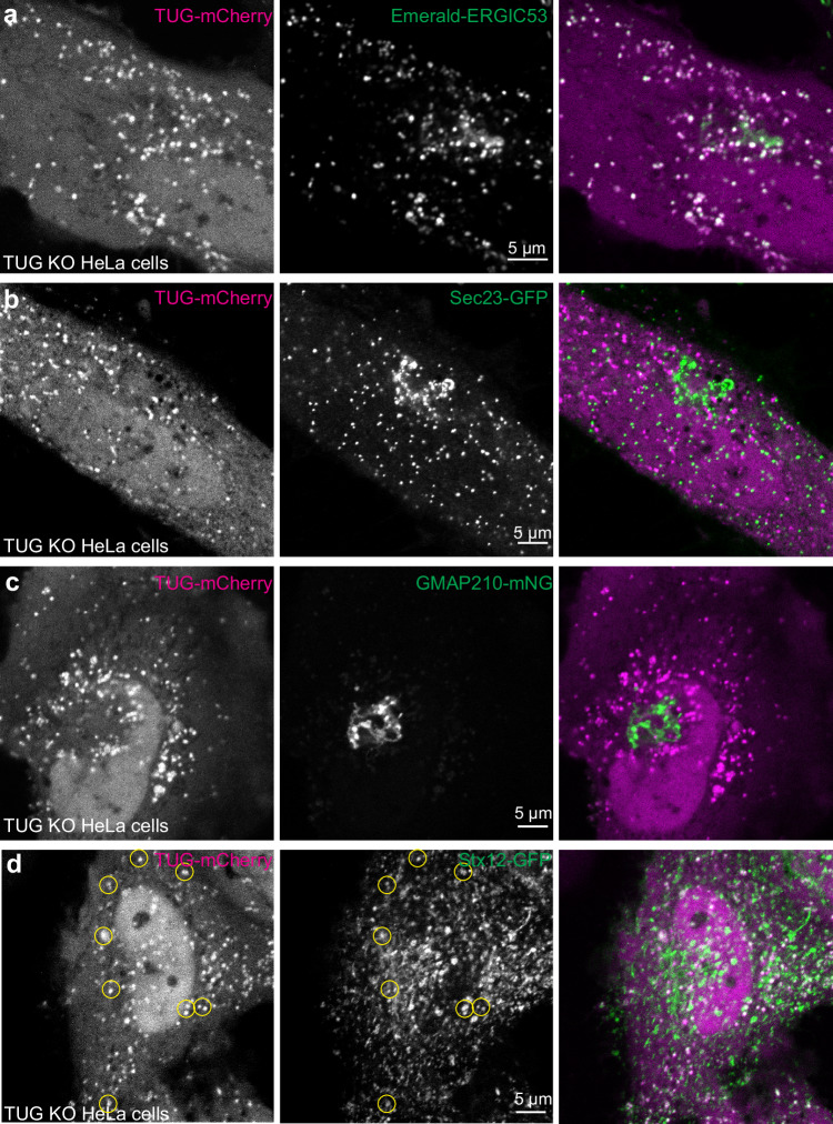
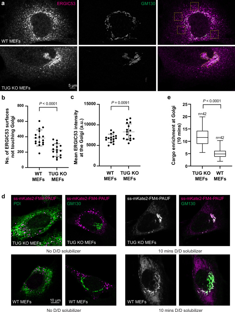
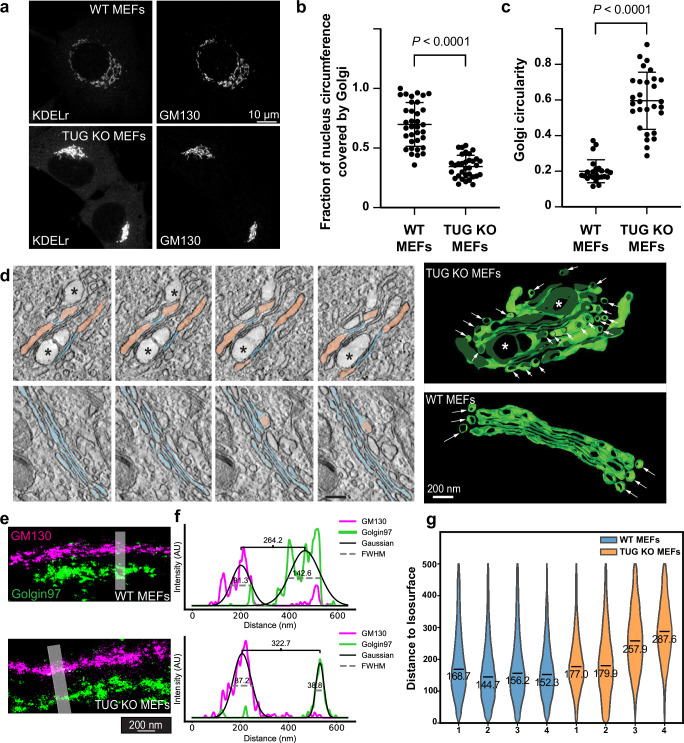
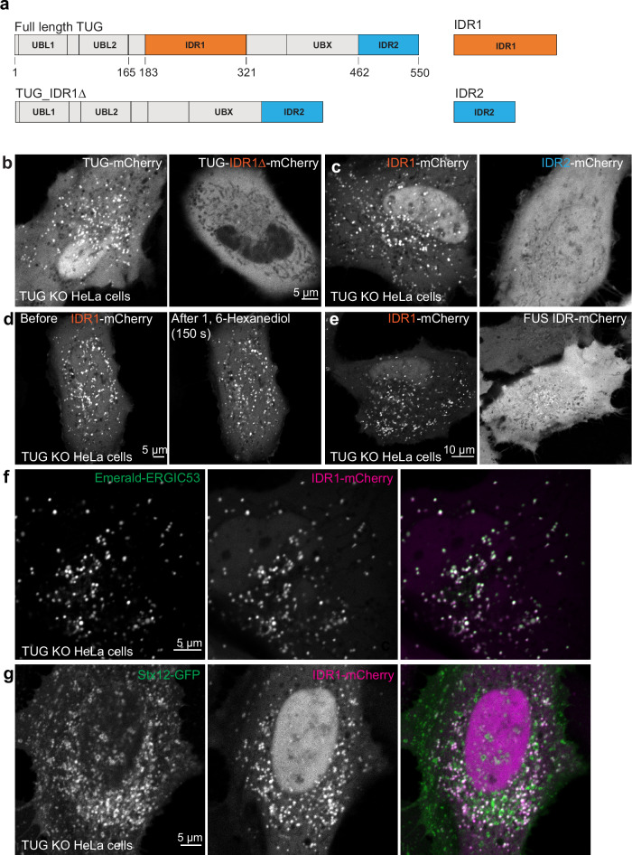
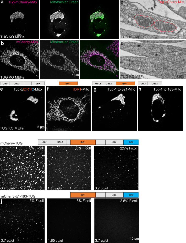
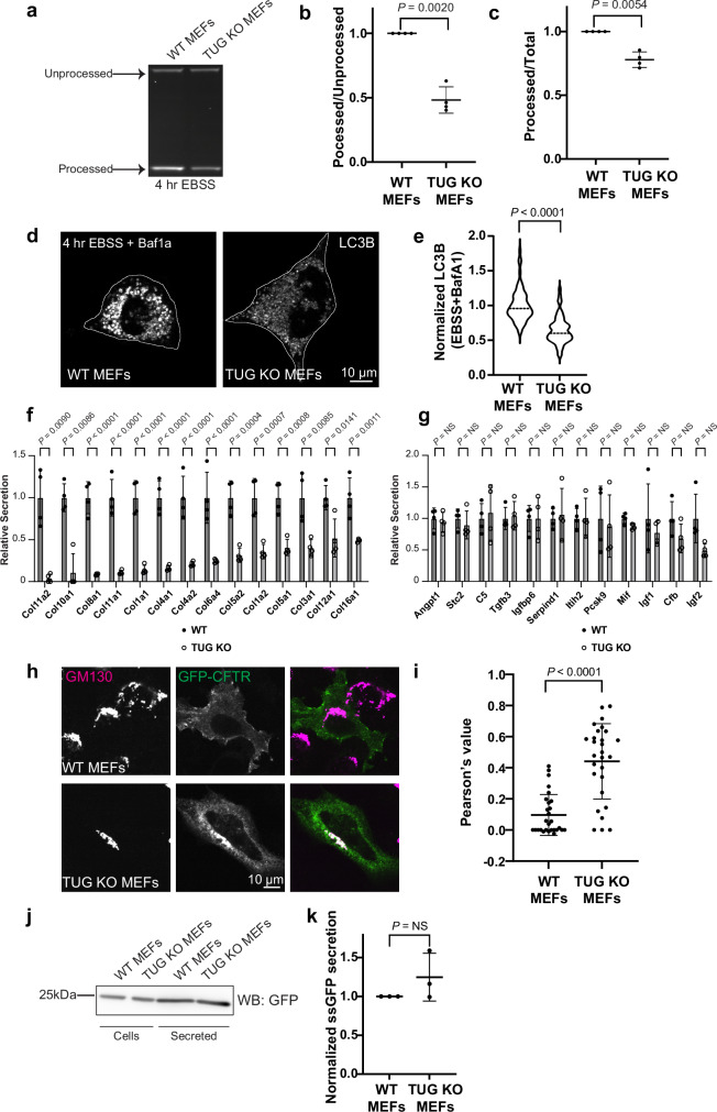
The Endoplasmic Reticulum (ER)-Golgi Intermediate Compartment (ERGIC) is a network of tubules and vesicles known for producing COPI vesicles and receiving COPII vesicles from the ER. Much about its identity, stability, and regulation remains unknown. Here, we show that TUG (UBXN9, Aspscr1) protein, a central regulator of GLUT4 trafficking, localizes to the ERGIC, and that its deletion enhances anterograde flux of a model soluble cargo protein. TUG deletion redistributes ERGIC markers to the cis-Golgi and alters Golgi morphology. TUG forms biomolecular condensates in vitro and contains a central disordered region that mediates its recruitment to ERGIC membranes. A distinct N-terminal region mediates its oligomerization in cells. TUG deletion disrupts ERGIC-dependent processes, including autophagy and collagen secretion, and alters the targeting of the CFTR chloride channel. We conclude that TUG organizes and stabilizes ERGIC membranes to support their roles in diverse secretory and degradative membrane trafficking pathways.
Overview
The claims and statistics of this paper haven’t been checked yet.
Run the check — free, no account needed. It reads the open-access text and checks the sources it cites.
Integrity signals
Registry
Cites
Statistics
Figures
Verification
Claims the paper makes
The claims and statistics of this paper haven’t been checked yet.
Run the check — free, no account needed. It reads the open-access text and checks the sources it cites.
Self-check
Percentages, sums and reported differences re-computed from the paper’s own numbers.
The self-check needs the paper’s full text.
Citations
Cites
Highly accurate protein structure prediction with AlphaFold
Jumper, J., Evans, R., Pritzel, A., Green, T., Figurnov, M., Ronneberger, O., Tunyasuvunakool, K., Bates, R., Žídek, A., Potapenko, A., et al. (2021) · Nature · doi:10.1038/s41586-021-03819-2 · Cited by 43,764
McBride, H. M., Rybin, V., Murphy, C., Giner, A., Teasdale, R., & Zerial, M. (1999) · Cell · doi:10.1016/s0092-8674(00)81966-2 · Cited by 520
Research works printed but not linked: 88
- Barlowe, C. & Helenius, A. Cargo capture and bulk flow in the early secretory pathway. Annu. Rev. Cell Dev. Biol. 32, 197–222 (2016). · DOI
- Appenzeller-Herzog, C. & Hauri, H. P. The ER-Golgi intermediate compartment (ERGIC): in search of its identity and function. J. Cell Sci. 119, 2173–2183 (2006). · DOI
- Saraste, J. & Marie, M. Intermediate compartment (IC): from pre-Golgi vacuoles to a semi-autonomous membrane system. Histochem Cell Biol. 150, 407–430 (2018). · DOI
- Weigel, A. V. et al. ER-to-Golgi protein delivery through an interwoven, tubular network extending from ER. Cell 184, 2412–2429.e2416 (2021). · DOI
- Tojima, T., Suda, Y., Jin, N., Kurokawa, K. & Nakano, A. Spatiotemporal dissection of the Golgi apparatus and the ER-Golgi intermediate compartment in budding yeast. Elife 13 (2024). · DOI
- Han, Y., Li, S. & Ge, L. Biogenesis of autophagosomes from the ERGIC membrane system. J. Genet. Genomics 50, 3–6 (2023). · DOI
- Ge, L., Melville, D., Zhang, M. & Schekman, R. The ER-Golgi intermediate compartment is a key membrane source for the LC3 lipidation step of autophagosome biogenesis. Elife 2, e00947 (2013). · DOI
- Chieregatti, E. & Meldolesi, J. Regulated exocytosis: new organelles for non-secretory purposes. Nat. Rev. Mol. Cell Biol. 6, 181–187 (2005). · DOI
- Grieve, A. G. & Rabouille, C. Golgi bypass: skirting around the heart of classical secretion. Cold Spring Harb. Perspect. Biol. 3 (2011). · DOI
- Gee, H. Y., Noh, S. H., Tang, B. L., Kim, K. H. & Lee, M. G. Rescue of DeltaF508-CFTR trafficking via a GRASP-dependent unconventional secretion pathway. Cell 146, 746–760 (2011). · DOI
- Yoo, J. S. et al. Non-conventional trafficking of the cystic fibrosis transmembrane conductance regulator through the early secretory pathway. J. Biol. Chem. 277, 11401–11409 (2002). · DOI
- Bogan, J. S. Regulation of glucose transporter translocation in health and diabetes. Annu. Rev. Biochem. 81, 507–532 (2012). · DOI · Paper page
- Li, D. T. et al. GLUT4 Storage vesicles: specialized organelles for regulated trafficking. Yale J. Biol. Med. 92, 453–470 (2019).
- Gould, G. W., Brodsky, F. M. & Bryant, N. J. Building GLUT4 vesicles: CHC22 Clathrin’s human touch. Trends Cell Biol. 30, 705–719 (2020). · DOI
- Greig, J. et al. CHC22 clathrin recruitment to the early secretory pathway requires two-site interaction with SNX5 and p115. EMBO J. 43, 4298–4323 (2024). · DOI
- Bogan, J. S., Hendon, N., McKee, A. E., Tsao, T. S. & Lodish, H. F. Functional cloning of TUG as a regulator of GLUT4 glucose transporter trafficking. Nature 425, 727–733 (2003). · DOI
- Yu, C., Cresswell, J., Loffler, M. G. & Bogan, J. S. The glucose transporter 4-regulating protein TUG is essential for highly insulin-responsive glucose uptake in 3T3-L1 adipocytes. J. Biol. Chem. 282, 7710–7722 (2007). · DOI
- Orme, C. M. & Bogan, J. S. The ubiquitin regulatory X (UBX) domain-containing protein TUG regulates the p97 ATPase and resides at the endoplasmic reticulum-golgi intermediate compartment. J. Biol. Chem. 287, 6679–6692 (2012). · DOI
- Bogan, J. S. et al. Endoproteolytic cleavage of TUG protein regulates GLUT4 glucose transporter translocation. J. Biol. Chem. 287, 23932–23947 (2012). · DOI
- Belman, J. P. et al. Acetylation of TUG protein promotes the accumulation of GLUT4 glucose transporters in an insulin-responsive intracellular compartment. J. Biol. Chem. 290, 4447–4463 (2015). · DOI
- Habtemichael, E. N. et al. Usp25m protease regulates ubiquitin-like processing of TUG proteins to control GLUT4 glucose transporter translocation in adipocytes. J. Biol. Chem. 293, 10466–10486 (2018). · DOI
- Bogan, J. S. Ubiquitin-like processing of TUG proteins as a mechanism to regulate glucose uptake and energy metabolism in fat and muscle. Front. Endocrinol. (Lausanne) 13, 1019405 (2022). · DOI
- Habtemichael, E. N. et al. Insulin-stimulated endoproteolytic TUG cleavage links energy expenditure with glucose uptake. Nat. Metab. 3, 378–393 (2021). · DOI
- Camus, S. M. et al. CHC22 clathrin mediates traffic from early secretory compartments for human GLUT4 pathway biogenesis. J Cell Biol, 219 (2020). · DOI
- Xu, Y. et al. Dual-mode of insulin action controls GLUT4 vesicle exocytosis. J. Cell Biol. 193, 643–653 (2011). · DOI
- Ladanyi, M. et al. The der(17)t(X;17)(p11;q25) of human alveolar soft part sarcoma fuses the TFE3 transcription factor gene to ASPL, a novel gene at 17q25. Oncogene 20, 48–57 (2001). · DOI
- Yan, R., Chen, K., Wang, B. & Xu, K. SURF4-induced tubular ERGIC selectively expedites ER-to-Golgi transport. Dev. Cell 57, 512–525.e518 (2022). · DOI
- Larance, M. et al. Characterization of the role of the Rab GTPase-activating protein AS160 in insulin-regulated GLUT4 trafficking. J. Biol. Chem. 280, 37803–37813 (2005). · DOI
- Jedrychowski, M. P. et al. Proteomic analysis of GLUT4 storage vesicles reveals LRP1 to be an important vesicle component and target of insulin signaling. J. Biol. Chem. 285, 104–114 (2010). · DOI
- Song, X., Lichti, C. F., Townsend, R. R. & Mueckler, M. Single point mutations result in the miss-sorting of Glut4 to a novel membrane compartment associated with stress granule proteins. PLoS ONE 8, e68516 (2013). · DOI
- Fazakerley, D. J. et al. Proteomic analysis of GLUT4 storage vesicles reveals tumor suppressor candidate 5 (TUSC5) as a novel regulator of insulin action in adipocytes. J. Biol. Chem. 290, 23528–23542 (2015). · DOI
- Ray, A. et al. Regulated dynamic subcellular GLUT4 localization revealed by proximal proteome mapping in human muscle cells. J. Cell Sci. 136 (2023). · DOI
- Wakana, Y. et al. Kinesin-5/Eg5 is important for transport of CARTS from the trans-Golgi network to the cell surface. J. Cell Biol. 202, 241–250 (2013). · DOI
- Lujan, P. et al. Sorting of secretory proteins at the trans-Golgi network by human TGN46. Elife 12 (2024). · DOI
- Saxena, S. et al. Endoplasmic reticulum exit sites are segregated for secretion based on cargo size. Dev. Cell (2024). · DOI
- Rivera, V. M. et al. Regulation of protein secretion through controlled aggregation in the endoplasmic reticulum. Science 287, 826–830 (2000). · DOI
- Ernst, A. M. et al. S-Palmitoylation sorts membrane cargo for anterograde transport in the golgi. Dev. Cell 47, 479 (2018). · DOI
- Huang, F. et al. Ultra-high resolution 3d imaging of whole cells. Cell 166, 1028–1040 (2016). · DOI
- Xue, B., Dunbrack, R. L., Williams, R. W., Dunker, A. K. & Uversky, V. N. PONDR-FIT: a meta-predictor of intrinsically disordered amino acids. Biochim. Biophys. Acta 1804, 996–1010 (2010). · DOI
- Liang, Q. et al. MolPhase, an advanced prediction algorithm for protein phase separation. EMBO J. 43, 1898–1918 (2024). · DOI
- Park, D. et al. Cooperative function of synaptophysin and synapsin in the generation of synaptic vesicle-like clusters in non-neuronal cells. Nat. Commun. 12, 263 (2021). · DOI
- Kato, M. & McKnight, S. L. A solid-state conceptualization of information transfer from gene to message to protein. Annu Rev. Biochem 87, 351–390 (2018). · DOI
- Lin, Y. et al. Toxic PR poly-dipeptides encoded by the C9orf72 repeat expansion target LC domain polymers. Cell 167, 789–802.e712 (2016). · DOI
- Patel, A. et al. A liquid-to-solid phase transition of the ALS protein FUS accelerated by disease mutation. Cell 162, 1066–1077 (2015). · DOI
- De La Cruz, N. et al. Disorder-mediated interactions target proteins to specific condensates. Mol. Cell 84, 3497–3512 e3499 (2024). · DOI
- Lyons, H. et al. Functional partitioning of transcriptional regulators by patterned charge blocks. Cell 186, 327–345 e328 (2023). · DOI
- Wang, J. et al. A molecular grammar governing the driving forces for phase separation of prion-like RNA binding proteins. Cell 174, 688–699 e616 (2018). · DOI
- Wong, M. & Munro, S. Membrane trafficking. The specificity of vesicle traffic to the Golgi is encoded in the golgin coiled-coil proteins. Science 346, 1256898 (2014). · DOI
- Su, X. et al. Phase separation of signaling molecules promotes T cell receptor signal transduction. Science 352, 595–599 (2016). · DOI
- Yim, W. W., Yamamoto, H. & Mizushima, N. A pulse-chasable reporter processing assay for mammalian autophagic flux with HaloTag. Elife 11 (2022). · DOI
- Aridor, M. A tango for coats and membranes: New insights into ER-to-Golgi traffic. Cell Rep. 38, 110258 (2022). · DOI
- Raote, I. & Malhotra, V. Tunnels for protein export from the endoplasmic reticulum. Annu. Rev. Biochem. 90, 605–630 (2021). · DOI
- Santos, A. J., Raote, I., Scarpa, M., Brouwers, N. & Malhotra, V. TANGO1 recruits ERGIC membranes to the endoplasmic reticulum for procollagen export. Elife 4 (2015). · DOI
- Gee, H. Y., Kim, J. & Lee, M. G. Unconventional secretion of transmembrane proteins. Semin Cell Dev. Biol. 83, 59–66 (2018). · DOI
- Cheng, J. et al. A Golgi-associated PDZ domain protein modulates cystic fibrosis transmembrane regulator plasma membrane expression. J. Biol. Chem. 277, 3520–3529 (2002). · DOI
- Cheng, J., Cebotaru, V., Cebotaru, L. & Guggino, W. B. Syntaxin 6 and CAL mediate the degradation of the cystic fibrosis transmembrane conductance regulator. Mol. Biol. Cell 21, 1178–1187 (2010). · DOI
- Rebane, A. A. et al. Liquid-liquid phase separation of the Golgi matrix protein GM130. FEBS Lett. 594, 1132–1144 (2020). · DOI
- Ziltener, P., Rebane, A. A., Graham, M., Ernst, A. M. & Rothman, J. E. The golgin family exhibits a propensity to form condensates in living cells. FEBS Lett. 594, 3086–3094 (2020). · DOI
- Zhang, Y. & Seemann, J. RNA scaffolds the Golgi ribbon by forming condensates with GM130. Nat. Cell Biol. 26, 1139–1153 (2024). · DOI
- Su, M. et al. The golgi rim is a precise tetraplex of golgin proteins that can self-assemble into filamentous bands. bioRxiv https://doi.org/10.1101/2025.03.27.645134 (2025). · DOI
- Gallo, R., Rai, A. K., McIntyre, A. B. R., Meyer, K. & Pelkmans, L. DYRK3 enables secretory trafficking by maintaining the liquid-like state of ER exit sites. Dev. Cell 58, 1880–1897.e1811 (2023). · DOI
- Wegeng, W. R. et al. A hollow TFG condensate spatially compartmentalizes the early secretory pathway. bioRxiv (2024). · DOI · Paper page
- Hanna, M. G., Peotter, J. L., Frankel, E. B. & Audhya, A. Membrane transport at an organelle interface in the early secretory pathway: take your coat off and stay a while: evolution of the metazoan early secretory pathway. Bioessays 40, e1800004 (2018). · DOI
- Peotter, J. L. et al. TFG regulates secretory and endosomal sorting pathways in neurons to promote their activity and maintenance. Proc. Natl Acad. Sci. USA 119, e2210649119 (2022). · DOI
- Johnson, A. et al. TFG clusters COPII-coated transport carriers and promotes early secretory pathway organization. EMBO J. 34, 811–827 (2015). · DOI
- Loffler, M. G. et al. Enhanced fasting glucose turnover in mice with disrupted action of TUG protein in skeletal muscle. J. Biol. Chem. 288, 20135–20150 (2013). · DOI
- Habtemichael, E. N. et al. Coordinated regulation of vasopressin inactivation and glucose uptake by action of TUG protein in muscle. J. Biol. Chem. 290, 14454–14461 (2015). · DOI
- Semiz, S. et al. Conventional kinesin KIF5B mediates insulin-stimulated GLUT4 movements on microtubules. EMBO J. 22, 2387–2399 (2003). · DOI
- Wang, Y. et al. Trans-Golgi network syntaxin 10 functions distinctly from syntaxins 6 and 16. Mol. Membr. Biol. 22, 313–325 (2005). · DOI
- Nielsen, E. et al. Rabenosyn-5, a novel Rab5 effector, is complexed with hVPS45 and recruited to endosomes through a FYVE finger domain. J. Cell Biol. 151, 601–612 (2000). · DOI
- Koike, S. & Jahn, R. SNAREs define targeting specificity of trafficking vesicles by combinatorial interaction with tethering factors. Nat. Commun. 10, 1608 (2019). · DOI
- Bisio, H. et al. The ZIP code of vesicle trafficking in apicomplexa: SEC1/Munc18 and SNARE proteins. mBio 11, (2020). · DOI
- Milovanovic, D., Wu, Y., Bian, X. & De Camilli, P. A liquid phase of synapsin and lipid vesicles. Science 361, 604–607 (2018). · DOI
- Park, D. et al. Synaptic vesicle proteins and ATG9A self-organize in distinct vesicle phases within synapsin condensates. Nat. Commun. 14, 455 (2023). · DOI
- Park, D. et al. Overlapping role of synaptophysin and synaptogyrin family proteins in determining the small size of synaptic vesicles. Proc. Natl Acad. Sci. USA 121, e2409605121 (2024). · DOI
- Leung, A. K. L. Poly(ADP-ribose): a dynamic trigger for biomolecular condensate formation. Trends Cell Biol. 30, 370–383 (2020). · DOI
- Rhine, K. et al. Poly(ADP-ribose) drives condensation of FUS via a transient interaction. Mol. Cell 82, 969–985 e911 (2022). · DOI
- Hendriks, I. A., Larsen, S. C. & Nielsen, M. L. An advanced strategy for comprehensive profiling of ADP-ribosylation sites using mass spectrometry-based proteomics. Mol. Cell Proteom. 18, 1010–1026 (2019). · DOI
- Liu, X. et al. Generation of mammalian cells stably expressing multiple genes at predetermined levels. Anal. Biochem 280, 20–28 (2000). · DOI
- Parchure, A. et al. Liquid-liquid phase separation facilitates the biogenesis of secretory storage granules. J Cell Biol. 221 (2022). · DOI
- Amick, J., Roczniak-Ferguson, A. & Ferguson, S. M. C9orf72 binds SMCR8, localizes to lysosomes, and regulates mTORC1 signaling. Mol. Biol. Cell 27, 3040–3051 (2016). · DOI
- Devereaux, K. et al. Regulation of mammalian autophagy by class II and III PI 3-kinases through PI3P synthesis. PLoS ONE 8, e76405 (2013). · DOI · Paper page
- Mastronarde, D. N. Correction for non-perpendicularity of beam and tilt axis in tomographic reconstructions with the IMOD package. J. Microsc. 230, 212–217 (2008). · DOI
- Zhang, Y. et al. Nanoscale subcellular architecture revealed by multicolor three-dimensional salvaged fluorescence imaging. Nat. Methods 17, 225–231 (2020). · DOI
- Naviaux, R. K., Costanzi, E., Haas, M. & Verma, I. M. The pCL vector system: rapid production of helper-free, high-titer, recombinant retroviruses. J. Virol. 70, 5701–5705 (1996). · DOI
- Marin, Z. et al. PYMEVisualize: an open-source tool for exploring 3D super-resolution data. Nat. Methods 18, 582–584 (2021). · DOI
- Marin, Z., Fuentes, L. A., Bewersdorf, J. & Baddeley, D. Extracting nanoscale membrane morphology from single-molecule localizations. Biophys. J. 122, 3022–3030 (2023). · DOI
- Cignoni, P. et al. MeshLab: an open-source mesh processing tool. In: Sixth Eurographics Italian Chapter Conference (eds Scarano, V., De Chiara, R. & Erra, U.) 129–136 (The Eurographics Association, 2008).
Metadata
- Authors
- Parchure, A., Tejada, H., Xi, Z., Kim, Y., Su, M., Yan, Y., Julca-Zevallos, O., Alcázar-Román, A. R., Villemeur, M., Liu, X., Toomre, D., Raote, I., & Bogan, J. S.
- Affiliations
- Yale University (US)
- Centre National de la Recherche Scientifique (FR)
- Corresponding author
- Parchure, Anup
- Bogan, Jonathan S.
- ORCID
- Venue
- Nature Communications
- Published
- 2025-07-01
- Volume
- 16
- Issue
- 1
- Pages
- 5518-5518
- ISSN
- 2041-1723 (ISSN-L)
- PMC
- PMC12218103
- OpenAlex
- W4411886980
- Cited by
- 5
- References
- 2
- Citations by year
- 2026: 3 · 2025: 1
- FWCI
- 1.99
- Type
- journal article
- Language
- en
- Open access
- gold
- doi.org
- Locations
- Nature Communications (publishedVersion, cc-by)
- bioRxiv (Cold Spring Harbor Laboratory) (acceptedVersion, cc-by-nc-nd)
- PubMed Central (publishedVersion, cc-by)
- PubMed (publishedVersion)
- HAL (submittedVersion, cc-by)
- DOAJ (DOAJ: Directory of Open Access Journals) (submittedVersion, cc-by-sa)
- Funders
- U.S. Department of Health & Human Services | NIH | National Institute of Diabetes and Digestive and Kidney Diseases (R01 DK129466, P30 DK045735)
- Blavatnik Family Foundation
- U.S. Department of Health & Human Services | NIH | National Institute of General Medical Sciences (R01 GM151829, R01 GM134148)
- Fondation pour la Recherche Médicale (AJE202210016216)
- Agence Nationale de la Recherche (MatSec)
- Fondation ARC pour la Recherche sur le Cancer
- Topics
- Life Sciences › Biochemistry, Genetics and Molecular Biology › Cell Biology › Cellular transport and secretion
- Life Sciences › Biochemistry, Genetics and Molecular Biology › Cell Biology › Autophagy in Disease and Therapy
- Life Sciences › Biochemistry, Genetics and Molecular Biology › Cell Biology › Endoplasmic Reticulum Stress and Disease
- Keywords
- ASPSCR1, ERGIC, early secretory pathway, intrinsically disordered regions, biomolecular condensates, protein oligomerization, Golgi morphology, cis-Golgi, autophagy, collagen secretion, GLUT4 trafficking, membrane trafficking
- MeSH terms
- Animals, Autophagy, Collagen, Endoplasmic Reticulum, Golgi Apparatus, Humans, Cystic Fibrosis Transmembrane Conductance Regulator, Protein Transport, COP-Coated Vesicles, Mice, Secretory Pathway
About these checks
Registry, retraction, Cites and statistics results are computed, not judged, with one exception: a printed reference that no index lists is read once by a model before it counts as a citation error. Claims are extracted by a model and kept only when their quote appears verbatim in the text; each cited source is then checked, with a model’s help, for whether it says what the claim relies on. Those verdicts can be wrong — read the claim’s quote and the cited source before relying on one.
Statistics and arithmetic are recomputed, not judged.
Confidence bands are shown only for fields calibrated on our annotated test set. None is calibrated yet, so no band is shown.
What counts as a claim ›How we measure confidence ›How we check this ›
Are you an author, or do you see a problem with this page? Request a review
How to cite
Cite CoChat for the checks on this page, and the paper for its own findings.
- This page
- CoChat. (2026). Citation check: TUG protein acts through a disordered region to organize the early secretory pathway. https://app.cochat.ai/papers/10.1038/s41467-025-60691-8
- The paper
- Parchure, A., Tejada, H., Xi, Z., Kim, Y., Su, M., Yan, Y., Julca-Zevallos, O., Alcázar-Román, A. R., Villemeur, M., Liu, X., Toomre, D., Raote, I., & Bogan, J. S. (2025). TUG protein acts through a disordered region to organize the early secretory pathway. Nature Communications. https://doi.org/10.1038/s41467-025-60691-8
