

The DOI matches its Crossref record; 1 of the 1 references it cites were found in a registry or an index.
Cryo-EM captures early intermediate steps in dynein activation by LIS1
Open accessPublished version of a preprint
Abstract
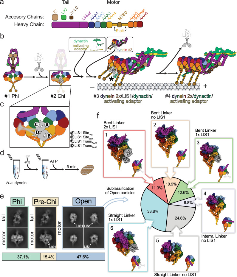
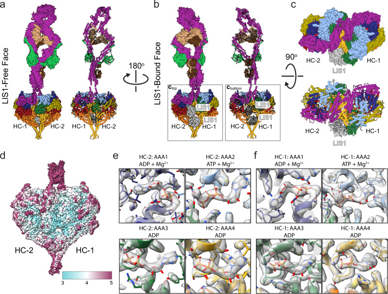
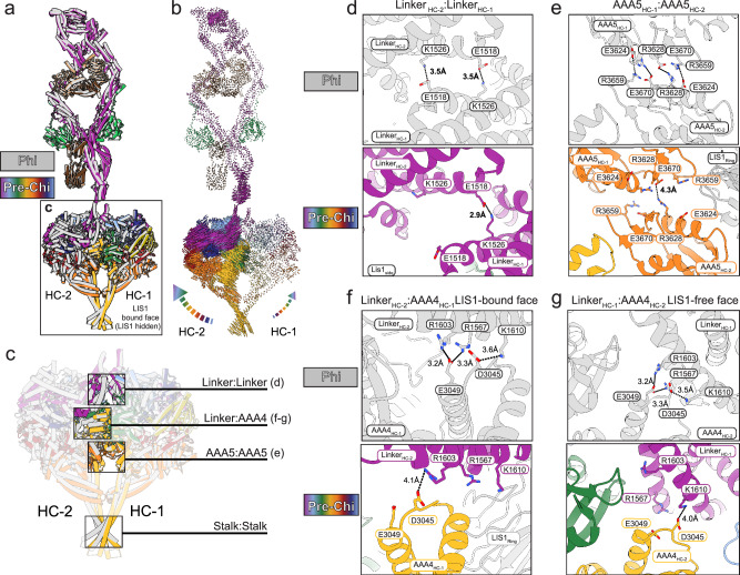
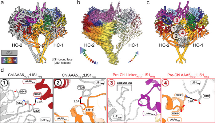
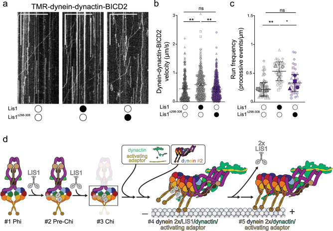
Cytoplasmic dynein-1 (dynein) is an essential molecular motor in eukaryotic cells. Dynein primarily exists in an autoinhibited Phi state and requires conformational changes to assemble with its cofactors and form active transport complexes. LIS1, a key dynein regulator, enhances dynein activation and assembly. Using cryo-EM and a human dynein-LIS1 sample incubated with ATP, we map the conformational landscape of dynein activation by LIS1 and identify an early intermediate state that we propose precedes the previously identified dynein-LIS1 Chi state. Mutations that disrupt this species, which we termed “Pre-Chi”, lead to motility defects in vitro, emphasizing its functional importance. Together, our findings provide insights into how LIS1 relieves dynein autoinhibition during the activation pathway.
Overview
The claims and statistics of this paper haven’t been checked yet.
Run the check — free, no account needed. It reads the open-access text and checks the sources it cites.
Integrity signals
Registry
Cites
Statistics
Figures
Verification
Claims the paper makes
The claims and statistics of this paper haven’t been checked yet.
Run the check — free, no account needed. It reads the open-access text and checks the sources it cites.
Self-check
Percentages, sums and reported differences re-computed from the paper’s own numbers.
The self-check needs the paper’s full text.
Citations
Cites
Highly accurate protein structure prediction with AlphaFold
Jumper, J., Evans, R., Pritzel, A., Green, T., Figurnov, M., Ronneberger, O., Tunyasuvunakool, K., Bates, R., Žídek, A., Potapenko, A., et al. (2021) · Nature · doi:10.1038/s41586-021-03819-2 · Cited by 43,764
Research works printed but not linked: 59
- Reck-Peterson, S. L., Redwine, W. B., Vale, R. D. & Carter, A. P. The cytoplasmic dynein transport machinery and its many cargoes. Nat. Rev. Mol. Cell Biol. 19, 382–398 (2018). · DOI
- Lipka, J., Kuijpers, M., Jaworski, J. & Hoogenraad, C. C. Mutations in cytoplasmic dynein and its regulators cause malformations of cortical development and neurodegenerative diseases. Biochem. Soc. Trans. 41, 1605–1612 (2013). · DOI
- Singh, K. et al. Molecular mechanism of dynein-dynactin complex assembly by LIS1. Science 383, eadk8544 (2024). · DOI
- Carter, A. P., Diamant, A. G. & Urnavicius, L. How dynein and dynactin transport cargos: a structural perspective. Curr. Opin. Struct. Biol. 37, 62–70 (2016). · DOI
- Canty, J. T., Tan, R., Kusakci, E., Fernandes, J. & Yildiz, A. Structure and mechanics of dynein motors. Annu. Rev. Biophys. 50, 549–574 (2021). · DOI
- Cianfrocco, M. A., DeSantis, M. E., Leschziner, A. E. & Reck-Peterson, S. L. Mechanism and regulation of cytoplasmic dynein. Annu. Rev. Cell Dev. Biol. 31, 83–108 (2015). · DOI
- Gibbons, I. R., Gibbons, B. H. & Mocz, G. Multiple nucleotide-binding sites in the sequence of dynein. 352, https://doi.org/10.1038/352640a0 (1991). · DOI
- Kon, T., Mogami, T., Ohkura, R., Nishiura, M. & Sutoh, K. ATP hydrolysis cycle–dependent tail motions in cytoplasmic dynein. Nat. Struct. Mol. Biol. 12, 513–519 (2005). · DOI
- Kon, T. et al. The 2.8 Å crystal structure of the dynein motor domain. Nature 484, 345–350 (2012). · DOI
- Cho, C., Reck-Peterson, S. L. & Vale, R. D. Regulatory ATPase sites of cytoplasmic dynein affect processivity and force generation. J. Biol. Chem. 283, 25839–25845 (2008). · DOI
- Schmidt, H., Gleave, E. S. & Carter, A. P. Insights into dynein motor domain function from a 3.3-Å crystal structure. Nat. Struct. Mol. Biol. 19, 492–497 (2012). · DOI
- Schmidt, H. & Carter, A. P. Review: Structure and mechanism of the dynein motor ATPase. Biopolymers 105, 557–567 (2016). · DOI
- Bhabha, G. et al. Allosteric communication in the dynein motor domain. Cell 159, 857–868 (2014). · DOI
- DeWitt, M. A., Cypranowska, C. A., Cleary, F. B., Belyy, V. & Yildiz, A. The AAA3 domain of cytoplasmic dynein acts as a switch to facilitate microtubule release. Nat. Struct. Mol. Biol. 22, 73–80 (2015). · DOI
- Amos, L. A. Brain dynein crossbridges microtubules into bundles. J. Cell Sci. 93, 19–28 (1989). · DOI
- Torisawa, T. et al. Autoinhibition and cooperative activation mechanisms of cytoplasmic dynein. Nat. Cell Biol. 16, 1118–1124 (2014). · DOI
- Zhang, K. et al. Cryo-EM reveals how human cytoplasmic dynein is auto-inhibited and activated. Cell 169, 1303–1314 (2017). · DOI
- Olenick, M. A. & Holzbaur, E. L. F. Dynein activators and adaptors at a glance. J. Cell Sci. 132, jcs227132 (2019). · DOI
- Xiang, X., Osmani, A. H., Osmani, S. A., Xin, M. & Morris, N. R. NudF, a nuclear migration gene in Aspergillus nidulans, is similar to the human LIS-1 gene required for neuronal migration. MBoC 6, 297–310 (1995). · DOI
- Markus, S. M., Marzo, M. G. & McKenney, R. J. New insights into the mechanism of dynein motor regulation by lissencephaly-1. ELife 9, e59737 (2020). · DOI
- Parrini, E., Conti, V., Dobyns, W. B. & Guerrini, R. Genetic basis of brain malformations. Mol. Syndromol. 7, 220–233 (2016). · DOI
- Kim, M. H. et al. The structure of the N-terminal domain of the product of the lissencephaly gene Lis1 and its functional implications. Structure 12, 987–998 (2004). · DOI
- Tarricone, C. et al. Coupling PAF signaling to dynein regulation: Structure of LIS1 in complex with PAF-acetylhydrolase. Neuron 44, 809–821 (2004).
- Huang, J., Roberts, A. J., Leschziner, A. E. & Reck-Peterson, S. L. Lis1 Acts as a “Clutch” between the ATPase and microtubule-binding domains of the dynein motor. Cell 150, 975–986 (2012). · DOI
- Kormanec, J., Schaaff-Gerstenschläger, I., Zimmermann, F. K., Perečko, D. & Küntzel, H. Nuclear migration in Saccbaromyces cerevisiae is controlled by the highly repetitive 313 kDa NUM1 protein. Mol. Gen. Genet. 230, 277–787 (1991). · DOI
- Elshenawy, M. M. et al. Lis1 activates dynein motility by modulating its pairing with dynactin. Nat. Cell Biol. 22, 570–578 (2020). · DOI
- Gillies, J. P. et al. Structural basis for cytoplasmic dynein-1 regulation by Lis1. ELife 11, e71229 (2022). · DOI
- Htet, Z. M. et al. LIS1 promotes the formation of activated cytoplasmic dynein-1 complexes. Nat. Cell Biol. 22, 518–525 (2020). · DOI
- Ton, W. D. et al. Microtubule-binding-induced allostery triggers LIS1 dissociation from dynein prior to cargo transport. Nat. Struct. Mol. Biol. https://doi.org/10.1038/s41594-023-01010-x (2023). · DOI
- Marzo, M. G., Griswold, J. M. & Markus, S. M. Pac1/LIS1 stabilizes an uninhibited conformation of dynein to coordinate its localization and activity. Nat. Cell Biol. 22, 559–569 (2020). · DOI
- Qiu, R., Zhang, J. & Xiang, X. LIS1 regulates cargo-adapter–mediated activation of dynein by overcoming its autoinhibition in vivo. J. Cell Biol. 218, 3630–3646 (2019). · DOI
- Qiu, R., Zhang, J., Rotty, J. D. & Xiang, X. Dynein activation in vivo is regulated by the nucleotide states of its AAA3 domain. Curr. Biol. 31, 4486–4498 (2021). · DOI
- Karasmanis, E. P. et al. Lis1 relieves cytoplasmic dynein-1 autoinhibition by acting as a molecular wedge. Nat. Struct. Mol. Biol. 30, 1357–1364 (2023). · DOI
- Toropova, K. et al. Lis1 regulates dynein by sterically blocking its mechanochemical cycle. ELife 3, e03372 (2014). · DOI
- DeSantis, M. E. et al. Lis1 has two opposing modes of regulating cytoplasmic dynein. Cell 170, 1197–1208 (2017). · DOI
- Reimer, J. M., DeSantis, M. E., Reck-Peterson, S. L. & Leschziner, A. E. Structures of human dynein in complex with the lissencephaly 1 protein, LIS1. ELife 12, e84302 (2023). · DOI · Paper page
- Kendrick, A. A. et al. Multiple steps of dynein activation by Lis1 visualized by cryo-EM. Nat. Struct. Mol. Biol. https://doi.org/10.1038/s41594-025-01558-w (2025). · DOI
- Geohring, I. C. et al. A nucleotide code governs Lis1’s ability to relieve dynein autoinhibition. Preprint at https://doi.org/10.1101/2024.12.30.630615 (2025). · DOI
- Punjani, A., Rubinstein, J. L., Fleet, D. J. & Brubaker, M. A. cryoSPARC: algorithms for rapid unsupervised cryo-EM structure determination. Nat. Methods 14, 290–296 (2017). · DOI · Paper page
- Zheng, S. Q. et al. MotionCor2: anisotropic correction of beam-induced motion for improved cryo-electron microscopy. Nat. Methods 14, 331–332 (2017). · DOI
- Rohou, A. & Grigorieff, N. CTFFIND4: Fast and accurate defocus estimation from electron micrographs. J. Struct. Biol. 192, 216–221 (2015). · DOI
- Bepler, T., Kelley, K., Noble, A. J. & Berger, B. Topaz-Denoise: general deep denoising models for cryoEM and cryoET. Nat. Commun. 11, 5208 (2020). · DOI
- Zhong, E. D., Bepler, T., Berger, B. & Davis, J. H. CryoDRGN: reconstruction of heterogeneous cryo-EM structures using neural networks. Nat. Methods 18, 176–185 (2021). · DOI
- Kinman, L. F., Powell, B. M., Zhong, E. D., Berger, B. & Davis, J. H. Uncovering structural ensembles from single-particle cryo-EM data using cryoDRGN. Nat. Protoc. 18, 319–339 (2023). · DOI
- Scheres, S. H. W. RELION: Implementation of a Bayesian approach to cryo-EM structure determination. J. Struct. Biol. 180, 519–530 (2012). · DOI · Paper page
- Chai, P. et al. The mechanochemical cycle of reactive full-length human dynein 1. Nat. Struct. Mol. Biol. https://doi.org/10.1038/s41594-025-01543-3 (2025). · DOI
- Simoes, C. et al. Novel frameshift mutation in LIS1 gene is a probable cause of lissencephaly: a case report. BMC Pediatr. 22, 545 (2022). · DOI
- Wang, S. & Zheng, Y. Identification of a novel dynein binding domain in nudel essential for spindle pole organization in Xenopus egg extract. J. Biol. Chem. 286, 587–593 (2011). · DOI
- Zhao, Y., Oten, S. & Yildiz, A. Nde1 promotes Lis1-mediated activation of dynein. Nat. Commun. 14, 7221 (2023). · DOI
- Moon, H. M. et al. LIS1 controls mitosis and mitotic spindle organization via the LIS1–NDEL1–dynein complex. Hum. Mol. Genet. 23, 449–466 (2014). · DOI
- Zyłkiewicz, E. et al. The N-terminal coiled-coil of Ndel1 is a regulated scaffold that recruits LIS1 to dynein. J. Cell Biol. 192, 433–445 (2011). · DOI
- Schlager, M. A., Hoang, H. T., Urnavicius, L., Bullock, S. L. & Carter, A. P. In vitro reconstitution of a highly processive recombinant human dynein complex. EMBO J. 33, 1855–1868 (2014). · DOI
- Baumbach, J. et al. Lissencephaly-1 is a context-dependent regulator of the human dynein complex. ELife 6, e21768 (2017). · DOI
- Nguyen, H. P. M., McGuire, K. L., Cook, B. D. & Mark A. Herzik, J. Manual blot-and-plunge freezing of biological specimens for single-particle cryogenic electron microscopy. J. Vis. Exp. e62765 https://doi.org/10.3791/62765 (2022). · DOI
- Pettersen, E. F. et al. UCSF ChimeraX: Structure visualization for researchers, educators, and developers. Protein Sci. 30, 70–82 (2021). · DOI
- Afonine, P. V. et al. Real-space refinement in PHENIX for cryo-EM and crystallography. Acta Cryst. D. 74, 531–544 (2018). · DOI
- Emsley, P., Lohkamp, B., Scott, W. G. & Cowtan, K. Features and development of Coot. Acta Crystallogr. D. Biol. Crystallogr. 66, 486–501 (2010). · DOI · Paper page
- Mirdita, M. et al. ColabFold: making protein folding accessible to all. Nat. Methods 19, 679–682 (2022). · DOI · Paper page
- Roberts, A. J., Goodman, B. S. & Reck-Peterson, S. L. Reconstitution of dynein transport to the microtubule plus end by kinesin. ELife 3, e02641 (2014). · DOI
Metadata
- Authors
- Nguyen, K. H. V., Karasmanis, E. P., Kendrick, A. A., Reck-Peterson, S. L., & Leschziner, A. E.
- Affiliations
- University of California San Diego (US)
- Salk Institute for Biological Studies (US)
- ORCID
- Venue
- Nature Communications
- Published
- 2025-08-01
- Volume
- 16
- Issue
- 1
- Pages
- 7054-7054
- ISSN
- 2041-1723 (ISSN-L)
- PMC
- PMC12317079
- OpenAlex
- W4412818674
- Cited by
- 5
- References
- 1
- Citations by year
- 2026: 5
- FWCI
- 2.48
- Type
- journal article
- Language
- en
- Open access
- gold
- doi.org
- Locations
- Nature Communications (publishedVersion, cc-by)
- bioRxiv (Cold Spring Harbor Laboratory) (acceptedVersion, cc-by-nc-nd)
- PubMed Central (publishedVersion, cc-by)
- PubMed (publishedVersion)
- DOAJ (DOAJ: Directory of Open Access Journals) (submittedVersion, cc-by-sa)
- Funders
- U.S. Department of Health & Human Services | NIH | National Institute of General Medical Sciences (R35 GM145296, T32 GM008326, R35 GM141825)
- Jane Coffin Childs Memorial Fund for Medical Research
- American Cancer Society
- Howard Hughes Medical Institute
- Topics
- Life Sciences › Biochemistry, Genetics and Molecular Biology › Cell Biology › Microtubule and mitosis dynamics
- Life Sciences › Biochemistry, Genetics and Molecular Biology › Genetics › Genetic and Kidney Cyst Diseases
- Life Sciences › Biochemistry, Genetics and Molecular Biology › Molecular Biology › Protein Structure and Dynamics
- Keywords
- cytoplasmic dynein-1, LIS1, dynein activation, molecular motors, motility defects
- MeSH terms
- Adenosine Triphosphate, Dyneins, Humans, Microtubule-Associated Proteins, Models, Molecular, Mutation, Protein Conformation, Cryoelectron Microscopy, 1-Alkyl-2-acetylglycerophosphocholine Esterase, Cytoplasmic Dyneins
About these checks
Registry, retraction, Cites and statistics results are computed, not judged, with one exception: a printed reference that no index lists is read once by a model before it counts as a citation error. Claims are extracted by a model and kept only when their quote appears verbatim in the text; each cited source is then checked, with a model’s help, for whether it says what the claim relies on. Those verdicts can be wrong — read the claim’s quote and the cited source before relying on one.
Statistics and arithmetic are recomputed, not judged.
Confidence bands are shown only for fields calibrated on our annotated test set. None is calibrated yet, so no band is shown.
What counts as a claim ›How we measure confidence ›How we check this ›
Are you an author, or do you see a problem with this page? Request a review
