

The DOI matches its Crossref record; 107 of the 107 references it cites were found in a registry or an index; 44 of the 54 sources its claims cite were checked for support.
Lymphoid gene expression supports neuroprotective microglia function
Open accessAbstract
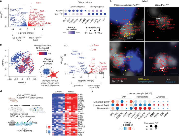
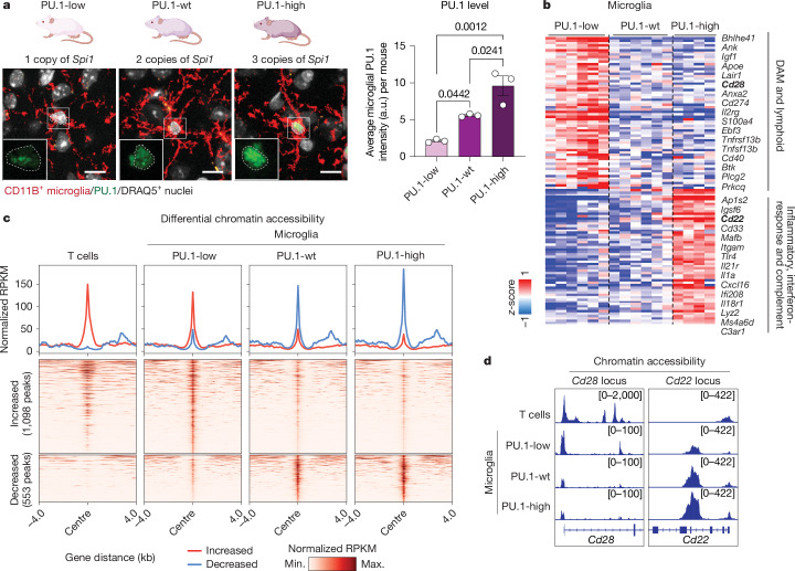
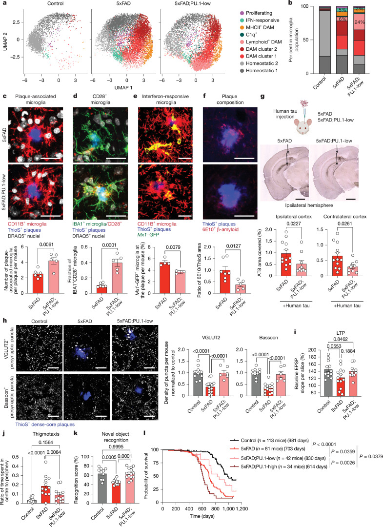
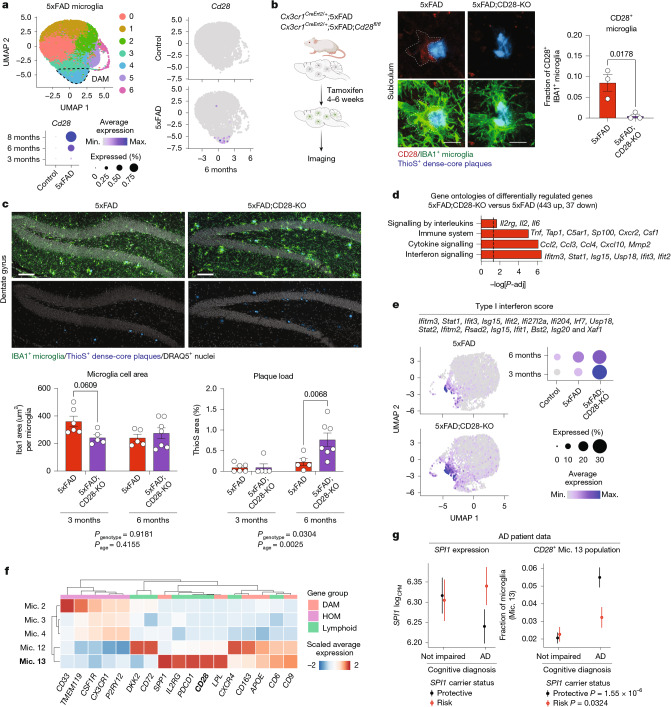
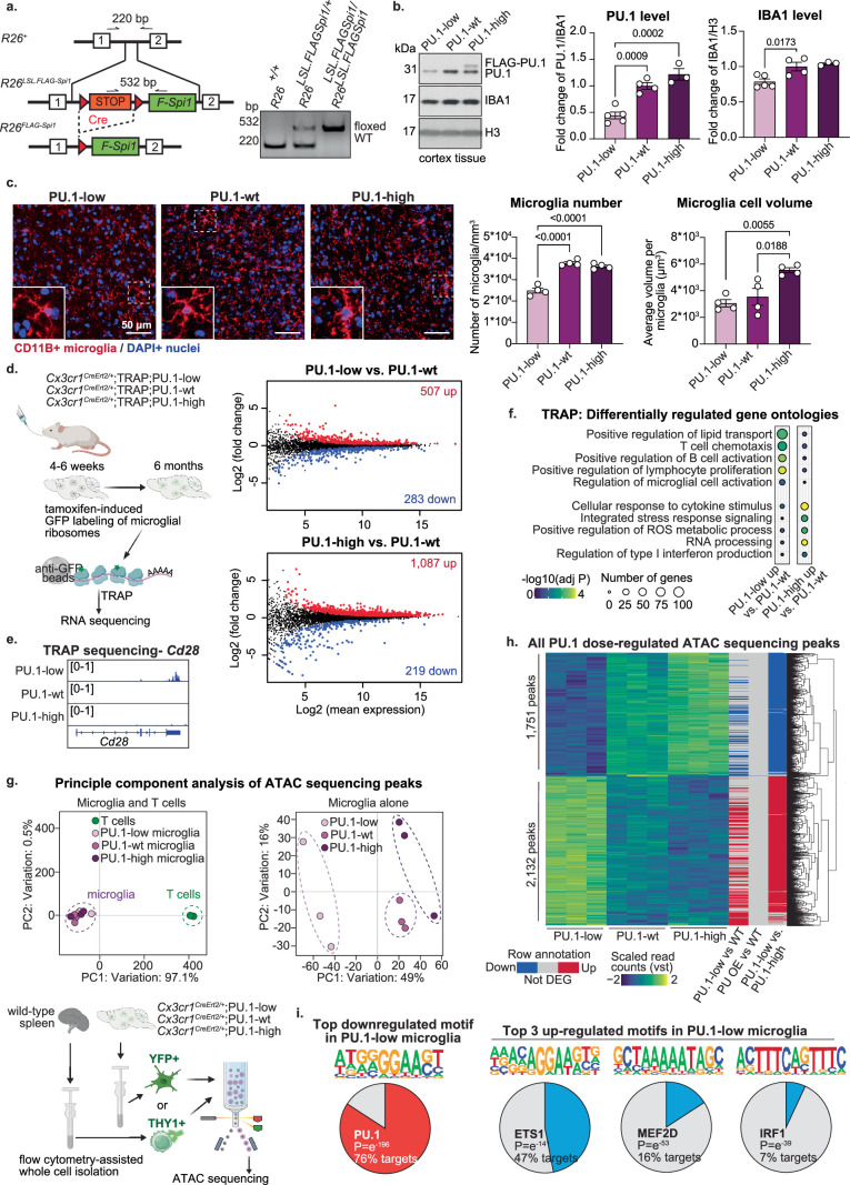
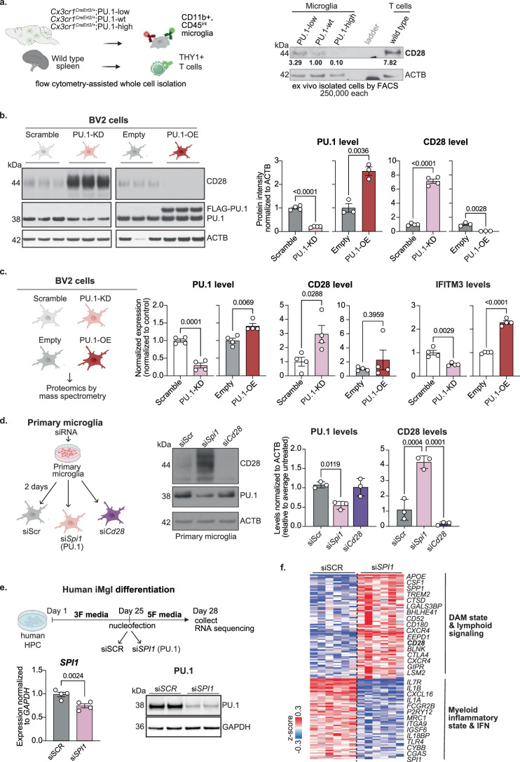
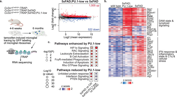
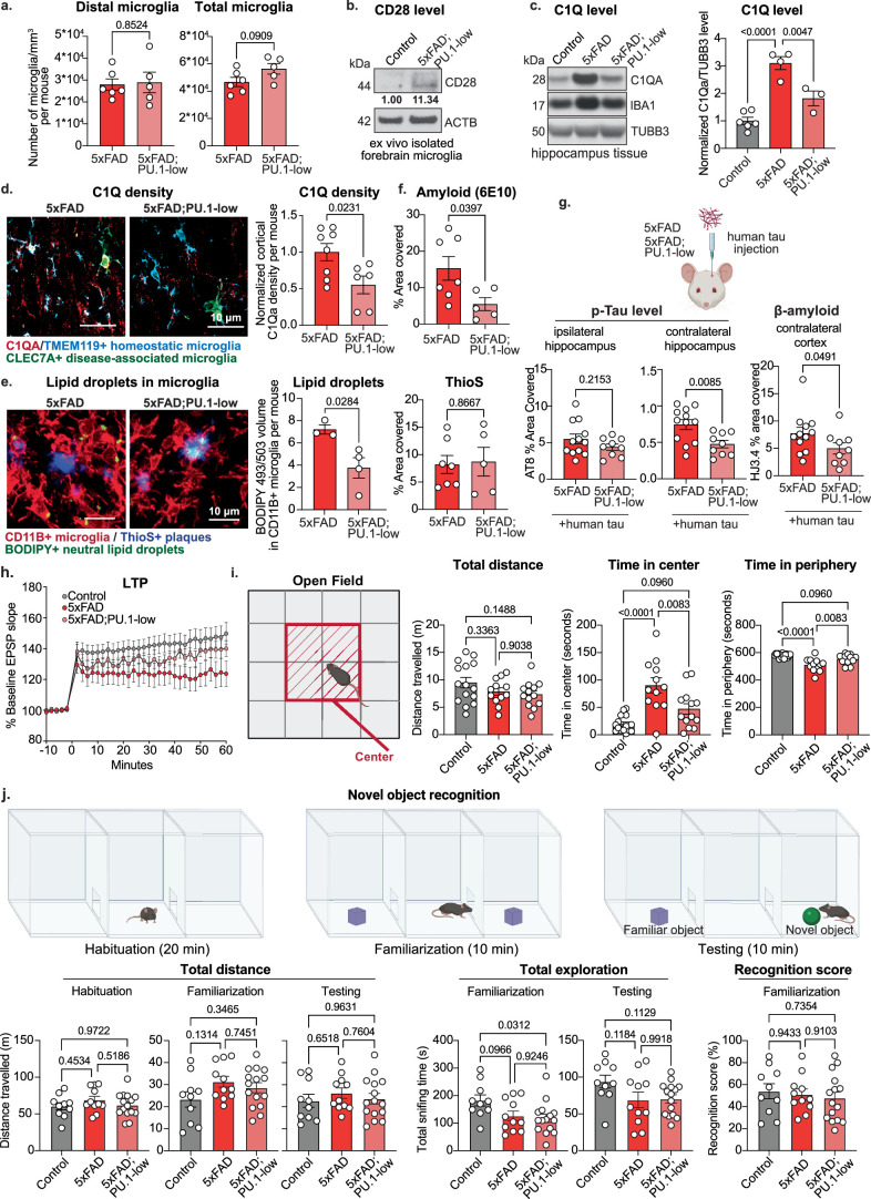
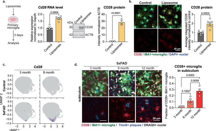
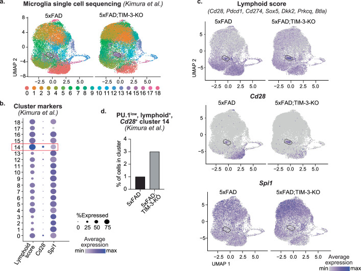
Abstract Microglia, the innate immune cells of the brain, play a defining role in the progression of Alzheimer’s disease (AD) 1 . The microglial response to amyloid plaques in AD can range from neuroprotective to neurotoxic 2 . Here we show that the protective function of microglia is governed by the transcription factor PU.1, which becomes downregulated following microglial contact with plaques. Lowering PU.1 expression in microglia reduces the severity of amyloid disease pathology in mice and is linked to the expression of immunoregulatory lymphoid receptor proteins, particularly CD28, a surface receptor that is critical for T cell activation 3,4 . Microglia-specific deficiency in CD28, which is expressed by a small subset of plaque-associated PU.1 low microglia, promotes a broad inflammatory microglial state that is associated with increased amyloid plaque load. Our findings indicate that PU.1 low CD28-expressing microglia may operate as suppressive microglia that mitigate the progression of AD by reducing the severity of neuroinflammation. This role of CD28 and potentially other lymphoid co-stimulatory and co-inhibitory receptor proteins in governing microglial responses in AD points to possible immunotherapy approaches for treating the disease by promoting protective microglial functions.
Overview
CoChat Summary
## TL;DR
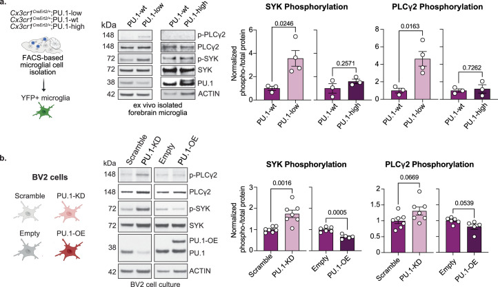
The paper argues that a plaque-associated PU.1-low microglial state is neuroprotective in Alzheimer’s disease and is linked to expression of lymphoid receptor genes, especially CD28. It shows that plaque contact downregulates PU.1 through SYK–PLCγ2-dependent signaling, and that disrupting CD28 in microglia shifts them toward inflammation and higher plaque burden.
## Key findings
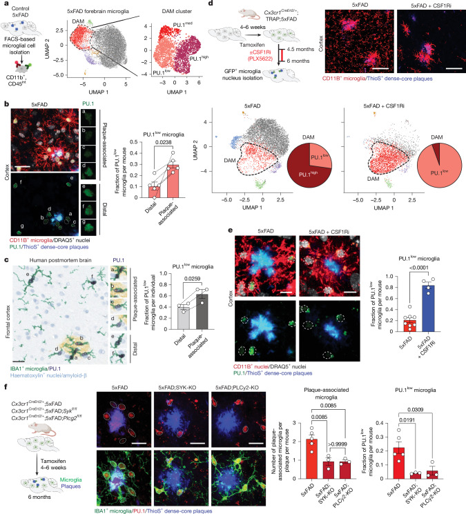
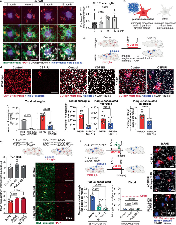
- In the 5xFAD mouse model and in human AD tissue, a distinct PU.1-low microglial subpopulation preferentially co-localized with amyloid plaques and increased with disease progression.
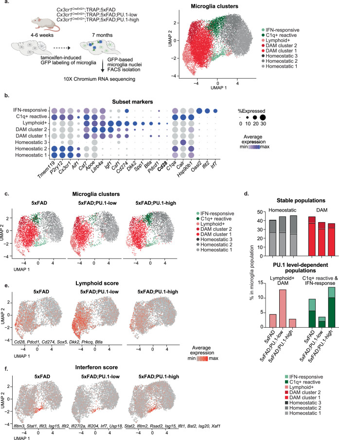
- Lowering PU.1 in microglia reduced the severity of amyloid disease pathology in mice; the paper also reports that microglia-specific deficiency in CD28 promoted a broad inflammatory microglial state associated with increased amyloid plaque load.
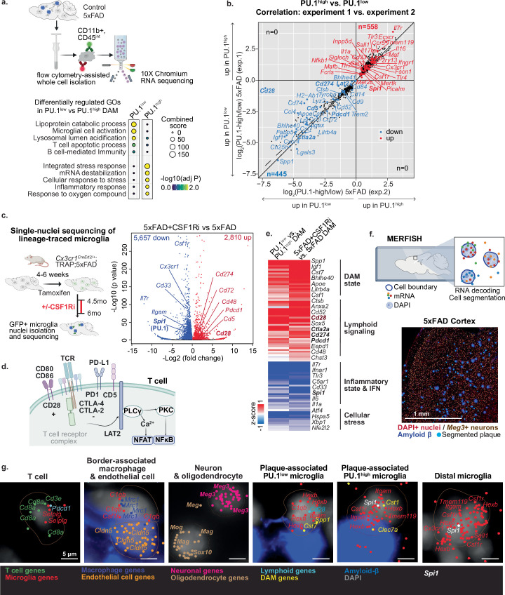
- Plaque-associated PU.1-low microglia expressed immunoregulatory lymphoid receptor and signaling genes, including CD28, Pdcd1, Cd274, Cd5, Ctla2a, Cd52, Lat2, and Cd72, and these transcripts were also found in microglial ribosome-associated RNA.
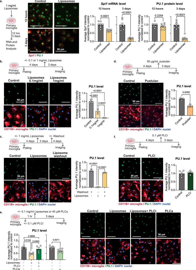
- Ex vivo ligands that engage TREM2 and CLEC7A induced PU.1 downregulation, and this effect was attenuated by phospholipase C inhibition; PLC activation alone was sufficient to downregulate PU.1.
- Microglia-specific inactivation of SYK or PLCγ2 reduced the number of PU.1-low microglia and prevented their CSF1R-independent survival at plaques.
## Methods
- Multi-modal design combining human post-mortem brain immunohistochemistry, mouse genetics in the 5xFAD AD model, ex vivo primary microglia culture, in vitro perturbations, and transcriptomic/epigenomic profiling.
- Mouse cohorts included microglia-specific Spi1/PU.1 manipulation, Cd28, Syk, and Plcg2 conditional knockouts, plus 5xFAD crosses; human tissue included 3 post-mortem frontal cortex AD samples for immunohistochemistry.
- Main analyses included single-cell and single-nucleus RNA-seq, TRAP sequencing, MERFISH spatial transcriptomics, ATAC-seq, proteomics, western blotting, and behavioral/survival assays, with standard group comparisons and clustering/differential-expression pipelines described in Methods.
## Limitations
- The paper states that it did not recruit participants and used only 3 post-mortem human brain samples for immunohistochemistry; broader human validation is therefore limited by sample availability.
- It states that sample size justification was not provided; instead, sample sizes were chosen pragmatically based on expected effect size, prior literature, and practical constraints.
- Evident from the text: several key human and mouse analyses rely on de-identified post-mortem tissue and model systems, so causality in human AD is not directly established by the human data alone.
Model-written summary of a shortened version of the paper’s full text (it is longer than we read). Check it against the paper before relying on it.
Integrity signals
Registry
Cites
Statistics
Figures

Verification
Claims the paper makes
25 Valid · 2 Not valid · 1 Not checked · 88 Own results and interpretations
At least one cited source doesn’t support this claim or points to a different paper, and none confirmed it — see the verdicts below.
In the paper
“which serves as a positive indicator of Spi1 downregulation.”
§ Lymphoid gene expression supports neuroprotective microglia function > Methods > Bioinformatics analysis of single-cell and single-nucleus sequencing
What the sources say
- Disputedcites [24] Huang, K. L. et al. A common haplotype lowers PU.1 expression in myeloid cells and delays onset of Alzheimer’s disease. Nat. Neurosci. 20, 1052–1061 (2017). sourceExperimental — accuracy not yet measured
The Results section on cis-eQTL associations reports a positive correlation between MYBPC3 and SPI1 expression in monocytes and macrophages. It also reports that the same allele is associated with lower expression of both genes. Thus, the paper does not support MYBPC3 upregulation as an indicator of SPI1 downregulation; its reported association runs in the opposite direction. Page numbers are unavailable in our copy. (LLM, conf=0.90)
Checked by a reasoning model — may be wrong.
hedgedstates an associationSingle case- Asserted: passes — The authors put it forward as true.
- Checkable: passes — It can be checked against evidence.
- Load-bearing: passes — The paper’s conclusions depend on it.
- Based: passes — The paper gives a reason why it holds.
- Disputed
Every source the paper cites for this claim was checked and supports it.
In the paper
“Microglia, the innate immune cells of the brain, play a defining role in the progression of Alzheimer’s disease (AD)1.”
§ Lymphoid gene expression supports neuroprotective microglia function > Abstract
What the sources say
- Supportedcites [1] Efthymiou, A. G. & Goate, A. M. Late onset Alzheimer’s disease genetics implicates microglial pathways in disease risk. Mol. Neurodegener. 12, 43–43 (2017). source
The Conclusion (‘Modeling microglial involvement in AD’) says genetic and network analyses support microglial involvement in AD development and progression. The Background identifies microglia as resident immune cells of the brain and says genetic studies implicate their function as a causal factor in late-onset AD. (LLM, conf=0.88)
plainstates a cause- Asserted: passes — The authors put it forward as true.
- Checkable: passes — It can be checked against evidence.
- Load-bearing: passes — The paper’s conclusions depend on it.
- Based: passes — The paper gives a reason why it holds.
- Supported
The cited sources support only part of this claim — read each source’s verdict below.
In the paper
“Human genetic data show that a common variant, located within the 3′ untranslated region of the PU.1-encoding gene SPI1 and associated with reduced PU.1 expression in myeloid cells, correlates with delayed disease onset and reduced severity24.”
§ Lymphoid gene expression supports neuroprotective microglia function > Main
What the sources say
- Partly supportedcites [24] Huang, K. L. et al. A common haplotype lowers PU.1 expression in myeloid cells and delays onset of Alzheimer’s disease. Nat. Neurosci. 20, 1052–1061 (2017). sourceExperimental — accuracy not yet measured
The Abstract reports that the rs1057233 G allele is associated with lower SPI1 expression in monocytes and macrophages and delayed Alzheimer’s disease onset. The Results (Conditional and SMR analysis; Cerebrospinal fluid biomarkers associations) also link the allele to AD risk and a CSF biomarker, but do not establish reduced disease severity. The passages found do not specify that rs1057233 lies in SPI1’s 3′ untranslated region. (LLM, conf=0.85)
plainstates an association- Asserted: passes — The authors put it forward as true.
- Checkable: passes — It can be checked against evidence.
- Load-bearing: passes — The paper’s conclusions depend on it.
- Based: passes — The paper gives a reason why it holds.
- Partly supported
Every source the paper cites for this claim was checked and supports it.
In the paper
“Lineage-tracing experiments using a microglia-specific genetic approach (translating ribosome affinity purification (TRAP) mice28; Fig. 1d, Supplementary Fig. 2 and Supplementary Table 3) confirmed that the plaque-associated CSF1R-independent PU.1low cells, despite showing low Csf1r gene expression (Supplementary Figs. 1h and 2f), are bona fide microglia.”
§ Lymphoid gene expression supports neuroprotective microglia function > Main
What the sources say
- Supportedcites [28] Ayata, P. et al. Epigenetic regulation of brain region-specific microglia clearance activity. Nat. Neurosci. 21, 1049–1060 (2018). source
The citation identifies the microglia-specific TRAP mouse approach used by the citing authors. The source’s Results section describes establishing that approach, and its Online Methods report microglia-specific Cre expression; the plaque-associated-cell conclusion is presented as the citing authors’ own finding. (LLM, conf=0.92)
boosted- Asserted: passes — The authors put it forward as true.
- Checkable: passes — It can be checked against evidence.
- Load-bearing: passes — The paper’s conclusions depend on it.
- Based: passes — The paper gives a reason why it holds.
- Supported
The cited sources support only part of this claim — read each source’s verdict below.
In the paper
“Unlike non-plaque-associated (distal microglia; Extended Data Fig. 1b) or PU.1high plaque-associated microglia, this subpopulation appears independent of the activity of the essential microglial survival receptor CSF1R26,27 (Fig. 1d,e and Extended Data Fig. 1b–d).”
§ Lymphoid gene expression supports neuroprotective microglia function > Main
What the sources say
- Supportedcites [26] Elmore, M. R. P. et al. Colony-stimulating factor 1 receptor signaling is necessary for microglia viability, unmasking a microglia progenitor cell in the adult brain. Neuron 82, 380–397 (2014). source
The citation is attached to the description of CSF1R as an essential microglial survival receptor, not to the citing paper’s finding about its PU.1low subpopulation. In the Results section (“Microglial death with CSF1R inhibition”), CSF1R inhibition reduced microglial numbers by more than 90%, supporting that description. (LLM, conf=0.91)
- Partly supportedcites [27] Stanley, E. R. & Chitu, V. CSF-1 receptor signaling in myeloid cells. Cold Spring Harbor Perspect. Biol. 6, a021857 (2014). sourceExperimental — accuracy not yet measured
The Survival section describes CSF-1R-mediated macrophage survival, and the CSF-1R Ligands section discusses maintenance of specific macrophage populations. These passages support CSF1R’s survival role broadly, but do not establish that it is essential for microglial survival specifically. The PU.1low population’s apparent independence is presented as the citing authors’ finding in their figures, not as a finding attributed to this review. (LLM, conf=0.78)
hedged- Asserted: passes — The authors put it forward as true.
- Checkable: passes — It can be checked against evidence.
- Load-bearing: passes — The paper’s conclusions depend on it.
- Based: passes — The paper gives a reason why it holds.
- Supported
35 more claims · 7 with their cited sources checked
Create a free account to see all 116 claims and to verify the 1 reference still open.
- Probably not a substantial claim. 59
- It may state two separate claims as one. 31
- The quote only partly states this claim. 1
Claims other papers make about this one
Each is checked against this paper’s own text: does it say what the citing paper says it does?
The source backs part of the claim, not all of it — or backs it for a different population, condition or comparison.
In the citing paper
“In this sense, PU.1-low CD28-positive microglia may represent one specific outcome within a wider transcription-factor network that includes PU.1, IRF8, SALL1, MEF2C, and other state-regulating factors.”
From Lymphoid-like Suppressive Microglia in Alzheimer’s Disease: A New Neuroimmune Regulatory Axis? (2026) · cites this paper as [20]
Experimental — accuracy not yet measuredThe source backs part of the claim, not all of it — or backs it for a different population, condition or comparison.
In the citing paper
“Many disease-associated microglial programs are spatially enriched around plaques, which suggests that microglial state transitions are not only disease-stage-dependent but also anatomically localized.”
From Lymphoid-like Suppressive Microglia in Alzheimer’s Disease: A New Neuroimmune Regulatory Axis? (2026) · cites this paper as [20]
Experimental — accuracy not yet measuredThe source backs part of the claim, not all of it — or backs it for a different population, condition or comparison.
In the citing paper
“CD28 and B7-family pathways are deeply involved in peripheral immune regulation, so systemic manipulation could affect T-cell responses, autoimmunity, infection risk, or immunosurveillance.”
From Lymphoid-like Suppressive Microglia in Alzheimer’s Disease: A New Neuroimmune Regulatory Axis? (2026) · cites this paper as [20]
Experimental — accuracy not yet measuredThe source backs part of the claim, not all of it — or backs it for a different population, condition or comparison.
In the citing paper
“In peripheral immunology, CD28 is a central T-cell co-stimulatory receptor, and interventions affecting CD28 or its B7-family ligand network can have major consequences for immune activation, tolerance, autoimmunity, and infection risk.”
From Lymphoid-like Suppressive Microglia in Alzheimer’s Disease: A New Neuroimmune Regulatory Axis? (2026) · cites this paper as [20]
Experimental — accuracy not yet measuredThe source backs part of the claim, not all of it — or backs it for a different population, condition or comparison.
In the citing paper
“No change in ThioS (+) amyloid burden, but anti‐Aβ antibody (+) amyloid burden↓, lipid droplet in microglia↓”
From Targeting Microglial Transcriptional Reprogramming as a Therapy Strategy for Alzheimer's Disease (2026) · cites this paper as [73]
Experimental — accuracy not yet measured
18 more claims about this paper
Create a free account to see every claim other papers make about this one.
Citations
Cites
Moderated estimation of fold change and dispersion for RNA-seq data with DESeq2
Love, M. I., Huber, W., & Anders, S. (2014) · Genome Biology · doi:10.1186/s13059-014-0550-8 · Cited by 95,106
Fiji: an open-source platform for biological-image analysis
Schindelin, J., Arganda-Carreras, I., Frise, E., Kaynig, V., Longair, M., Pietzsch, T., Preibisch, S., Rueden, C., Saalfeld, S., Schmid, B., et al. (2012) · Nature Methods · doi:10.1038/nmeth.2019 · Cited by 66,890
Fast gapped-read alignment with Bowtie 2
Langmead, B., & Salzberg, S. L. (2012) · Nature Methods · doi:10.1038/nmeth.1923 · Cited by 56,400
Robinson, M. D., McCarthy, D. J., & Smyth, G. K. (2010) · Bioinformatics · doi:10.1093/bioinformatics/btp616 · Cited by 41,364
limma powers differential expression analyses for RNA-sequencing and microarray studies
Ritchie, M. E., Phipson, B., Wu, D., Hu, Y., Law, C. W., Shi, W., & Smyth, G. K. (2015) · Nucleic Acids Research · doi:10.1093/nar/gkv007 · Cited by 40,458
featureCounts: an efficient general purpose program for assigning sequence reads to genomic features
Liao, Y., Smyth, G. K., & Shi, W. (2014) · Bioinformatics · doi:10.1093/bioinformatics/btt656 · Cited by 27,219
Model-based Analysis of ChIP-Seq (MACS)
Zhang, Y., Liu, T., Meyer, C. A., Eeckhoute, J., Johnson, D. S., Bernstein, B. E., Nusbaum, C., Myers, R. M., Brown, M., Li, W., et al. (2008) · Genome Biology · doi:10.1186/gb-2008-9-9-r137 · Cited by 18,386
Graph-based genome alignment and genotyping with HISAT2 and HISAT-genotype
Kim, D., Paggi, J. M., Park, C., Bennett, C., & Salzberg, S. L. (2019) · Nature Biotechnology · doi:10.1038/s41587-019-0201-4 · Cited by 15,022
Enrichr: a comprehensive gene set enrichment analysis web server 2016 update
Kuleshov, M. V., Jones, M. R., Rouillard, A. D., Fernandez, N. F., Duan, Q., Wang, Z., Koplev, S., Jenkins, S. L., Jagodnik, K. M., Lachmann, A., et al. (2016) · Nucleic Acids Research · doi:10.1093/nar/gkw377 · Cited by 10,799
The Perseus computational platform for comprehensive analysis of (prote)omics data
Tyanova, S., Temu, T., Sinitcyn, P., Carlson, A., Hein, M. Y., Geiger, T., Mann, M., & Cox, J. (2016) · Nature Methods · doi:10.1038/nmeth.3901 · Cited by 8,803
Research works printed but not linked: 1
- Samuels, J. D., Lukens, J. R. & Price, R. J. Emerging roles for ITAM and ITIM receptor signaling in microglial biology and Alzheimer’s disease-related amyloidosis. J. Neurochem. 168, 3558–3573 (2024).
Cited by
Immune dysfunction in Alzheimer disease
Butovsky, O., Rosenzweig, N., Kleemann, K. L., Jorfi, M., Kuchroo, V. K., Tanzi, R. E., & Weiner, H. L. (2025) · Nature reviews. Neuroscience · doi:10.1038/s41583-025-00997-0 · Cited by 47
“…SPI1 expression, paralleling the effect of a protective SPI1 3′UTR allele, promote a CD28⁺ immunoregulatory microglial state that restrains neuroinflammation and amyloid burden, whereas higher SPI1 levels bias microglia toward broadly pro-inflammatory, disease-exacerbating responses 177 .”
Microglia in systemic neuroimmune communication: functions beyond phagocytosis
Di Pietro, A. A., Powley, S., Perciballi, E., Stewart, M., Therrien, M., & Scott-Hewitt, N. (2026) · Trends in Immunology · doi:10.1016/j.it.2025.12.002 · Cited by 13
Varicella zoster virus and the central nervous system
Ogunjimi, B., Warren‐Gash, C., Ouwendijk, W. J. D., Breuer, J., Mogensen, T. H., & Koelle, D. M. (2026) · Nature Reviews Microbiology · doi:10.1038/s41579-026-01289-9 · Cited by 8
“Microglia are monocyte-like brain-resident cells that display an activated phenotype in Alzheimer disease 175 .”
PU.1: the conductor of Alzheimer’s symphony
Sun, C., & Liu, Q. (2026) · Molecular Neurodegeneration · doi:10.1186/s13024-026-00937-1 · Cited by 2
“A recent study published in Nature [2], Pinar Ayata, Jessica M. Crowley and colleagues identifies the transcription factor PU.1 (Spi1) as a master regulator that fine-tunes microglial identity and function, revealing how subtle modulation of its activity can pivot microglia from a pathogenic to a…”
Epigenetic noise in the aging brain: tuning neuronal vulnerability to neurodegeneration
Yin, X., Zhang, H., Zhang, R., Xue, J., & Hou, Y. (2026) · Trends in Neurosciences · doi:10.1016/j.tins.2026.04.006 · Cited by 3
Metadata
- Authors
- Ayata, P., Crowley, J. M., Challman, M. F., Sahasrabuddhe, V., Gratuze, M., Werneburg, S., Ribeiro, D., Hays, E. C., Durán-Laforet, V., Faust, T. E., Hwang, P., Mendes Lopes, F., Nikopoulou, C., Buchholz, S., Murphy, R. E., Mei, T., Pimenova, A. A., Romero-Molina, C., Garretti, F., Patel, T. A., De Sanctis, C., Ramirez Jimenez, A. V., Crow, M., Weiss, F. D., Ulrich, J. D., Marcora, E., Murray, J. W., Meissner, F., Beyer, A., Hasson, D., Crary, J. F., Schafer, D. P., Holtzman, D. M., Goate, A. M., Tarakhovsky, A., & Schaefer, A.
- Affiliations
- The Graduate Center, CUNY (US)
- Icahn School of Medicine at Mount Sinai (US)
- Centre National de la Recherche Scientifique (FR)
- University of Massachusetts Chan Medical School (US)
- University of Cologne (DE)
- Genentech (US)
- University of Bonn (DE)
- Washington University in St. Louis (US)
- Columbia University Irving Medical Center (US)
- Tisch Cancer Institute (US)
- Rockefeller University (US)
- Corresponding author
- Ayata, Pinar
- Schaefer, Anne
- ORCID
- Ayata, Pinar
- Sahasrabuddhe, Vinaya
- Gratuze, Maud
- Werneburg, Sebastian
- Ribeiro, Diogo
- Hays, Emma C.
- Faust, Travis E.
- Hwang, Philip
- Nikopoulou, Chrysa
- Buchholz, Sarah
- Mei, Taoyu
- Pimenova, Anna A.
- Garretti, Francesca
- Patel, Tulsi A.
- Crow, Megan
- Weiss, Felix D.
- Ulrich, Jason D.
- Marcora, Edoardo
- Murray, John W.
- Meissner, Felix
- Beyer, Andreas
- Hasson, Dan
- Crary, John F.
- Schafer, Dorothy P.
- Holtzman, David M.
- Goate, Alison M.
- Tarakhovsky, Alexander
- Schaefer, Anne
- Venue
- Nature
- Published
- 2025-11-05
- Volume
- 648
- Issue
- 8092
- Pages
- 157-165
- ISSN
- 0028-0836 (ISSN-L)
- 1476-4687
- PMC
- PMC12675299
- OpenAlex
- W4415929059
- Cited by
- 32
- References
- 105
- Citations by year
- 2026: 31 · 2025: 1
- FWCI
- 11.42
- Type
- journal article
- Language
- en
- Open access
- hybrid
- doi.org
- Licence
- cc-by
- creativecommons.org/licenses/by/4.0 (tdm)
- creativecommons.org/licenses/by/4.0 (vor)
- Locations
- Nature (publishedVersion, cc-by)
- PubMed (publishedVersion)
- Digital Commons@Becker (Washington University School of Medicine) (submittedVersion)
- HAL AMU (submittedVersion, other-oa)
- Funders
- Max-Planck-Gesellschaft
- National Institutes of Health (1s10od026880-01, 1s10od030463-01, UL1TR004419)
- National Institute on Aging (U01AG058635, R01AG068281, R01AG068558, RF1AG068558, U01AG066757, RF1AG068281, RF1AG047644, RF1AG054011, R01AG085404, F30AG079492, F30AG079491, P30AG066444, P30AG066514, U19AG069701, R01AG072489)
- National Institute of Mental Health (DP2MH100012, R01MH118329, R01MH113743)
- National Institute on Drug Abuse (P01DA047233)
- National Eye Institute (P30EY007003)
- National Cancer Institute (P30CA196521)
- National Institute of Neurological Disorders and Stroke (R01NS106721, R01NS117533, RF1NS090934)
- National Center for Advancing Translational Sciences
- Topics
- Life Sciences › Neuroscience › Neurology › Neuroinflammation and Neurodegeneration Mechanisms
- Life Sciences › Neuroscience › Cellular and Molecular Neuroscience › Alzheimer's disease research and treatments
- Life Sciences › Biochemistry, Genetics and Molecular Biology › Molecular Biology › Inflammasome and immune disorders
- Keywords
- microglia, Alzheimer's disease, PU.1, CD28, amyloid plaques, neuroinflammation, neuroprotection, co-inhibitory receptors, microglial heterogeneity, immunotherapy
- MeSH terms
- Neuroprotection, Proto-Oncogene Protein Spi-1, Alzheimer Disease, Animals, Female, Humans, Male, Mice, Inbred C57BL, Proto-Oncogene Proteins, Trans-Activators, Microglia, CD28 Antigens, Disease Progression, Mice, Plaque, Amyloid
About these checks
Registry, retraction, Cites and statistics results are computed, not judged, with one exception: a printed reference that no index lists is read once by a model before it counts as a citation error. Claims are extracted by a model and kept only when their quote appears verbatim in the text; each cited source is then checked, with a model’s help, for whether it says what the claim relies on. Those verdicts can be wrong — read the claim’s quote and the cited source before relying on one.
The claim verdicts this report counts are experimental: their accuracy is not yet measured.
A language model drew the claims from the paper on 2026-10-10, under version 3 of the claim definition (model deepseek-v4.1-flash, prompt extract-v3).
A second model labelled each claim on 2026-10-10 (model jev-1.13-20260917, definition revision gold-guideline-v0.2).
Cited sources are checked with a language model’s help (gpt-6-sol).
Statistics and arithmetic are recomputed, not judged.
Confidence bands are shown only for fields calibrated on our annotated test set. None is calibrated yet, so no band is shown.
What counts as a claim ›How we measure confidence ›How we check this ›
Are you an author, or do you see a problem with this page? Request a review
How to cite
Cite CoChat for the checks on this page, and the paper for its own findings.
- This page
- CoChat. (2026). Citation check: Lymphoid gene expression supports neuroprotective microglia function. https://app.cochat.ai/papers/10.1038/s41586-025-09662-z
- The paper
- Ayata, P., Crowley, J. M., Challman, M. F., Sahasrabuddhe, V., Gratuze, M., Werneburg, S., Ribeiro, D., Hays, E. C., Durán-Laforet, V., Faust, T. E., Hwang, P., Mendes Lopes, F., Nikopoulou, C., Buchholz, S., Murphy, R. E., Mei, T., Pimenova, A. A., Romero-Molina, C., Garretti, F., . . . Schaefer, A. (2025). Lymphoid gene expression supports neuroprotective microglia function. Nature. https://doi.org/10.1038/s41586-025-09662-z
Data sources
Bibliographic data: Semantic Scholar, OpenAlex, Crossref.
Contains information from the Semantic Scholar Academic Graph Datasets, made available under the ODC Attribution License (ODC-BY 1.0).
Semantic Scholar’s data is described in: Kinney, R., et al. (2023). The Semantic Scholar Open Data Platform. arXiv:2301.10140.
