

The DOI matches its Crossref record; 48 of the 49 references it cites were found in a registry or an index.
Nucleus segmentation across imaging experiments: the 2018 Data Science Bowl
Open accessAbstract
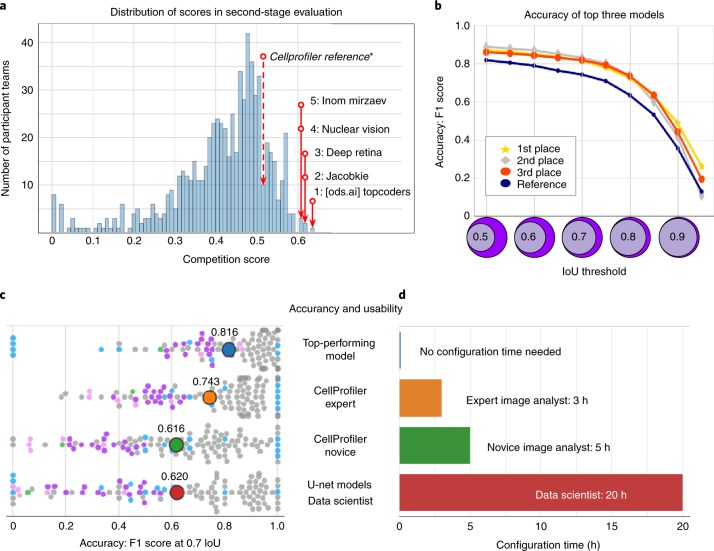
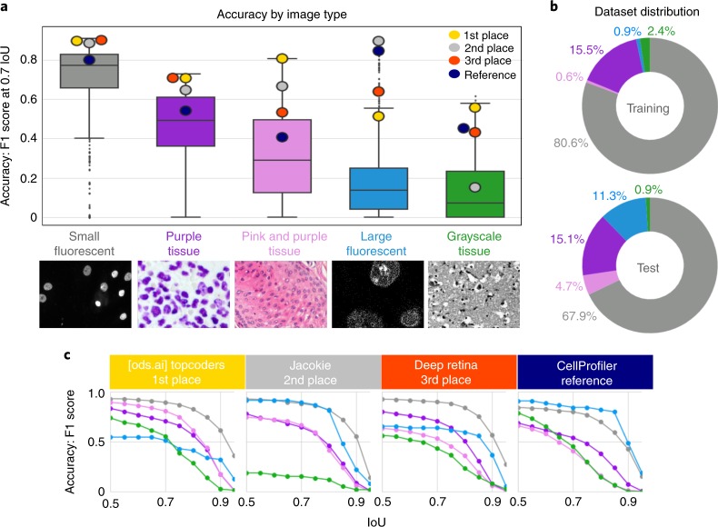
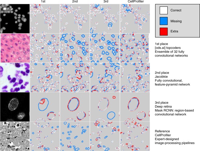
Segmenting the nuclei of cells in microscopy images is often the first step in the quantitative analysis of imaging data for biological and biomedical applications. Many bioimage analysis tools can segment nuclei in images but need to be selected and configured for every experiment. The 2018 Data Science Bowl attracted 3,891 teams worldwide to make the first attempt to build a segmentation method that could be applied to any two-dimensional light microscopy image of stained nuclei across experiments, with no human interaction. Top participants in the challenge succeeded in this task, developing deep-learning-based models that identified cell nuclei across many image types and experimental conditions without the need to manually adjust segmentation parameters. This represents an important step toward configuration-free bioimage analysis software tools.
Overview
The claims and statistics of this paper haven’t been checked yet.
Run the check — free, no account needed. It reads the open-access text and checks the sources it cites.
Integrity signals
Registry
Cites
Statistics
Figures
Verification
Claims the paper makes
The claims and statistics of this paper haven’t been checked yet.
Run the check — free, no account needed. It reads the open-access text and checks the sources it cites.
Claims other papers make about this one
Each is checked against this paper’s own text: does it say what the citing paper says it does?
The source backs part of the claim, not all of it — or backs it for a different population, condition or comparison.
In the citing paper
“However, we did not see an improvement for the ensemble of models compared to the generalist model (Extended Data Fig. 2), consistent with the results of ref. 27.”
From Cellpose 2.0: how to train your own model (2022) · cites this paper as [27]
Experimental — accuracy not yet measuredThe cited source says what the claim attributes to it.
In the citing paper
“Finally, we point out that the LiveCell study used a different average precision score from ours, which additionally requires a confidence score per ROI, while we used the average precision formulation from the Data Science Bowl challenge and other studies12,27,35.”
From Cellpose 2.0: how to train your own model (2022) · cites this paper as [27]
Self-check
Percentages, sums and reported differences re-computed from the paper’s own numbers.
The self-check needs the paper’s full text.
Citations
Cites
Deep Residual Learning for Image Recognition
He, K., Zhang, X., Ren, S., & Sun, J. (2016) · 2016 IEEE Conference on Computer Vision and Pattern Recognition (CVPR) · doi:10.1109/cvpr.2016.90 · Cited by 235,474
U-Net: Convolutional Networks for Biomedical Image Segmentation
Ronneberger, O., Fischer, P., & Brox, T. (2015) · Lecture notes in computer science · doi:10.1007/978-3-319-24574-4_28 · Cited by 99,404
LeCun, Y., Bengio, Y., & Hinton, G. (2015) · Nature · doi:10.1038/nature14539 · Cited by 84,126
Fiji: an open-source platform for biological-image analysis
Schindelin, J., Arganda-Carreras, I., Frise, E., Kaynig, V., Longair, M., Pietzsch, T., Preibisch, S., Rueden, C., Saalfeld, S., Schmid, B., et al. (2012) · Nature Methods · doi:10.1038/nmeth.2019 · Cited by 66,890
Microsoft COCO: Common Objects in Context
Lin, T.-Y., Maire, M., Belongie, S., Hays, J., Perona, P., Ramanan, D., Dollár, P., & Zitnick, C. L. (2014) · Lecture Notes in Computer Science · doi:10.1007/978-3-319-10602-1_48 · Cited by 45,460
A Threshold Selection Method from Gray-Level Histograms
Otsu, N. (1979) · IEEE Transactions on Systems, Man and Cybernetics · doi:10.1109/tsmc.1979.4310076 · Cited by 44,708
ImageNet Large Scale Visual Recognition Challenge
Russakovsky, O., Deng, J., Su, H., Krause, J., Satheesh, S., Ma, S., Huang, Z., Karpathy, A., Khosla, A., Bernstein, M., et al. (2015) · International Journal of Computer Vision · doi:10.1007/s11263-015-0816-y · Cited by 39,380
Feature Pyramid Networks for Object Detection
Lin, T.-Y., Dollar, P., Girshick, R., He, K., Hariharan, B., & Belongie, S. (2017) · 2017 IEEE Conference on Computer Vision and Pattern Recognition (CVPR) · doi:10.1109/cvpr.2017.106 · Cited by 30,939
He, K., Gkioxari, G., Dollar, P., & Girshick, R. (2017) · 2017 IEEE International Conference on Computer Vision (ICCV) · doi:10.1109/iccv.2017.322 · Cited by 29,137
Unpaired image-to-image translation using cycle-consistent adversarial networks
Zhu, J.-Y., Park, T., Isola, P., & Efros, A. A. (2017) · 2017 IEEE International Conference on Computer Vision (ICCV) · doi:10.1109/iccv.2017.244 · Cited by 23,602
Research works printed but not linked: 3
- Hollandi, R. et al. A deep learning framework for nucleus segmentation using image style transfer. Preprint at bioRxiv https://doi.org/10.1101/580605 (2019). · DOI
- Bolukbasi, T., Chang, K.-W., Zou, J. Y., Saligrama, V. & Kalai, A. T. in Advances in Neural Information Processing Systems (eds Lee, D. D. et al.) 4349–4357 (papers.nips.cc, 2016).
- Ren, S., He, K., Girshick, R. & Sun, J. in Advances in Neural Information Processing Systems 28 (eds. Cortes, C. et al.) 91–99 (Curran Associates, 2015).
Cited by
Sorted by citations
Kirillov, A., Mintun, E., Ravi, N., Mao, H., Rolland, C., Gustafson, L., Xiao, T., Whitehead, S., Berg, A. C., Lo, W.-Y., et al. (2023) · 2023 IEEE/CVF International Conference on Computer Vision (ICCV) · doi:10.1109/iccv51070.2023.00371 · Cited by 11,953
“ADE20K[117] BBBC038v1[12] Cityscapes[25] DOORS[80] DRAM[24] EgoHOS[113] GTEA[34,63] Hypersim[86] IBD[17] iShape[111] LVIS[44] NDD20[100] NDISPark[22,23] OVIS[81] PPDLS[74] Plittersdorf[46] STREETS[91] TimberSeg[38] TrashCan[52] VISOR[28,27] WoodScape[112] PIDRay[104] ZeroWaste-f[6] Figure 8: Samples from the 23 divers…”
BackgroundInfluentialCellpose: a generalist algorithm for cellular segmentation
Stringer, C., Wang, T., Michaelos, M., & Pachitariu, M. (2021) · Nature Methods · doi:10.1038/s41592-020-01018-x · Cited by 4,369
Albumentations: fast and flexible image augmentations
Buslaev, A., Iglovikov, V. I., Khvedchenya, E., Parinov, A., Druzhinin, M., & Kalinin, A. A. (2020) · Information · doi:10.3390/info11020125 · Cited by 2,714
U-Net and Its Variants for Medical Image Segmentation: A Review of Theory and Applications
Siddique, N. A., Paheding, S., Elkin, C. P., & Devabhaktuni, V. (2021) · IEEE Access · doi:10.1109/access.2021.3086020 · Cited by 2,185
Cellpose 2.0: how to train your own model
Pachitariu, M., & Stringer, C. (2022) · Nature Methods · doi:10.1038/s41592-022-01663-4 · Cited by 1,381
Metadata
- Authors
- Caicedo, J. C., Goodman, A., Karhohs, K. W., Cimini, B. A., Ackerman, J., Haghighi, M., Heng, C., Becker, T., Doan, M., McQuin, C., Rohban, M., Singh, S., & Carpenter, A. E.
- Affiliations
- Broad Institute (US)
- Corresponding author
- Carpenter, Anne E.
- ORCID
- Venue
- Nature Methods
- Published
- 2019-10-21
- Volume
- 16
- Issue
- 12
- Pages
- 1247-1253
- ISSN
- 1548-7091 (ISSN-L)
- 1548-7105
- PMC
- PMC6919559
- OpenAlex
- W2980998394
- Cited by
- 809
- References
- 49
- Citations by year
- 2026: 86 · 2025: 142 · 2024: 150 · 2023: 144 · 2022: 124 · 2021: 114 · 2020: 44 · 2019: 4
- FWCI
- 34.63
- Type
- journal article
- Language
- en
- Update notices
- correction (2020-01-22)
- Open access
- hybrid
- doi.org
- Locations
- Nature Methods (publishedVersion, cc-by)
- Europe PMC (PubMed Central) (publishedVersion, cc-by)
- PubMed Central (publishedVersion, cc-by)
- PubMed (publishedVersion)
- Funders
- Eli Lilly and Company
- Nvidia
- Broad Institute
- National Institutes of Health
- National Institute of General Medical Sciences (R35GM122547)
- Topics
- Life Sciences › Biochemistry, Genetics and Molecular Biology › Biophysics › Cell Image Analysis Techniques
- Physical Sciences › Computer Science › Computer Vision and Pattern Recognition › Digital Imaging for Blood Diseases
- Physical Sciences › Computer Science › Computer Vision and Pattern Recognition › Advanced Neural Network Applications
- Keywords
- nuclei segmentation, bioimage analysis, deep learning, microscopy image analysis, automated image analysis
- MeSH terms
- Data Science, Cell Nucleus, Humans, Image Processing, Computer-Assisted, Microscopy, Fluorescence
About these checks
Registry, retraction, Cites and statistics results are computed, not judged, with one exception: a printed reference that no index lists is read once by a model before it counts as a citation error. Claims are extracted by a model and kept only when their quote appears verbatim in the text; each cited source is then checked, with a model’s help, for whether it says what the claim relies on. Those verdicts can be wrong — read the claim’s quote and the cited source before relying on one.
Statistics and arithmetic are recomputed, not judged.
Confidence bands are shown only for fields calibrated on our annotated test set. None is calibrated yet, so no band is shown.
What counts as a claim ›How we measure confidence ›How we check this ›
Are you an author, or do you see a problem with this page? Request a review
