

The DOI matches its Crossref record; 29 of the 29 references it cites were found in a registry or an index.
LIVECell—A large-scale dataset for label-free live cell segmentation
Open accessAbstract
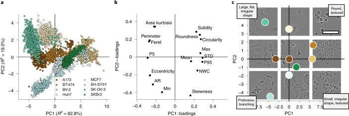
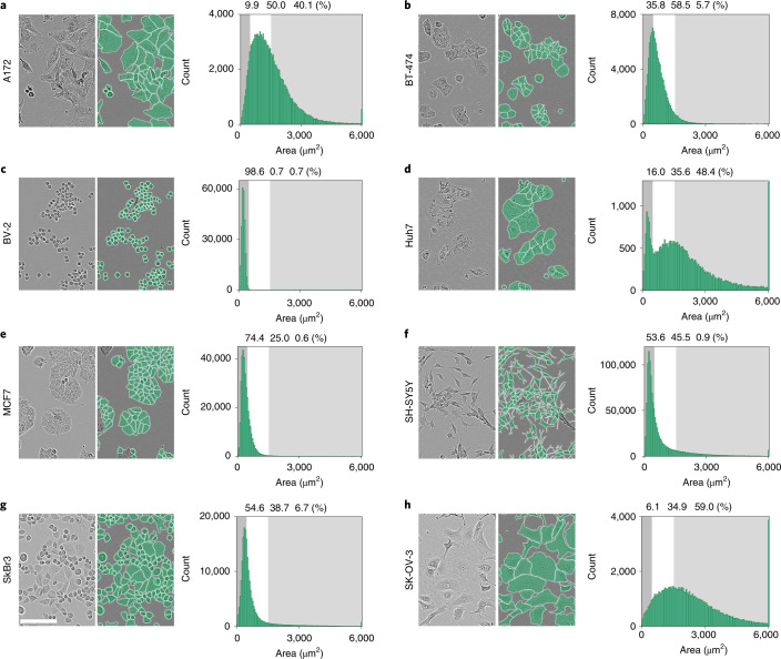
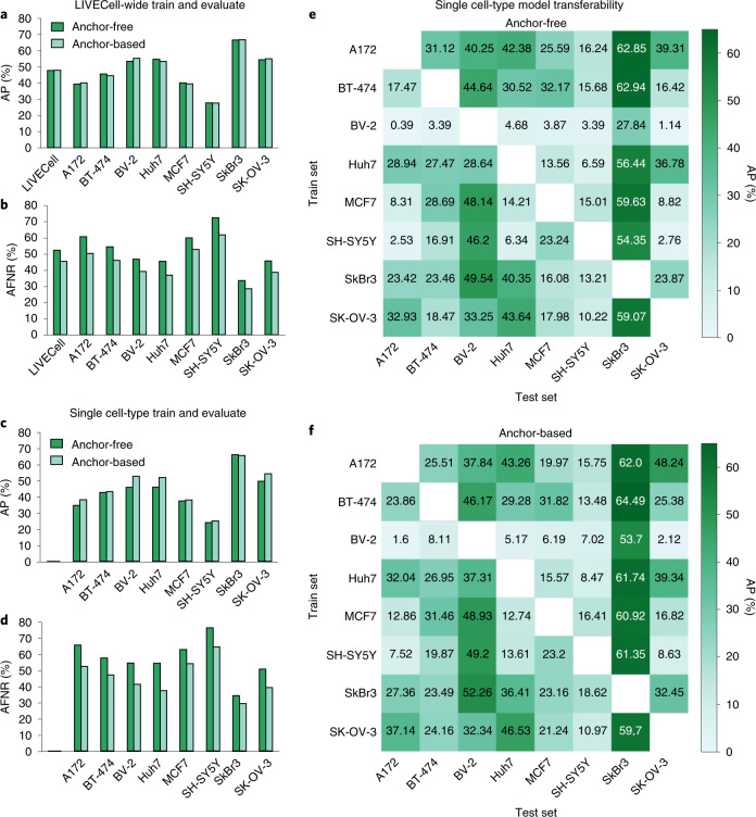
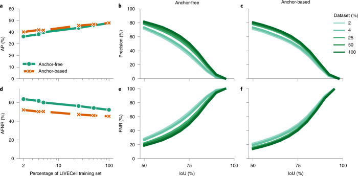
Light microscopy combined with well-established protocols of two-dimensional cell culture facilitates high-throughput quantitative imaging to study biological phenomena. Accurate segmentation of individual cells in images enables exploration of complex biological questions, but can require sophisticated imaging processing pipelines in cases of low contrast and high object density. Deep learning-based methods are considered state-of-the-art for image segmentation but typically require vast amounts of annotated data, for which there is no suitable resource available in the field of label-free cellular imaging. Here, we present LIVECell, a large, high-quality, manually annotated and expert-validated dataset of phase-contrast images, consisting of over 1.6 million cells from a diverse set of cell morphologies and culture densities. To further demonstrate its use, we train convolutional neural network-based models using LIVECell and evaluate model segmentation accuracy with a proposed a suite of benchmarks.
Overview
The claims and statistics of this paper haven’t been checked yet.
Run the check — free, no account needed. It reads the open-access text and checks the sources it cites.
Integrity signals
Registry
Cites
Statistics
Figures
Verification
Claims the paper makes
The claims and statistics of this paper haven’t been checked yet.
Run the check — free, no account needed. It reads the open-access text and checks the sources it cites.
Claims other papers make about this one
Each is checked against this paper’s own text: does it say what the citing paper says it does?
The source backs part of the claim, not all of it — or backs it for a different population, condition or comparison.
In the citing paper
“The LiveCell dataset consists of 3,188 training and 1,516 test images of eight different cell lines collected using phase-contrast microscopy, and each image has manual segmentations of the cells (https://sartorius-research.github.io/LIVECell/)7.”
From Cellpose 2.0: how to train your own model (2022) · cites this paper as [7]
Experimental — accuracy not yet measured
Self-check
Percentages, sums and reported differences re-computed from the paper’s own numbers.
The self-check needs the paper’s full text.
Citations
Cites
U-Net: Convolutional Networks for Biomedical Image Segmentation
Ronneberger, O., Fischer, P., & Brox, T. (2015) · Lecture notes in computer science · doi:10.1007/978-3-319-24574-4_28 · Cited by 99,404
An Image is Worth 16x16 Words: Transformers for Image Recognition at Scale
Dosovitskiy, A., Beyer, L., Kolesnikov, A., Weissenborn, D., Zhai, X., Unterthiner, T., Dehghani, M., Minderer, M., Heigold, G., Gelly, S., et al. (2020) · arXiv · doi:10.48550/arxiv.2010.11929 · Cited by 47,472
Microsoft COCO: Common Objects in Context
Lin, T.-Y., Maire, M., Belongie, S., Hays, J., Perona, P., Ramanan, D., Dollár, P., & Zitnick, C. L. (2014) · Lecture Notes in Computer Science · doi:10.1007/978-3-319-10602-1_48 · Cited by 45,460
PyTorch: An Imperative Style, High-Performance Deep Learning Library
Paszke, A., Gross, S., Massa, F., Lerer, A., Bradbury, J., Chanan, G., Killeen, T., Lin, Z., Gimelshein, N., Antiga, L., et al. (2019) · arXiv · doi:10.48550/arxiv.1912.01703 · Cited by 31,473
FCOS: Fully Convolutional One-Stage Object Detection
Tian, Z., Shen, C., Chen, H., & He, T. (2019) · 2019 IEEE/CVF International Conference on Computer Vision (ICCV) · doi:10.1109/iccv.2019.00972 · Cited by 7,013
CellProfiler: image analysis software for identifying and quantifying cell phenotypes
Carpenter, A. E., Jones, T. R., Lamprecht, M. R., Clarke, C., Kang, I. H., Friman, O., Guertin, D. A., Chang, J. H., Lindquist, R. A., Moffat, J., et al. (2006) · Genome biology · doi:10.1186/gb-2006-7-10-r100 · Cited by 5,212
Cellpose: a generalist algorithm for cellular segmentation
Stringer, C., Wang, T., Michaelos, M., & Pachitariu, M. (2021) · Nature Methods · doi:10.1038/s41592-020-01018-x · Cited by 4,369
Kuznetsova, A., Rom, H., Alldrin, N., Uijlings, J., Krasin, I., Pont-Tuset, J., Kamali, S., Popov, S., Malloci, M., Kolesnikov, A., et al. (2018) · arXiv (Cornell University) · doi:10.48550/arxiv.1811.00982 · Cited by 2,194
ResNeSt: Split-Attention Networks
Zhang, H., Wu, C., Zhang, Z., Zhu, Y., Lin, H., Zhang, Z., Sun, Y., He, T., Mueller, J. W., Manmatha, R., et al. (2022) · IEEE/CVF Conference on Computer Vision and Pattern Recognition Workshops (CVPRW) · doi:10.1109/cvprw56347.2022.00309 · Cited by 2,115
Cascade R-CNN: High Quality Object Detection and Instance Segmentation
Cai, Z., & Vasconcelos, N. M. (2019) · IEEE Transactions on Pattern Analysis and Machine Intelligence · doi:10.1109/tpami.2019.2956516 · Cited by 2,062
Research works printed but not linked: 1
- Levene, H. in Contributions to Probability and Statistics: Essays in Honor of Harold Hotelling (eds Olkin, I. et al.) 279–292 (Stanford Univ. Press, 1961).
Cited by
Sorted by citations
Cellpose 2.0: how to train your own model
Pachitariu, M., & Stringer, C. (2022) · Nature Methods · doi:10.1038/s41592-022-01663-4 · Cited by 1,381
Cellpose3: one-click image restoration for improved cellular segmentation
Stringer, C., & Pachitariu, M. (2025) · Nature Methods · doi:10.1038/s41592-025-02595-5 · Cited by 557
Segment Anything for Microscopy
Archit, A., Freckmann, L., Nair, S., Khalid, N., Hilt, P., Rajashekar, V., Freitag, M., Teuber, C., Spitzner, M., Contreras, C. T., et al. (2025) · Nature Methods · doi:10.1038/s41592-024-02580-4 · Cited by 426
Self-citationCellpose-SAM: superhuman generalization for cellular segmentationPreprint
Pachitariu, M., Rariden, M., & Stringer, C. (2025) · bioRxiv (Cold Spring Harbor Laboratory) · doi:10.1101/2025.04.28.651001 · Cited by 402
A guide to artificial intelligence for cancer researchers
Pérez-López, R., Laleh, N. G., Mahmood, F., & Kather, J. N. (2024) · Nature reviews. Cancer · doi:10.1038/s41568-024-00694-7 · Cited by 227
Metadata
- Authors
- Edlund, C., Jackson, T. R., Khalid, N., Bevan, N., Dale, T., Dengel, A., Ahmed, S., Trygg, J., & Sjögren, R.
- Affiliations
- Sartorius (United Kingdom) (GB)
- German Research Centre for Artificial Intelligence (DE)
- Umeå University (SE)
- Corresponding author
- Sjögren, Rickard
- ORCID
- Venue
- Nature Methods
- Published
- 2021-08-30
- Volume
- 18
- Issue
- 9
- Pages
- 1038-1045
- ISSN
- 1548-7091 (ISSN-L)
- 1548-7105
- PMC
- PMC8440198
- OpenAlex
- W3196890347
- Cited by
- 304
- References
- 29
- Citations by year
- 2026: 56 · 2025: 88 · 2024: 63 · 2023: 65 · 2022: 31 · 2021: 1
- FWCI
- 18.09
- Type
- journal article
- Language
- en
- Open access
- hybrid
- doi.org
- Locations
- Nature Methods (publishedVersion, cc-by)
- Europe PMC (PubMed Central) (publishedVersion, other-oa)
- PubMed Central (publishedVersion, cc-by)
- PubMed (publishedVersion)
- DiVA at Umeå University (Umeå University) (submittedVersion, other-oa)
- Topics
- Life Sciences › Biochemistry, Genetics and Molecular Biology › Biophysics › Cell Image Analysis Techniques
- Physical Sciences › Computer Science › Computer Vision and Pattern Recognition › Advanced Neural Network Applications
- Physical Sciences › Computer Science › Computer Vision and Pattern Recognition › Medical Image Segmentation Techniques
- Keywords
- cell segmentation, label-free imaging, phase contrast microscopy, convolutional neural networks, segmentation benchmark, cell culture, high-throughput imaging, cell morphology
- MeSH terms
- Humans, Image Processing, Computer-Assisted, Microscopy, Models, Biological, Databases, Factual, Neural Networks, Computer, Cell Culture Techniques
About these checks
Registry, retraction, Cites and statistics results are computed, not judged, with one exception: a printed reference that no index lists is read once by a model before it counts as a citation error. Claims are extracted by a model and kept only when their quote appears verbatim in the text; each cited source is then checked, with a model’s help, for whether it says what the claim relies on. Those verdicts can be wrong — read the claim’s quote and the cited source before relying on one.
Statistics and arithmetic are recomputed, not judged.
Confidence bands are shown only for fields calibrated on our annotated test set. None is calibrated yet, so no band is shown.
What counts as a claim ›How we measure confidence ›How we check this ›
Are you an author, or do you see a problem with this page? Request a review
