

The DOI matches its Crossref record; 68 of the 69 references it cites were found in a registry or an index.
Estrogen Regulates Bone Turnover by Targeting RANKL Expression in Bone Lining Cells
Open accessAbstract
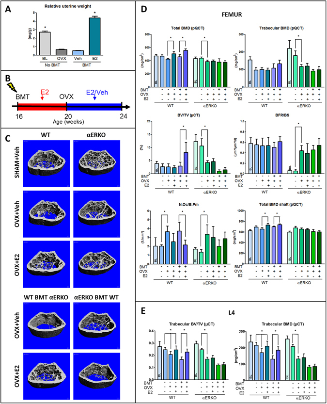
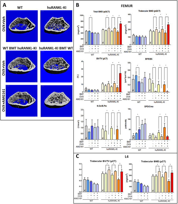
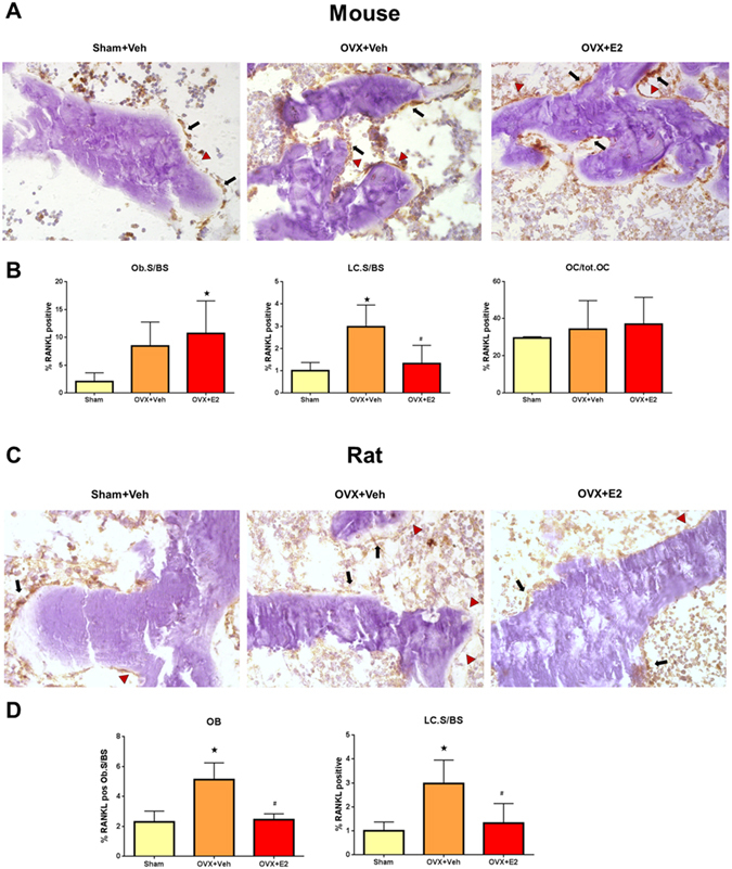
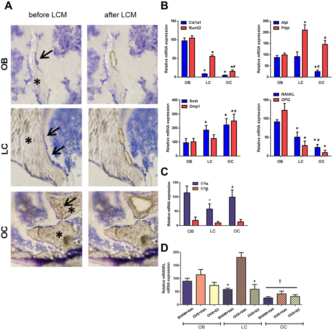
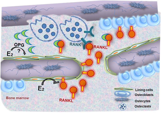
Estrogen is critical for skeletal homeostasis and regulates bone remodeling, in part, by modulating the expression of receptor activator of NF-κB ligand (RANKL), an essential cytokine for bone resorption by osteoclasts. RANKL can be produced by a variety of hematopoietic (e.g. T and B-cell) and mesenchymal (osteoblast lineage, chondrocyte) cell types. The cellular mechanisms by which estrogen acts on bone are still a matter of controversy. By using murine reconstitution models that allow for selective deletion of estrogen receptor-alpha (ERα) or selective inhibition of RANKL in hematopoietic vs. mesenchymal cells, in conjunction with in situ expression profiling in bone cells, we identified bone lining cells as important gatekeepers of estrogen-controlled bone resorption. Our data indicate that the increase in bone resorption observed in states of estrogen deficiency in mice is mainly caused by lack of ERα-mediated suppression of RANKL expression in bone lining cells.
Overview
The claims and statistics of this paper haven’t been checked yet.
Run the check — free, no account needed. It reads the open-access text and checks the sources it cites.
Integrity signals
Registry
Cites
Statistics
Figures
Verification
Claims the paper makes
The claims and statistics of this paper haven’t been checked yet.
Run the check — free, no account needed. It reads the open-access text and checks the sources it cites.
Claims other papers make about this one
Each is checked against this paper’s own text: does it say what the citing paper says it does?
In the citing paper
“Fusion also requires RANKL, which is highly expressed by BLCs;161 osteoblasts in the growing skeleton; and osteocyte arborizations that reach the bone surface in adults.51,52”
From Bone remodeling: an operational process ensuring survival and bone mechanical competence (2022) · cites this paper as [161]
Self-check
Percentages, sums and reported differences re-computed from the paper’s own numbers.
The self-check needs the paper’s full text.
Citations
Cites
A new mathematical model for relative quantification in real-time RT-PCR
Pfaffl, M. W. (2001) · Nucleic Acids Research · doi:10.1093/nar/29.9.e45 · Cited by 34,610
Osteoprotegerin ligand is a cytokine that regulates osteoclast differentiation and activation.
Lacey, D. L., Timms, E., Tan, H.-L., Kelley, M. J., Dunstan, C. R., Burgess, T., Elliott, R., Colombero, A., Elliott, G., Scully, S., et al. (1998) · Cell · doi:10.1016/s0092-8674(00)81569-x · Cited by 5,439
Osteoprotegerin: a novel secreted protein involved in the regulation of bone density.
Simonet, W. S., Lacey, D. L., Dunstan, C. R., Kelley, M., Chang, M.-S., Lüthy, R., Nguyen, H. Q., Wooden, S., Bennett, L., Boone, T., et al. (1997) · Cell · doi:10.1016/s0092-8674(00)80209-3 · Cited by 5,130
OPGL is a key regulator of osteoclastogenesis, lymphocyte development and lymph node organogenesis.
Kong, Y.-Y., Yoshida, H., Sarosi, I., Tan, H.-L., Timms, E., Capparelli, C., Morony, S., Oliveira-dos-Santos, A. J., Van, G., Itie, A., et al. (1999) · Nature · doi:10.1038/16852 · Cited by 3,351
Osteoprotegerin-deficient mice develop early onset osteoporosis and arterial calcification.
Bucay, N., Sarosi, I., Dunstan, C. R., Morony, S., Tarpley, J., Capparelli, C., Scully, S., Tan, H. L., Xu, W., Lacey, D. L., et al. (1998) · Genes & Development · doi:10.1101/gad.12.9.1260 · Cited by 2,483
Pathogenesis of osteoporosis: concepts, conflicts, and prospects
Raisz, L. G. (2005) · Journal of Clinical Investigation · doi:10.1172/jci27071 · Cited by 1,779
Evidence for osteocyte regulation of bone homeostasis through RANKL expression
Nakashima, T., Hayashi, M., Fukunaga, T., Kurata, K., Oh-hora, M., Feng, J. Q., Bonewald, L. F., Kodama, T., Wutz, A., Wagner, E. F., et al. (2011) · Nature Medicine · doi:10.1038/nm.2452 · Cited by 1,778
Matrix-embedded cells control osteoclast formation
Xiong, J., Onal, M., Jilka, R. L., Weinstein, R. S., Manolagas, S. C., & O'Brien, C. A. (2011) · Nature Medicine · doi:10.1038/nm.2448 · Cited by 1,304
Denosumab in postmenopausal women with low bone mineral density.
McClung, M. R., Lewiecki, E. M., Cohen, S. B., Bolognese, M. A., Woodson, G. C., Moffett, A. H., Peacock, M., Miller, P. D., Lederman, S. N., Chesnut, C. H., et al. (2006) · New England Journal of Medicine · doi:10.1056/nejmoa044459 · Cited by 1,228
Li, J., Sarosi, I., Yan, X.-Q., Morony, S., Capparelli, C., Tan, H.-L., McCabe, S., Elliott, R., Scully, S., Van, G., et al. (2000) · Proceedings of the National Academy of Sciences · doi:10.1073/pnas.97.4.1566 · Cited by 1,119
Self-citation
Cited by
Sorted by citations
Items 101–200 of 251
Jörg, D. J., Fuertinger, D. H., Cherif, A., Bushinsky, D. A., Mermelstein, A., Raimann, J. G., & Kotanko, P. (2022) · eLife · doi:10.7554/elife.76228 · Cited by 15
Huang, X., Li, Y., Liao, H., Luo, X., Zhao, Y., Huang, Y., Zhou, Z., & Xiang, Q. (2023) · Journal of Functional Biomaterials · doi:10.3390/jfb14040193 · Cited by 15
Consumption of Phytoestrogens Affects Bone Health by Regulating Estrogen Metabolism
Tomczyk‐Warunek, A., Winiarska‐Mieczan, A., Blicharski, T., Blicharski, R., Kowal, F., Pano, I. T., Tomaszewska, E., & Muszyński, S. (2024) · Journal of Nutrition · doi:10.1016/j.tjnut.2024.05.026 · Cited by 17
Efferocytosis and Bone Dynamics
Batoon, L., Hawse, J. R., McCauley, L. K., Weivoda, M. M., & Roca, H. (2024) · Current Osteoporosis Reports · doi:10.1007/s11914-024-00878-y · Cited by 16
Ormeloxifene, a selective estrogen receptor modulator, protects against pulmonary hypertension
Abdulkareem, A. O., Tiwari, P., Lone, Z. R., Iqbal, H., Gupta, S. C., Jha, R. K., Chanda, D., Jagavelu, K., & Hanif, K. (2023) · European Journal of Pharmacology · doi:10.1016/j.ejphar.2023.175558 · Cited by 15
Xu, J., Cao, B., Li, C., & Li, G. (2023) · Frontiers in Oncology · doi:10.3389/fonc.2023.1218206 · Cited by 14
The Possible Protective Role of Powder Cuttlefish Bone, Crabshell and Eggshell on Osteoporotic Rats
Negm, S. H. (2018) · Journal of Food and Dairy Sciences · doi:10.21608/jfds.2018.77765 · Cited by 14
Bone matrix quality in a developing high-fat diet mouse model is altered by RAGE deletion
Stephen, S. J., Bailey, S., D'Erminio, D. N., Krishnamoorthy, D., Iatridis, J. C., & Vashishth, D. (2022) · Bone · doi:10.1016/j.bone.2022.116470 · Cited by 14
Livingstone, K. M., Tan, M. H., Abbott, G., Duckham, R. L., Croft, L., Ward, J., McEvoy, M., Keske, M. A., Austin, C. M., & Bowe, S. J. (2021) · Frontiers in Endocrinology · doi:10.3389/fendo.2021.692677 · Cited by 13
Kuiper, J. R., Vuong, A. M., Lanphear, B. P., Calafat, A. M., Ospina, M., Cecil, K. M., Xu, Y. Y., Yolton, K. A., Kalkwarf, H. J., Braun, J. M., et al. (2022) · The Science of The Total Environment · doi:10.1016/j.scitotenv.2022.158246 · Cited by 15
Activation of NF-κB signaling regulates ovariectomy-induced bone loss and weight gain
Huang, F., Gao, J., Li, A., Mizokami, A., Matsuda, M., Aoki, K., Katagiri, T., Kawakubo‐Yasukochi, T., & Jimi, E. (2024) · Biochimica et Biophysica Acta (BBA) - Molecular Basis of Disease · doi:10.1016/j.bbadis.2024.167320 · Cited by 15
Sensing the future: A review on emerging technologies for assessing and monitoring bone health
Naik, A., Kale, A., & Rajwade, J. M. (2024) · Biomaterials Advances · doi:10.1016/j.bioadv.2024.214008 · Cited by 14
Normal bone physiology, remodelling and its hormonal regulation
Mallorie, A., & Shine, B. S. F. (2022) · Surgery (Oxford) · doi:10.1016/j.mpsur.2021.12.001 · Cited by 30
Progesterone in gender-affirming therapy of trans women
Milionis, C., Ilias, Ι., & Koukkou, E. G. (2022) · World Journal of Biological Chemistry · doi:10.4331/wjbc.v13.i3.66 · Cited by 13
Effects of Loganin on Bone Formation and Resorption In Vitro and In Vivo
Lee, C.-G., Kim, D.-W., Kim, J., Uprety, L. P., Oh, K.-I., Singh, S., Yoo, J., Jin, H., Choi, T. H., Park, E., et al. (2022) · International Journal of Molecular Sciences · doi:10.3390/ijms232214128 · Cited by 13
Osteoclasts at Bone Remodeling: Order from Order
Takito, J., & Nonaka, N. (2023) · Results and problems in cell differentiation · doi:10.1007/978-3-031-37936-9_12 · Cited by 14
Fatima, S., Abrar, M., Shahid, A., Moin, H., & Majeed, S. (2025) · Expert Review of Endocrinology & Metabolism · doi:10.1080/17446651.2025.2510595 · Cited by 12
Rossini, A., Frigerio, S., Dozio, E., Trevisan, R., Perseghin, G., & Corbetta, S. (2020) · International Journal of Endocrinology · doi:10.1155/2020/1809150 · Cited by 12
Küchler, É. C., de Lara, R. M., Omori, M. A., Marañón‐Vásquez, G. A., Baratto‐Filho, F., Nelson‐Filho, P., Stuani, M. B. S., Blanck‐Lubarsch, M., Schroeder, A., Proff, P., et al. (2021) · Head & Face Medicine · doi:10.1186/s13005-021-00265-3 · Cited by 12
Kottmann, V., Nienhaus, M., Drees, P., Gercek, E., & Ritz, U. (2026) · Biochimica et Biophysica Acta (BBA) - Reviews on Cancer · doi:10.1016/j.bbcan.2026.189551 · Cited by 14
The potential link between the development of Alzheimer’s disease and osteoporosis
Nasme, F., Behera, J., Tyagi, P., Debnath, N., Falcone, J. C., & Tyagi, N. (2025) · Biogerontology · doi:10.1007/s10522-024-10181-z · Cited by 12
Sims, N. A., & Martin, T. J. (2019) · Elsevier eBooks · doi:10.1016/b978-0-12-814841-9.00004-x · Cited by 11
Role of Mast-Cell-Derived RANKL in Ovariectomy-Induced Bone Loss in Mice
Fischer, V., Bülow, J. M., Krüger, B. T., Ragipoglu, D., Vikman, A., Haffner‐Luntzer, M., Katsoulis‐Dimitriou, K., Dudeck, A., & Ignatius, A. A. (2023) · International Journal of Molecular Sciences · doi:10.3390/ijms24119135 · Cited by 11
Idiosyncratic bone responses to blood flow restriction exercise: new insights and future directions
Hughes, L., & Centner, C. (2023) · Journal of Applied Physiology · doi:10.1152/japplphysiol.00723.2022 · Cited by 11
Корокин, М. В., Soldatov, V. O., Гудырев, О. С., Koklin, I. S., Taran, E. I., Mishenin, M. O., Korokina, L. V., Kochkarov, A. A., Покровский, М. В., Вараксин, М. В., et al. (2022) · RESEARCH RESULTS IN BIOMEDICINE · doi:10.18413/2658-6533-2022-8-4-0-5 · Cited by 15
Slavić, S., Andrukhova, O., Ford, K., Handschuh, S., Latic, N., Reichart, U., Sasgary, S., Bergow, C., Hofbauer, L. C., Kostenuik, P. J., et al. (2018) · Journal of Molecular Medicine · doi:10.1007/s00109-018-1641-x · Cited by 10
Self-citationModulation of aromatase by natural compounds—A pharmacophore guided molecular modelling simulations
Rampogu, S. D., Park, C., Son, M., Baek, A., Zeb, A., Lee, G., & Lee, K. W. (2018) · South African Journal of Botany · doi:10.1016/j.sajb.2018.06.019 · Cited by 10
Genistein Supplementation and Bone Health in Breast Cancer in Rats
Skrajnowska, D., Bielecki, W., Szterk, A., Ofiara, K., & Bobrowska‐Korczak, B. (2024) · Nutrients · doi:10.3390/nu16060912 · Cited by 9
Exposure to BPA and BPS during pregnancy disrupts the bone mineralization in the offspring
Varma, S., Molangiri, A., Mudavath, S., Rajendran, A., Ajumeera, R., Duttaroy, A. K., & Basak, S. (2024) · Food and Chemical Toxicology · doi:10.1016/j.fct.2024.114772 · Cited by 9
Kwon, M., Kim, B., Min, S. Y., & Chae, S. (2025) · Journal of Clinical Medicine · doi:10.3390/jcm14030732 · Cited by 9
Role of Polyphenols in the Metabolism of the Skeletal System in Humans and Animals – A Review
Skiba, G., Raj, S., Sobol, M., Kowalczyk, P., & GRELA, E. R. (2021) · Annals of Animal Science · doi:10.2478/aoas-2021-0040 · Cited by 9
Shim, K., Gu, D. R., Hwang, Y., Yang, H. J., Ryuk, J.-A., & Ha, H. (2021) · Molecules · doi:10.3390/molecules27010169 · Cited by 9
Konar, M., & Sahoo, H. (2020) · International Journal of Biological Macromolecules · doi:10.1016/j.ijbiomac.2020.02.113 · Cited by 9
The interaction of the active compounds of Labisia pumila on RANK–RANKL–OPG system
Noor, Z., Kania, N., Azharuddin, A., Budhiparama, N. C., Nurdiana, N., Hidajat, N. N., Fatmawati, F., & Setiawan, B. (2017) · Clinical Nutrition Experimental · doi:10.1016/j.yclnex.2017.09.001 · Cited by 9
Tissue-specific mechanism of estrogen in osteoporosis and vascular calcification
Wu, W., Qiao, A., & Du, T. (2025) · European Journal of Pharmacology · doi:10.1016/j.ejphar.2025.177783 · Cited by 9
Cvetko, E. D., Nešić, N., Matić, A., Ahić, J. M., & Drenjančević, I. (2022) · European Journal of Applied Physiology · doi:10.1007/s00421-022-04900-y · Cited by 9
RANK/RANKL Signaling Pathway in Breast Development and Cancer
Pérez-Chacón, G., Santamarı́a, P. G., Redondo-Pedraza, J., & González‐Suárez, E. (2025) · Advances in experimental medicine and biology · doi:10.1007/978-3-031-70875-6_16 · Cited by 9
Influence of extract derived cell cultures of broccoli against osteoporosis in ovariectomized rats
Salama, A., Abdelhameed, M. F., Mostafa, S. A., Nada, S. A., Taha, H. S., & Amer, A. A. (2021) · Egyptian Journal of Chemistry · doi:10.21608/ejchem.2021.64803.3419 · Cited by 8
Wendy Saltzman, A., Abbott, D. H., Binkley, N., & Colman, R. J. (2018) · American Journal of Primatology · doi:10.1002/ajp.22905 · Cited by 8
Cheng, J. N., Frye, J. B., Whitman, S. A., Kunihiro, A. G., Brickey, J. A., & Funk, J. L. (2021) · Journal of Cancer Metastasis and Treatment · doi:10.20517/2394-4722.2021.27 · Cited by 9
In Vitro Cell Culture Model for Osteoclast Activation during Estrogen Withdrawal
Gandhi, N., Omer, S., & Harrison, R. E. (2024) · International Journal of Molecular Sciences · doi:10.3390/ijms25116134 · Cited by 8
A Laser Capture Microdissection Protocol That Yields High Quality RNA from Fresh-frozen Mouse Bones
Marek, A., Schüler, C., Satué, M., Haigl, B., & Erben, R. G. (2019) · Journal of Visualized Experiments · doi:10.3791/60197 · Cited by 7
Self-citationHobeika, E. M., Armouti, M., Kala, H. S., & Stocco, C. O. (2019) · Elsevier eBooks · doi:10.1016/b978-0-12-813814-4.00025-0 · Cited by 7
Oster, M., Qasir, H., Reyer, H., Ponsuksili, S., Trakooljul, N., Sommerfeld, V., Rodehutscord, M., & Wimmers, K. (2025) · Poultry Science · doi:10.1016/j.psj.2025.105798 · Cited by 6
Mazzoli, R., Filippini, T., Iamandii, I., Pasquale, L. D., Veneri, F., Birnbaum, L. S., Rothman, K. J., & Vinceti, M. (2025) · Environmental Health · doi:10.1186/s12940-025-01226-y · Cited by 6
Lu, L., Wen, Q., Zhang, X., Lv, J., Zhang, L., Liu, L., Yu, X., & Li, N. (2022) · BMJ Open · doi:10.1136/bmjopen-2022-062677 · Cited by 6
Yim, E.-J., Jo, S.-W., Kang, H. J., Park, S. K., Yu, K. Y., Jeong, D., & Park, S. (2022) · Fermentation · doi:10.3390/fermentation8050211 · Cited by 6
Rossetti, B. R., Lemos, A. F., Só, B. B., Weissheimer, T., Martins, M. D., & Só, M. V. R. (2022) · Archives of Oral Biology · doi:10.1016/j.archoralbio.2022.105496 · Cited by 7
Cheng, J. N., Frye, J. B., Whitman, S. A., & Funk, J. L. (2019) · Clinical & Experimental Metastasis · doi:10.1007/s10585-019-10012-3 · Cited by 6
Yoshida, T., Wang, J., & Stern, P. H. (2019) · Handbook of experimental pharmacology · doi:10.1007/164_2019_327 · Cited by 6
Cathepsin K inhibitors promote osteoclast-osteoblast communication and engagement of osteogenesis
Panwar, P., Olesen, J. B., Delaissé, J., Søe, K., & Brömme, D. (2025) · JBMR Plus · doi:10.1093/jbmrpl/ziaf079 · Cited by 5
Suppression of bone resorption in ovariectomized mice using estrogen-immobilized polyphosphodiesters
Iwasaki, Y., Fukaura, S., Mabuchi, S., Okuno, Y., Yokota, A., & Neo, M. (2024) · Materialia · doi:10.1016/j.mtla.2024.102166 · Cited by 5
Hung, Y.-T., Wu, W., Lee, R., Yao, T.-K., & Yeh, K. (2025) · Biomedicines · doi:10.3390/biomedicines13030732 · Cited by 4
Inflammaging-Driven Osteoporosis: Is a Galectin-Targeted Approach Needed?
Russo, M., Lepre, C. C., Itro, A., Martin, G., Conza, G., Trotta, M. C., Puticiu, M. I., Hermenean, A. O., Gimigliano, F., D’Amico, M., et al. (2025) · International Journal of Molecular Sciences · doi:10.3390/ijms26136473 · Cited by 7
Thomsen, L. K., Christiansen, P. V. D., Andreasen, C. M., & Andersen, T. L. (2025) · Osteoporosis International · doi:10.1007/s00198-025-07612-z · Cited by 4
A novel miRNA-TF-mRNA regulatory network associated with cellular senescence in osteoporosis
Zhang, X., Xu, N., Zhou, Y., & Xu, J. (2025) · Stem Cell Reports · doi:10.1016/j.stemcr.2025.102672 · Cited by 4
Yang, N., Zhang, W., Cui, X., Xi, L., Zhou, Y., & Feng, H. (2026) · Naunyn-Schmiedeberg s Archives of Pharmacology · doi:10.1007/s00210-026-05301-0 · Cited by 4
Estrogenic active Ecklonia cava extract improves bone loss and depressive behaviour in OVX mice
Lim, H. J., Cho, C. H., Lee, S., Won, Y., Bak, S. G., Kim, M., Kim, S., Yoon, M., Ha, H. J., Jang, J. T., et al. (2023) · Journal of Functional Foods · doi:10.1016/j.jff.2023.105423 · Cited by 4
Baek, D.-C., Hwang, S., Lee, J., Wang, J., Son, C., & Lee, E. J. (2023) · International Journal of Molecular Sciences · doi:10.3390/ijms24054876 · Cited by 4
Igwe, A. A., Onyeso, O. K., Adandom, I. I., Onyeso, K. M., Anyachukwu, C. C., Awosoga, O., Ezema, C. I., & Okoye, C. G. (2023) · Bulletin of Faculty of Physical Therapy · doi:10.1186/s43161-023-00141-y · Cited by 4
Heo, K. W., Noh, M. H., Hur, D. Y., Hong, T. U., Park, S. Y., & Kim, W. J. (2023) · Science Progress · doi:10.1177/00368504231199204 · Cited by 4
Palmier, M., Maître, M., Doat, H., Lesté-Lasserre, T., Maurel, D. B., & Boiziau, C. (2024) · JBMR Plus · doi:10.1093/jbmrpl/ziae078 · Cited by 4
Canadian Urologic Association best practice report: Bone health in prostate cancer
Lavallée, L. T., McLarty, R., Tran, C. S., Breauj, R. H., Richard, P. O., Shayegan, B., Danielson, B. L., Jammal, M.-P., & Fred Saad, C. (2021) · Canadian Urological Association Journal · doi:10.5489/cuaj.7660 · Cited by 4
Bone histomorphometry in rodents
Ma, Y. L., Burr, D. B., & Erben, R. G. (2019) · Elsevier eBooks · doi:10.1016/b978-0-12-814841-9.00082-8 · Cited by 4
Self-citationThe role of postnatal estrogen deficiency on cranium dimensions
de Lara, R. M., dos Santos, M. C., Omori, M. A., Baratto‐Filho, F., Brancher, J. A., Nelson‐Filho, P., Schroeder, A., Küchler, É. C., Torres, M. F., & Kirschneck, C. (2020) · Clinical Oral Investigations · doi:10.1007/s00784-020-03655-0 · Cited by 4
Osteoporosis: Possible Pathways Involved and the Role of Natural Phytoestrogens in Bone Metabolism
Thent, Z. C., Das, S., Mahakkanukrauh, P., & Lanzotti, V. (2019) · Sains Malaysiana · doi:10.17576/jsm-2019-4809-22 · Cited by 5
Circulating miR-19b in Serum Exosomes: A Novel Biomarker for Estrogen-Associated Osteoporosis
Renuka, C., Palanichamy, K., Muthu, S., & Vadivalagan, C. (2025) · Indian Journal of Orthopaedics · doi:10.1007/s43465-025-01568-2 · Cited by 3
Xu, H., Settakorn, K., Khantamat, O., Li, J., Charoenphandhu, N., Chanpaisaeng, K., Paradee, N., Srichairatanakool, S., & Koonyosying, P. (2025) · npj Science of Food · doi:10.1038/s41538-025-00601-w · Cited by 4
Fan, Y., Yu, Y., & Yan, P. (2025) · Toxicology and Applied Pharmacology · doi:10.1016/j.taap.2025.117345 · Cited by 3
Ouyang, S., Cao, D., Yin, Y., Ma, F., & Ren, G. (2025) · Scientific Reports · doi:10.1038/s41598-025-01807-4 · Cited by 3
Landwehr, V. C., Fretwurst, T., Heinen, J., Vach, K., Nelson, K., Nahles, S., & Iglhaut, G. (2022) · International Journal of Implant Dentistry · doi:10.1186/s40729-022-00447-x · Cited by 3
Effect of resistance exercise on bone health of old aged individuals: Review
Khanna, A. (2022) · Science & Sports · doi:10.1016/j.scispo.2022.09.003 · Cited by 4
Karim, K., Giribabu, N., & Salleh, N. B. (2023) · Applied Biochemistry and Biotechnology · doi:10.1007/s12010-023-04515-9 · Cited by 3
Cheng, J. N., Frye, J. B., Whitman, S. A., Ehsani, S., Ali, S., & Funk, J. L. (2024) · Endocrinology · doi:10.1210/endocr/bqae038 · Cited by 3
Influence of Osteoporosis on the Course of Apical Periodontitis
Stanev, E., & Vassileva, R. (2024) · European Journal of Dentistry · doi:10.1055/s-0044-1785533 · Cited by 3
Park, H. K., Ha, H., Lee, H., Lee, G., Go, G., Yoon, T. M., Kim, T., & Kim, W. (2024) · Molecular Nutrition & Food Research · doi:10.1002/mnfr.202400158 · Cited by 3
Bone—From macrophage to osteoclast and osteolytic diseases
Ramos‐Junior, E. S., Taira, T. M., & Fukada, S. Y. (2022) · Elsevier eBooks · doi:10.1016/b978-0-12-821385-8.00011-1 · Cited by 3
Ehnert, S., Nussler, A. K., Schulz, K. A., Cardenas, A. K., Meneses‐Lorente, G., McCallum, S., Fürst‐Recktenwald, S., & Roth, A. B. (2025) · Archives of Toxicology · doi:10.1007/s00204-025-04111-2 · Cited by 2
Camassa, J. A. D. A., BARROS, V. A., Babo, P. S., Pereira, F. A. M., Morais, J. J. L., Fertuzinhos, A., de Azevedo, J. M. T., Reis, R. L., Gomes, M. E., Martins‐Bessa, A., et al. (2025) · Biology · doi:10.3390/biology14081031 · Cited by 2
Li, G., Wang, C., Tian, C., Chen, L., Jiang, W., Gao, L., Ai, Q. Y. H., Liu, S., & Wang, J. (2025) · Archives of Physical Medicine and Rehabilitation · doi:10.1016/j.apmr.2025.10.017 · Cited by 2
Zhou, Z., Wei, X., Zhang, X., Ainsworth, B. E., Lü, J., & Liu, Y. (2026) · Journal of sport and health science/Journal of Sport and Health Science · doi:10.1016/j.jshs.2026.101127 · Cited by 3
Bone mineral density and bone turnover changes with advanced menstrual recovery: The REFUEL study
Salamunes, A. C. C., Williams, N. I., Ricker, E. A., Mallinson, R. J., Koltun, K. J., & Souza, M. J. D. (2026) · Bone · doi:10.1016/j.bone.2026.117834 · Cited by 2
Puspita, R. M., Agustiningsih, D., & Purwanti, N. (2026) · Physiology International · doi:10.1556/2060.2026.00710 · Cited by 2
Osteoclast activity in osteoporosis mandibular bone based on RANKL and osteoprotegerin ratio
Kuntjoro, M., Hendrijantini, N., Rantam, F. A., Hong, G., Salim, S., & Ari, M. D. A. (2021) · Journal of Pharmacy & Pharmacognosy Research · doi:10.56499/jppres21.1094_9.6.870 · Cited by 2
Suzuki, R., Shirataki, Y., Tomomura, A., Bandow, K., Sakagami, H., & Tomomura, M. (2023) · International Journal of Molecular Sciences · doi:10.3390/ijms242417479 · Cited by 2
Song, J., Han, S., Choi, S., Lee, J., Jeong, Y., Lee, H. M., Son, J., Jeong, D. Y., Yu, S., & Lee, W. (2024) · Heliyon · doi:10.1016/j.heliyon.2024.e24842 · Cited by 2
Li, J., Li, Y., Li, S., Lu, Y., & Rai, P. (2024) · Journal of Orthopaedic Surgery and Research · doi:10.1186/s13018-024-04579-1 · Cited by 2
The antiosteoporotic effect of oxymatrine compared to testosterone in orchiectomized rats
Shaban, A. M., Ali, E. A., Tayel, S. G., Rizk, S. K., & Agamy, D. F. E. (2025) · Journal of Orthopaedic Surgery and Research · doi:10.1186/s13018-024-05344-0 · Cited by 2
Badran, S., Wahab, A., Fayyaz, S., Muhammad, B. T., & Choudhary, M. I. (2020) · Letters in Drug Design & Discovery · doi:10.2174/1570180817666200204110859 · Cited by 2
Upmanyu, V., Sapra, L., & Srivastava, R. K. (2022) · Studies in natural products chemistry · doi:10.1016/b978-0-323-91250-1.00005-7 · Cited by 2
Pei, S., Wasi, M., Wang, S., Chu, T., Guerra, R. M., & Wang, L. (2023) · Elsevier eBooks · doi:10.1016/b978-0-323-96123-3.00010-5 · Cited by 3
Osteoporosis in men: what is similar and what is different?
Laurent, M. R., Gielen, E., ORWOLL, E. S., & Vanderschueren, D. (2020) · Elsevier eBooks · doi:10.1016/b978-0-12-813073-5.00025-3 · Cited by 2
Genetic contributions to premenstrual symptoms: revisiting the role of the ESR1 genePreprint
Lahnsteiner, A., Hidalgo‐Lopez, E., Noachtar, I. A., Hausinger, T., Gnaiger, A. M., Griesbach, K., Scutelnic, D., Risch, A., & Pletzer, B. A. (2025) · medRxiv · doi:10.1101/2025.01.13.25320449 · Cited by 3
The role of vitamin D and physical activity on osteopenia and osteoporosis
Holubiac, I. Ș. (2022) · Elsevier eBooks · doi:10.1016/b978-0-323-85174-9.00019-4 · Cited by 1
Khloussy, H. M., Badawy, A. D., Mohamed, Y. S. I. E., & Maher, M. (2023) · The Egyptian Journal of Hospital Medicine · doi:10.21608/ejhm.2023.279677 · Cited by 1
Feng, Z., Chen, X., Xu, F., Liu, Y., & Zheng, Y. (2025) · Frontiers in Psychiatry · doi:10.3389/fpsyt.2025.1629900 · Cited by 1
Anti-osteoporotic effects of cuttlebone powder in an ovariectomy-induced osteoporosis model in mice
Sreemak, K., Chonpathompikunlert, P., Sukketsiri, W., Klaypradit, W., & Koyama, T. (2025) · Discover Food · doi:10.1007/s44187-025-00656-4 · Cited by 1
Arcand, P.-H., Peresuh, S. J., Confessore, J., Testa, E. J., Quinn, M. S., Singh, M., Avellino, G., Daniels, A. H., & Arcand, M. A. (2025) · JSES Reviews Reports and Techniques · doi:10.1016/j.xrrt.2025.100631 · Cited by 1
Sex differences in risk factors among Alzheimer's dementia patients with metabolic encephalopathy
Ojo, D. T., Broughton, P. X., Imeh‐Nathaniel, A., Faulstich, N. G., Patterson, J. W., Bucci, K. J., Pham, D., Brewer, P. C., Omar, S. H., Roley, L. T., et al. (2025) · Archives of Gerontology and Geriatrics Plus · doi:10.1016/j.aggp.2025.100213 · Cited by 1
Thatcher, T. B. C., Sharrad, S., & Bobrovskaya, L. (2026) · Neurological Sciences · doi:10.1007/s10072-025-08650-5 · Cited by 1
Metadata
- Authors
- Streicher, C., Heyny, A., Andrukhova, O., Haigl, B., Slavic, S., Schüler, C., Kollmann, K., Kantner, I., Sexl, V., Kleiter, M., Hofbauer, L. C., Kostenuik, P. J., & Erben, R. G.
- Affiliations
- University of Veterinary Medicine Vienna (AT)
- Technische Universität Dresden (DE)
- Amgen (United States) (US)
- Corresponding author
- Erben, Reinhold G.
- ORCID
- Venue
- Scientific Reports
- Published
- 2017-07-25
- Volume
- 7
- Issue
- 1
- Pages
- 6460-6460
- ISSN
- 2045-2322 (ISSN-L)
- PMC
- PMC5527119
- OpenAlex
- W2738919786
- Cited by
- 251
- References
- 69
- Citations by year
- 2026: 30 · 2025: 36 · 2024: 32 · 2023: 29 · 2022: 40 · 2021: 26 · 2020: 25 · 2019: 18 · 2018: 13 · 2017: 1
- FWCI
- 6.37
- Type
- journal article
- Language
- en
- Open access
- gold
- doi.org
- Locations
- Scientific Reports (publishedVersion, cc-by)
- PubMed Central (publishedVersion, cc-by)
- PubMed (publishedVersion)
- DOAJ (DOAJ: Directory of Open Access Journals) (submittedVersion, cc-by-sa)
- Funders
- Austrian Science Fund (FWF P 21904-B11)
- Topics
- Life Sciences › Biochemistry, Genetics and Molecular Biology › Molecular Biology › Bone Metabolism and Diseases
- Life Sciences › Biochemistry, Genetics and Molecular Biology › Endocrinology › Estrogen and related hormone effects
- Health Sciences › Medicine › Orthopedics and Sports Medicine › Bone health and osteoporosis research
- Keywords
- estrogen, estrogen receptor alpha, RANKL, bone lining cells, bone resorption, bone turnover, bone remodeling, estrogen deficiency, osteoclasts, skeletal homeostasis, hematopoietic cells, mesenchymal cells
- MeSH terms
- Alkaline Phosphatase, Animals, Bone and Bones, Estrogens, Female, Gene Expression Regulation, Humans, Isoenzymes, Mice, Transgenic, Rats, Inbred F344, Bone Density, Bone Marrow Transplantation, Bone Remodeling, Mice, Knockout, Estrogen Receptor alpha, RANK Ligand, GPI-Linked Proteins, Mesenchymal Stem Cells
About these checks
Registry, retraction, Cites and statistics results are computed, not judged, with one exception: a printed reference that no index lists is read once by a model before it counts as a citation error. Claims are extracted by a model and kept only when their quote appears verbatim in the text; each cited source is then checked, with a model’s help, for whether it says what the claim relies on. Those verdicts can be wrong — read the claim’s quote and the cited source before relying on one.
Statistics and arithmetic are recomputed, not judged.
Confidence bands are shown only for fields calibrated on our annotated test set. None is calibrated yet, so no band is shown.
What counts as a claim ›How we measure confidence ›How we check this ›
Are you an author, or do you see a problem with this page? Request a review
