

The DOI matches its Crossref record; 1 of the 1 references it cites were found in a registry or an index.
A surfactant polymer dressing potentiates antimicrobial efficacy in biofilm disruption
Abstract
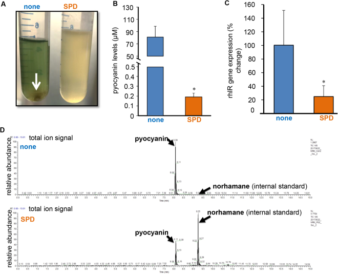
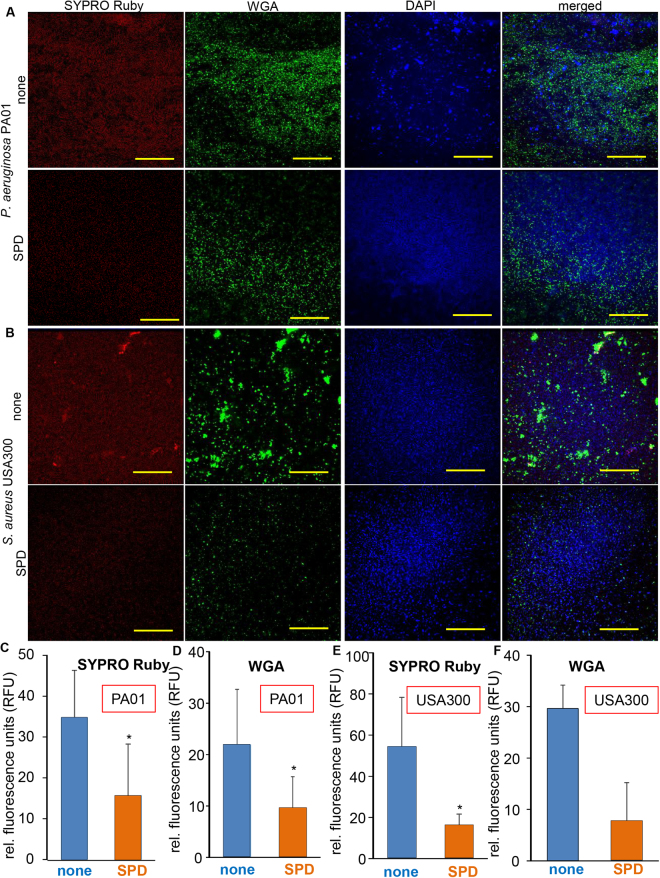
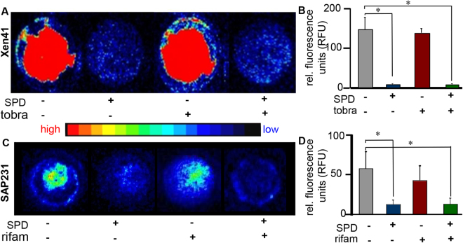
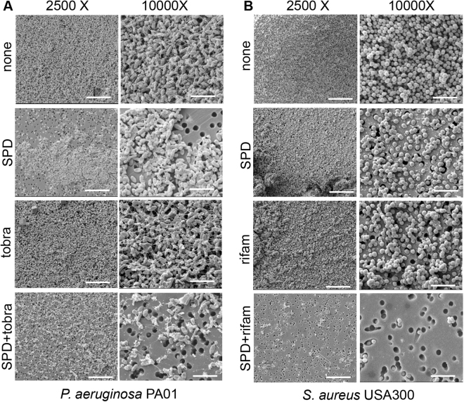
A 100% water-soluble surfactant polymer dressing (SPD) that is bio-compatible and non-ionic has been reported to improve wound closure in preliminary clinical studies. The mechanism of action of SPD in wound healing remains unclear. Biofilm infection is a significant problem that hinders proper wound closure. The objective of this study was to characterize the mechanism of action of SPD inhibition of bacterial biofilm development. Static biofilms (48 h) of the primary wound pathogens Pseudomonas aeruginosa (PA01), Staphylococcus aureus (USA300) were grown on polycarbonate membranes and treated with SPD with and without antibiotics for an additional 24 h. The standard antibiotics – tobramycin (10 μg/ml) for PA01 and rifampicin (10 μg/ml) for USA300, were used in these studies. Following 24 h treatment with and without antibiotics, the biofilms were characterized using scanning electron microscopy (SEM) structural imaging, in vitro imaging system (IVIS) proliferation imaging, colony forming units (CFU), viability assay, quantitative PCR (qPCR) for virulence gene expression. Because SPD is a surfactant based dressing, it potentially has a direct effect on Gram negative bacteria such as Pseudomonas primarily due to the lipid-based outer membrane of the bacteria. SPD is a surfactant based dressing that has potent anti-biofilm properties directly or in synergy with antibiotics.
Overview
The claims and statistics of this paper haven’t been checked yet.
Run the check — free, no account needed. It reads the open-access text and checks the sources it cites.
Integrity signals
Registry
Cites
Statistics
Figures
Verification
Claims the paper makes
The claims and statistics of this paper haven’t been checked yet.
Run the check — free, no account needed. It reads the open-access text and checks the sources it cites.
Claims other papers make about this one
Each is checked against this paper’s own text: does it say what the citing paper says it does?
In the citing paper
“WGA, a widely used lectin-based stain, is often used for the detection of EPS-encasing biofilm aggregates.42,43,70”
From SEMTWIST Quantification of Biofilm Infection in Human Chronic Wound Using Scanning Electron Microscopy and Machine Learning (2025) · cites this paper as [43]
In the citing paper
“To further strengthen our findings on the concordance between human evaluations and SEM data, this work relied on the highly specific PNA-FISH molecular technique widely recognized for its application to BFI detection.43”
From SEMTWIST Quantification of Biofilm Infection in Human Chronic Wound Using Scanning Electron Microscopy and Machine Learning (2025) · cites this paper as [43]
In the citing paper
“Fluorescence-based techniques such as wheat germ agglutinin (WGA) staining have shown promising results for in vitro biofilm experiments with limited efficacy for detecting biofilms within infected wound tissue samples.42,43”
From SEMTWIST Quantification of Biofilm Infection in Human Chronic Wound Using Scanning Electron Microscopy and Machine Learning (2025) · cites this paper as [43]
Self-check
Percentages, sums and reported differences re-computed from the paper’s own numbers.
The self-check needs the paper’s full text.
Citations
Cites
Mixed‐species biofilm compromises wound healing by disrupting epidermal barrier function
Roy, S., Elgharably, H., Sinha, M., Ganesh, K., Chaney, S., Mann, E., Miller, C., Khanna, S., Bergdall, V. K., Powell, H. M., et al. (2014) · The Journal of Pathology · doi:10.1002/path.4360 · Cited by 208
Self-citation
Research works printed but not linked: 70
- Frykberg, R. G. & Banks, J. Challenges in the treatment of chronic wounds. Advances in wound care 4, 560–582 (2015). · DOI
- Sen, C. K. et al. Human skin wounds: a major and snowballing threat to public health and the economy. Wound Repair and Regeneration 17, 763–771 (2009). · DOI · Paper page
- Jones, C. E. & Kennedy, J. P. Treatment options to manage wound biofilm. Advances in wound care 1, 120–126 (2012). · DOI
- Vert, M. et al. Terminology for biorelated polymers and applications (IUPAC Recommendations 2012). Pure and Applied Chemistry 84, 377–410 (2012). · DOI
- Flemming, H.-C. & Wingender, J. The biofilm matrix. Nature reviews. Microbiology 8, 623 (2010). · DOI · Paper page
- Dufour, D., Leung, V. & Lévesque, C. M. Bacterial biofilm: structure, function, and antimicrobial resistance. Endodontic Topics 22, 2–16 (2010). · DOI
- Parsek, M. R. & Singh, P. K. Bacterial biofilms: an emerging link to disease pathogenesis. Annual Reviews in Microbiology 57, 677–701 (2003). · DOI
- Donné, J. & Dewilde, S. Chapter Five-The Challenging World of Biofilm Physiology. Advances in microbial physiology 67, 235–292 (2015). · DOI
- Bjarnsholt, T. et al. The in vivo biofilm. Trends in microbiology 21, 466–474 (2013). · DOI · Paper page
- Singh, R., Ray, P., Das, A. & Sharma, M. Penetration of antibiotics through Staphylococcus aureus and Staphylococcus epidermidis biofilms. Journal of antimicrobial chemotherapy 65, 1955–1958 (2010). · DOI
- Cos, P., Tote, K., Horemans, T. & Maes, L. Biofilms: an extra hurdle for effective antimicrobial therapy. Current pharmaceutical design 16, 2279–2295 (2010). · DOI
- Steed, D. L., Donohoe, D., Webster, M. W. & Lindsley, L. Effect of extensive debridement and treatment on the healing of diabetic foot ulcers. Diabetic Ulcer Study Group. Journal of the American College of Surgeons 183, 61–64 (1996).
- Falanga, V. Wound healing and its impairment in the diabetic foot. The Lancet 366, 1736–1743 (2005). · DOI
- Steed, D. L. Debridement. The American journal of surgery 187, S71–S74 (2004). · DOI
- Ayello, E. A. & Cuddigan, J. E. Debridement: controlling the necrotic/cellular burden. Advances in skin & wound care 17, 66–75 (2004). · DOI
- Ousey, K. & McIntosh, C. Understanding wound bed preparation and wound debridement. British journal of community nursing 15 (2010). · DOI
- Stephen-Haynes, J. & Thompson, G. The different methods of wound debridement. British journal of community nursing 12 (2007). · DOI
- Falabella, A. F. Debridement and wound bed preparation. Dermatologic therapy 19, 317–325 (2006). · DOI
- Marberry, K. M. et al. Surfactant wound irrigation for the treatment of staphylococcal clinical isolates. Clinical orthopaedics and related research 403, 73–79 (2002). · DOI
- Anglen, J., Gainor, B., Simpson, W. & Christensen, G. The use of detergent irrigation for musculoskeletal wounds. International orthopaedics 27, 40–46 (2003). · DOI
- Bryant, C. A. et al. Search for a nontoxic surgical scrub solution for periorbital lacerations. Annals of emergency medicine 13, 317–321 (1984). · DOI
- Dire, D. J. & Welsh, A. P. A comparison of wound irrigation solutions used in the emergency department. Annals of emergency medicine 19, 704–708 (1990). · DOI
- Edlich, R. F. et al. Physical and chemical configuration of sutures in the development of surgical infection. Annals of surgery 177, 679 (1973). · DOI
- Howell, J. M., Bresnahan, K. A., Stair, T. O., Dhindsa, H. S. & Edwards, B. A. Comparison of effects of suture and cyanoacrylate tissue adhesive on bacterial counts in contaminated lacerations. Antimicrobial agents and chemotherapy 39, 559–560 (1995). · DOI
- Rodeheaver, G. Controversies in topical wound management. Wounds 1, 19–27 (1989).
- Magee, C. et al. Potentiation of wound infection by surgical drains. The American Journal of Surgery 131, 547–549 (1976). · DOI
- Faulkner, D. M. et al. A new stable pluronic® F68 gel carrier for antibiotics in contaminated wound treatment. The American journal of emergency medicine 15, 20–24 (1997). · DOI
- Black, J. S. & Drake, D. B. A prospective randomized trial comparing silver sulfadiazine cream with a water-soluble polyantimicrobial gel in partial-thickness burn wounds. Plastic Surgical Nursing 35, 46–49 (2015). · DOI
- Zölß, C. & Cech, J. D. Efficacy of a new multifunctional surfactant‐based biomaterial dressing with 1% silver sulphadiazine in chronic wounds. International wound journal 13, 738–743 (2016). · DOI
- Rodeheaver, G. T., Smith, S. L., Thacker, J. G., Edgerton, M. T. & Edlich, R. F. Mechanical cleansing of contaminated wounds with a surfactant. The American Journal of Surgery 129, 241–245 (1975). · DOI
- Fowler, E. B. et al. Evaluation of pluronic polyols as carriers for grafting materials: study in rat calvaria defects. Journal of periodontology 73, 191–197 (2002). · DOI
- Kant, V. et al. Topical pluronic F-127 gel application enhances cutaneous wound healing in rats. Acta Histochemica 116, 5–13, https://doi.org/10.1016/j.acthis.2013.04.010 (2014). · DOI
- Cho, H.-Y., Tsuchido, T., Ono, H. & Takano, M. Cell death of Bacillus subtilis caused by surfactants at low concentrations results from induced cell autolysis. Journal of Fermentation and Bioengineering 70, 11–14 (1990). · DOI
- Tsuchido, T., Ahn, Y.-H. & Takano, M. Lysis of Bacillus subtilis cells by glycerol and sucrose esters of fatty acids. Applied and environmental microbiology 53, 505–508 (1987). · DOI
- Tsuchido, T., Svarachorn, A., Soga, H. & Takano, M. Lysis and aberrant morphology of Bacillus subtilis cells caused by surfactants and their relation to autolysin activity. Antimicrobial agents and chemotherapy 34, 781–785 (1990). · DOI
- Majtan, V., Majtanova, L., Hostacka, A., Hybenova, D. & Mlynarcik, D. Effect of quaternary ammonium salts and amine oxides on Pseudomonas aeruginosa. Microbios 84, 41–51 (1995).
- Fitzgerald, K. A., Davies, A. & Russell, A. D. Uptake of 14C-chlorhexidine diacetate to Escherichia coli and Pseudomonas aeruginosa and its release by azolectin. FEMS Microbiol Lett 51, 327–332 (1989). · DOI
- Denyer, S. P. Mechanisms of action of antibacterial biocides. International biodeterioration & biodegradation 36, 227–245 (1995). · DOI
- Cowles, P. B. Alkyl sulfates: their selective bacteriostatic action. The Yale journal of biology and medicine 11, 33 (1938).
- Birkeland, J. M. & Steinhaus, E. A. Selective bacteriostatic action of sodium lauryl sulfate and of “Dreft. Proceedings of the Society for Experimental Biology and Medicine 40, 86–88 (1939). · DOI
- Baker, Z., Harrison, R. W. & Miller, B. F. & Technical Assistance of Robert, W. The Bactericidal Action of Synthetic Detergents. J Exp Med 74, 611–620 (1941). · DOI
- Armstrong, W. M. Enhancement of the biological activity of sodium dodecyl sulphate by inorganic cations. Nature 179, 780–781 (1957). · DOI
- Kabara, J. J. Structure-function relationships of surfactants as antimicrobial agents. J Soc Cosmet Chem 29, 733–741 (1978).
- Pesci, E. C., Pearson, J. P., Seed, P. C. & Iglewski, B. H. Regulation of las and rhl quorum sensing in Pseudomonas aeruginosa. Journal of bacteriology 179, 3127–3132 (1997). · DOI
- Miller, M. B. & Bassler, B. L. Quorum sensing in bacteria. Annu Rev Microbiol 55, 165–199, https://doi.org/10.1146/annurev.micro.55.1.165 (2001). · DOI
- Rutherford, S. T. & Bassler, B. L. Bacterial quorum sensing: its role in virulence and possibilities for its control. Cold Spring Harb Perspect Med 2, https://doi.org/10.1101/cshperspect.a012427 (2012). · DOI
- de Kievit, T. R. & Iglewski, B. H. Bacterial quorum sensing in pathogenic relationships. Infect Immun 68, 4839–4849 (2000). · DOI
- Antunes, L. C. et al. Inhibition of Salmonella host cell invasion by dimethyl sulfide. Appl Environ Microbiol 76, 5300–5304, https://doi.org/10.1128/AEM.00851-10 (2010). · DOI
- Percival, S. et al. Surfactants and their role in wound cleansing and biofilm management. Journal of Wound Care 26, 680–690 (2017). · DOI
- Ganesh, A., Nagendrababu, V., John, A. & Deivanayagam, K. The Effect of Addition of an EPS Degrading Enzyme with and without Detergent to 2% Chlorhexidine on Disruption of Enterococcus faecalis Biofilm: A Confocal Laser Scanning Microscopic Study. Journal of Clinical and Diagnostic Research: JCDR 9, ZC61 (2015).
- Phillips, P. L. et al. Antimicrobial dressing efficacy against mature Pseudomonas aeruginosa biofilm on porcine skin explants. International wound journal 12, 469–483 (2015). · DOI
- Bonnichsen, L. et al. Lipopeptide biosurfactant viscosin enhances dispersal of Pseudomonas fluorescens SBW25 biofilms. Microbiology 161, 2289–2297 (2015). · DOI
- Parsek, M. R. & Greenberg, E. Sociomicrobiology: the connections between quorum sensing and biofilms. Trends in microbiology 13, 27–33 (2005). · DOI
- Costerton, J. W., Stewart, P. S. & Greenberg, E. P. Bacterial biofilms: a common cause of persistent infections. Science 284, 1318–1322 (1999). · DOI · Paper page
- Stewart, P. S. Mechanisms of antibiotic resistance in bacterial biofilms. International Journal of Medical Microbiology 292, 107–113 (2002). · DOI
- Hentzer, M. et al. Attenuation of Pseudomonas aeruginosa virulence by quorum sensing inhibitors. The EMBO journal 22, 3803–3815 (2003). · DOI
- Solano, C., Echeverz, M. & Lasa, I. Biofilm dispersion and quorum sensing. Current opinion in microbiology 18, 96–104 (2014). · DOI
- Petrova, O. E. & Sauer, K. Escaping the biofilm in more than one way: desorption, detachment or dispersion. Current opinion in microbiology 30, 67–78 (2016). · DOI
- Harris, T. L., Worthington, R. J. & Melander, C. Potent Small‐Molecule Suppression of Oxacillin Resistance in Methicillin‐Resistant Staphylococcus aureus. Angewandte Chemie International Edition 51, 11254–11257 (2012). · DOI
- Fischbach, M. A. Combination therapies for combating antimicrobial resistance. Current opinion in microbiology 14, 519–523 (2011). · DOI
- Tamma, P. D., Cosgrove, S. E. & Maragakis, L. L. Combination therapy for treatment of infections with gram-negative bacteria. Clinical microbiology reviews 25, 450–470 (2012). · DOI
- Yang, Q., Larose, C., Della Porta, A. C., Schultz, G. S. & Gibson, D. J. A surfactant‐based wound dressing can reduce bacterial biofilms in a porcine skin explant model. International wound journal 14, 408–413 (2017). · DOI
- Hidron, A. I., Low, C. E., Honig, E. G. & Blumberg, H. M. Emergence of community-acquired meticillin-resistant Staphylococcus aureus strain USA300 as a cause of necrotising community-onset pneumonia. The Lancet infectious diseases 9, 384–392 (2009). · DOI
- Baker, P. et al. Exopolysaccharide biosynthetic glycoside hydrolases can be utilized to disrupt and prevent Pseudomonas aeruginosa biofilms. Science advances 2, e1501632 (2016). · DOI
- Dusane, D. H. et al. Effects of loading concentration, blood and synovial fluid on antibiotic release and anti-biofilm activity of bone cement beads. Journal of Controlled Release 248, 24–32 (2017). · DOI
- Dietrich, L. E., Price‐Whelan, A., Petersen, A., Whiteley, M. & Newman, D. K. The phenazine pyocyanin is a terminal signalling factor in the quorum sensing network of Pseudomonas aeruginosa. Molecular microbiology 61, 1308–1321 (2006). · DOI
- Lau, G. W., Hassett, D. J., Ran, H. & Kong, F. The role of pyocyanin in Pseudomonas aeruginosa infection. Trends in molecular medicine 10, 599–606 (2004). · DOI · Paper page
- Banerjee, J. et al. Silver-zinc redox-coupled electroceutical wound dressing disrupts bacterial biofilm. PloS one 10, e0119531 (2015). · DOI
- Elgharably, H. et al. First evidence of sternal wound biofilm following cardiac surgery. PloS one 8, e70360 (2013). · DOI
- Arciola, C. R., Campoccia, D., Ravaioli, S. & Montanaro, L. Polysaccharide intercellular adhesin in biofilm: structural and regulatory aspects. Frontiers in cellular and infection microbiology 5 (2015). · DOI
Cited by
Sorted by citations
Singh, S., Muniz De Oliveira, F., Wang, C., Kumar, M., Xuan, Y., DeMazumder, D., Sen, C. K., & Roy, S. (2025) · Advances in Wound Care · doi:10.1089/wound.2024.0291 · Cited by 6
Self-citation
Metadata
- Authors
- Das Ghatak, P., Mathew-Steiner, S. S., Pandey, P., Roy, S., & Sen, C. K.
- Venue
- Scientific Reports
- Published
- 2018-01-17
- Volume
- 8
- Issue
- 1
- ISSN
- 2045-2322
- PMC
- PMC5772662
- Cited by
- 56
- References
- 1
- Language
- en
About these checks
Registry, retraction, Cites and statistics results are computed, not judged, with one exception: a printed reference that no index lists is read once by a model before it counts as a citation error. Claims are extracted by a model and kept only when their quote appears verbatim in the text; each cited source is then checked, with a model’s help, for whether it says what the claim relies on. Those verdicts can be wrong — read the claim’s quote and the cited source before relying on one.
Statistics and arithmetic are recomputed, not judged.
Confidence bands are shown only for fields calibrated on our annotated test set. None is calibrated yet, so no band is shown.
What counts as a claim ›How we measure confidence ›How we check this ›
Are you an author, or do you see a problem with this page? Request a review
