

The DOI matches its Crossref record; 14 of the 14 references it cites were found in a registry or an index.
Structures of Angptl3 and Angptl4, modulators of triglyceride levels and coronary artery disease
Abstract
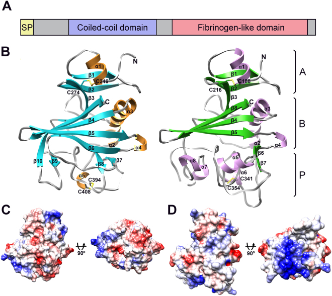
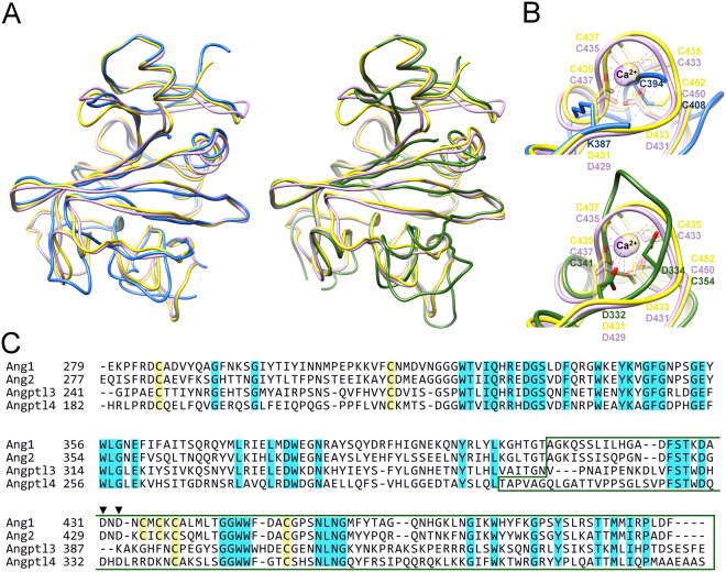
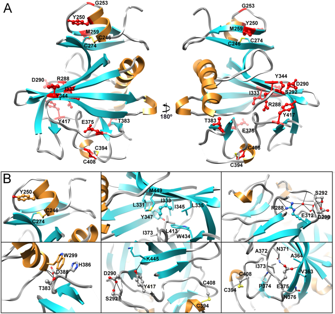
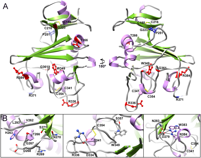
Coronary artery disease is the most common cause of death globally and is linked to a number of risk factors including serum low density lipoprotein, high density lipoprotein, triglycerides and lipoprotein(a). Recently two proteins, angiopoietin-like protein 3 and 4, have emerged from genetic studies as being factors that significantly modulate plasma triglyceride levels and coronary artery disease. The exact function and mechanism of action of both proteins remains to be elucidated, however, mutations in these proteins results in up to 34% reduction in coronary artery disease and inhibition of function results in reduced plasma triglyceride levels. Here we report the crystal structures of the fibrinogen-like domains of both proteins. These structures offer new insights into the reported loss of function mutations, the mechanisms of action of the proteins and open up the possibility for the rational design of low molecular weight inhibitors for intervention in coronary artery disease.
Overview
The claims and statistics of this paper haven’t been checked yet.
Run the check — free, no account needed. It reads the open-access text and checks the sources it cites.
Integrity signals
Registry
Cites
Statistics
Figures
Verification
Claims the paper makes
The claims and statistics of this paper haven’t been checked yet.
Run the check — free, no account needed. It reads the open-access text and checks the sources it cites.
Claims other papers make about this one
Each is checked against this paper’s own text: does it say what the citing paper says it does?
In the citing paper
“However, only the C-terminal fibrinogen domains of ANGPTL3 and ANGPTL4 have been solved (183)”
From Regulation of lipoprotein metabolism by ANGPTL3, ANGPTL4, and ANGPTL8 (2021) · cites this paper as [183]
Self-check
Percentages, sums and reported differences re-computed from the paper’s own numbers.
The self-check needs the paper’s full text.
Citations
Cites
Evolocumab and Clinical Outcomes in Patients with Cardiovascular Disease
Sabatine, M. S., Giugliano, R. P., Keech, A. C., Honarpour, N., Wiviott, S. D., Murphy, S. A., Kuder, J. F., Wang, H., Liu, T., Wasserman, S. M., et al. (2017) · New England Journal of Medicine · doi:10.1056/nejmoa1615664 · Cited by 6,138
Towards automated crystallographic structure refinement with phenix.refine
Afonine, P. V., Grosse-Kunstleve, R. W., Echols, N., Headd, J. J., Moriarty, N. W., Mustyakimov, M., Terwilliger, T. C., Urzhumtsev, A., Zwart, P. H., & Adams, P. D. (2012) · Acta Crystallographica Section D Biological Crystallography · doi:10.1107/s0907444912001308 · Cited by 5,511
Sequence Variations in PCSK9, Low LDL, and Protection against Coronary Heart Disease
Cohen, J. C., Boerwinkle, E., Mosley, T. H., & Hobbs, H. H. (2006) · New England Journal of Medicine · doi:10.1056/nejmoa054013 · Cited by 3,181
A comprehensive 1000 Genomes–based genome-wide association meta-analysis of coronary artery disease
the CARDIoGRAMplusC4D Consortium (2015) · Nature Genetics · doi:10.1038/ng.3396 · Cited by 2,447
Triglycerides and Cardiovascular Disease: A Scientific Statement From the American Heart Association
Miller, M., Stone, N. J., Ballantyne, C., Bittner, V., Criqui, M. H., Ginsberg, H. N., Goldberg, A. C., Howard, W. J., Jacobson, M. S., Kris-Etherton, P. M., et al. (2011) · Circulation · doi:10.1161/cir.0b013e3182160726 · Cited by 1,871
Loss-of-Function Mutations in APOC3, Triglycerides, and Coronary Disease
The TG and HDL Working Group of the Exome Sequencing Project, N. H. (2014) · New England Journal of Medicine · doi:10.1056/nejmoa1307095 · Cited by 1,071
Genetic and Pharmacologic Inactivation of ANGPTL3 and Cardiovascular Disease
Dewey, F. E., Gusarova, V., Dunbar, R. L., O’Dushlaine, C., Schurmann, C., Gottesman, O., McCarthy, S., Van Hout, C. V., Bruse, S., Dansky, H. M., et al. (2017) · New England Journal of Medicine · doi:10.1056/nejmoa1612790 · Cited by 885
Dysfunctional HDL and atherosclerotic cardiovascular disease
Rosenson, R. S., Brewer, H. B., Ansell, B. J., Barter, P., Chapman, M. J., Heinecke, J. W., Kontush, A., Tall, A. R., & Webb, N. R. (2016) · Nature Reviews Cardiology · doi:10.1038/nrcardio.2015.124 · Cited by 822
Exome sequencing identifies rare LDLR and APOA5 alleles conferring risk for myocardial infarction
Do, R., Stitziel, N. O., Won, H.-H., Jørgensen, A. B., Duga, S., Angelica Merlini, P., Kiezun, A., Farrall, M., Goel, A., Zuk, O., et al. (2015) · Nature · doi:10.1038/nature13917 · Cited by 650
Cardiovascular and Metabolic Effects of ANGPTL3 Antisense Oligonucleotides
Graham, M. J., Lee, R. G., Brandt, T. A., Tai, L.-J., Fu, W., Peralta, R., Yu, R., Hurh, E., Paz, E., McEvoy, B. W., et al. (2017) · New England Journal of Medicine · doi:10.1056/nejmoa1701329 · Cited by 573
Inactivating Variants in ANGPTL4 and Risk of Coronary Artery Disease
Dewey, F. E., Gusarova, V., O’Dushlaine, C., Gottesman, O., Trejos, J., Hunt, C., Van Hout, C. V., Habegger, L., Buckler, D., Lai, K.-M. V., et al. (2016) · New England Journal of Medicine · doi:10.1056/nejmoa1510926 · Cited by 497
Coding Variation in ANGPTL4, LPL, and SVEP1 and the Risk of Coronary Disease
Myocardial Infarction Genetics and CARDIoGRAM Exome Consortia Investigators (2016) · New England Journal of Medicine · doi:10.1056/nejmoa1507652 · Cited by 493
ANGPTL3 deficiency and protection against coronary artery disease
Stitziel, N. O., Khera, A. V., Wang, X., Bierhals, A. J., Vourakis, A. C., Sperry, A. E., Natarajan, P., Klarin, D., Emdin, C. A., Zekavat, S. M., et al. (2017) · Journal of the American College of Cardiology · doi:10.1016/j.jacc.2017.02.030 · Cited by 467
Angiopoietin-like 3 in lipoprotein metabolism
Kersten, S. (2017) · Nature Reviews Endocrinology · doi:10.1038/nrendo.2017.119 · Cited by 220
Research works printed but not linked: 42
- Wang, H. et al. Global, regional, and national life expectancy, all-cause mortality, and cause-specific mortality for 249 causes of death, 1980–2015: a systematic analysis for the Global Burden of Disease Study 2015. Lancet 388, 1775–1812 (2016). · DOI
- Nakanishi, R. et al. Current but not past smoking increases the risk of cardiac events: insights from coronary computed tomographic angiography. Eur Heart J 36, 1031–1040 (2015). · DOI
- Lehrke, M. & Marx, N. Diabetes mellitus and heart failure. Am J Med 130, S40–S50 (2017). · DOI
- Khera, A. V. & Kathiresan, S. Genetics of coronary artery disease: discovery, biology and clinical translation. Nat Rev Genet 18, 331–344 (2017). · DOI · Paper page
- Kivimäki, M. et al. Job strain as a risk factor for coronary heart disease: a collaborative meta-analysis of individual participant data. Lancet 380, 1491–1497 (2012). · DOI
- Cosselman, K. E., Navas-Acien, A. & Kaufman, J. D. Environmental factors in cardiovascular disease. Nat Rev Cardiol 12, 627–642 (2015). · DOI · Paper page
- Nordestgaard, B. D. & Varbo, A. Triglycerides and cardiovascular disease. Lancet 384, 626–635 (2014). · DOI · Paper page
- Reiner, Ž. Hypertriglycerideaemia and risk of coronary artery disease. Nat Rev Cardiol 14, 401–411 (2017). · DOI
- Natarajan, P. & Kathiresan, S. PCSK9 inhibitors. Cell 165, 1037 (2016). · DOI
- Thomas, M. & Augustin, H. G. The role of the angiopoietins in vascular morphogenesis. Angiogenesis 12, 125–137 (2009). · DOI
- Ono, M. et al. Protein region important for regulation of lipid metabolism in Angiopoietin-like 3 (ANGPTL3): ANGPTL3 is cleaved and activated in vivo. J Biol Chem 278, 41804–41809 (2003). · DOI · Paper page
- Quagliarini, F. et al. Atypical angiopoietin-like protein that regulates ANGPTL3. Proc Natl Acad Sci USA 109, 19751–19756 (2012). · DOI · Paper page
- Chomel, C. et al. Interaction of the coiled-coil domain with glycosaminoglycans protects angiopoietin-like 4 from proteolysis and regulates its antiangiogenic activity. FASEB J 23, 940–949 (2009). · DOI
- Arca, M., Minicocci, I. & Maranghi, M. The angiopoietin-like protein 3: a hepatokine with expanding role in metabolism. Curr Opin Lipidol 24, 313–320 (2013). · DOI
- Grootaert, C., Van de Wiele, T., Verstraete, W., Bracke, M. & Vanhoecke, B. Angiopoietin-like protein 4: health effects, modulating agents and structure-function relationships. Expert Rev Proteomics 9, 181–199 (2012). · DOI
- Goh, Y. Y. et al. Angiopoietin-like 4 interacts with matrix proteins to modulate wound healing. J Biol Chem 285, 32999–33009 (2010). · DOI
- Chong, H. C. et al. Angiopoietin-like 4 stimulates STAT3-mediated iNOS expression and enhances angiogenesis to accelerate would healing in diabetic mice. Mol Ther 22, 1593–1604 (2014). · DOI
- Musunuru, K. & Kathiresan, S. Is ANGPLT3 the next PCSK9? Nat Rev Endocrinol 13, 503–504 (2017). · DOI
- Romeo, S. et al. Rare loss-of-function mutations in ANGPTL family members contribute to plasma triglyceride levels in humans. J Clin Invest 119, 70–79 (2009).
- Pisciotta, L. et al. Characterization of three kindreds with familial combined hypolipidemia caused by loss-of-function mutations in ANGPTL3. Circ Cardiovasc Genet 5, 42–50 (2012). · DOI · Paper page
- Wang, X., Wang, D. & Shan, Z. Clinical and genetic analysis of a family diagnosed with familial hypobetalipoproteinemia in which the proband was diagnosed with diabetes mellitus. Atherosclerosis 239, 552–556 (2017). · DOI
- Romeo, S. et al. Population-based resequencing of ANGPTL4 uncovers variations that reduce triglycerides and increase HDL. Nat Genet 39, 513–516 (2007). · DOI · Paper page
- Smart-Halajko, M. C. et al. ANGPTL4 variants E40K and T266M are associated with lower fasting triglyceride levels in non-Hispanic white Americans from the Look AHEAD clinical trial. BMC Med Genet 12, 89 (2011). · DOI
- Shimizugawa, T. et al. ANGPTL3 decrease very low density lipoprotein triglyceride clearance by inhibition of lipoprotein lipase. J Biol Chem 277, 33742–33748 (2002). · DOI · Paper page
- Yoshida, K., Shimizugawa, T., Ono, M. & Furukawa, H. Angiopoietin-like protein 4 is a potent hyperlipidemia-inducing factor in mice and inhibitor of lipoprotein lipase. J Lipid Res 43, 1770–1772 (2002). · DOI · Paper page
- Holm, L. & Rosenström, P. Dali server: conservation mapping in 3D. Nucl Acids Res 38, W545–549 (2010). · DOI · Paper page
- Yu, X. et al. Structural basis for angiopoietin-1 mediated signaling initiation. Proc Natl Acad Sci USA 110, 7205–7210 (2013). · DOI
- Barton, W. A., Tzvetkova, D. & Nikolov, D. B. Structure of the angiopoietin-2 receptor binding domain and identification of surfaces involved in Tie2 recognition. Structure 13, 825–832 (2005). · DOI
- Garlatti, V. et al. Structural Basis for innate immune sensing by M-Ficolin and its control by a pH-dependent conformational switch. J Biol Chem 282, 35814–35820 (2007). · DOI
- Garlatti, V. et al. Structural insights into the innate immune recognition specificities of L- and H-Ficolins. EMBO J 26, 623–633 (2007). · DOI
- Kairies, N. et al. The 2.0-A crystal structure of tachylectin 5A provides evidence for the common origin of the innate immunity and the blood coagulation systems. Proc Natl Acad Sci USA 98, 13519–13524 (2001). · DOI
- Koopman, J., Haverkate, F., Briet, E. & Lord, S. T. A congenitally abnormal fibrinogen (Vlissingen) with 6-base deletion of γ-chain gene, causing defective calcium binding and impaired fibrin polymerization. J Biol Chem 266, 13456–13461 (1991). · DOI
- Kawabata, S. & Tsuda, R. Molecular basis of non-self recognition by horseshoe crab tachylectins. Biochim Biophys Acta 1572, 414–421 (2002). · DOI
- Pósfai, G. et al. Emergent properties of reduced-genome Escherichia coli. Science 312, 1044–1046 (2006). · DOI
- Gaciarz, A. et al. Systematic screening of soluble expression of antibody fragments in the cytoplasm of E. coli. Micro Cell Fact 15, 22 (2016). · DOI · Paper page
- Gaciarz, A. & Ruddock, L.W. Complementary determining regions and frameworks contribute to the disulfide bond independent folding of intrinsically stable scFv. PLoS One 12, e0189964 (2017). · DOI
- Svensson, O., Monaco, S., Popov, A. N., Nurizzo, D. & Bowler, M. W. Fully automatic characterization and data collection from crystals of biological macromolecules. Acta Cryst D71, 1757–1767 (2015).
- Kabsch, W. XDS. Acta Cryst D66, 125–132 (2010).
- McCoy, A. J. et al. Phaser crystallographic software. J Appl Crystallogr 40, 658–674 (2007). · DOI · Paper page
- Emsley, P., Lohkamp, B., Scott, W. & Cowtan, K. Features and development of Coot. Acta Crystallogr D Biol Crystallogr 66, 486–501 (2010). · DOI · Paper page
- Chen, V. B. et al. MolProbity: all-atom structure validation for macromolecular crystallography. Acta Cryst D66, 16–21 (2010).
- Pettersen, E. F. et al. UCSF Chimera–a visualization system for exploratory research and analysis. J Comput Chem. 25, 1605–12 (2004). · DOI
Cited by
Sorted by citations
Lipoprotein lipase and its regulators: an unfolding story
Wu, S. A., Kersten, S., & Qi, L. (2021) · Trends in Endocrinology & Metabolism · doi:10.1016/j.tem.2020.11.005 · Cited by 225
Regulation of lipoprotein metabolism by ANGPTL3, ANGPTL4, and ANGPTL8
Sylvers-Davie, K. L., & Davies, B. S. J. (2021) · American Journal of Physiology-Endocrinology and Metabolism · doi:10.1152/ajpendo.00195.2021 · Cited by 154
Metadata
- Authors
- Biterova, E., Esmaeeli, M., Alanen, H. I., Saaranen, M., & Ruddock, L. W.
- ORCID
- Saaranen, Mirva
- Venue
- Scientific Reports
- Published
- 2018-04-30
- Volume
- 8
- Issue
- 1
- ISSN
- 2045-2322
- PMC
- PMC5928061
- Cited by
- 53
- References
- 14
- Language
- en
About these checks
Registry, retraction, Cites and statistics results are computed, not judged, with one exception: a printed reference that no index lists is read once by a model before it counts as a citation error. Claims are extracted by a model and kept only when their quote appears verbatim in the text; each cited source is then checked, with a model’s help, for whether it says what the claim relies on. Those verdicts can be wrong — read the claim’s quote and the cited source before relying on one.
Statistics and arithmetic are recomputed, not judged.
Confidence bands are shown only for fields calibrated on our annotated test set. None is calibrated yet, so no band is shown.
What counts as a claim ›How we measure confidence ›How we check this ›
Are you an author, or do you see a problem with this page? Request a review
