Cited by 60RNA modifications and cancerRNA and protein synthesis mechanismsRNA regulation and disease

The DOI matches its Crossref record; 2 of the 2 references it cites were found in a registry or an index.

A single N1-methyladenosine on the large ribosomal subunit rRNA impacts locally its structure and the translation of key metabolic enzymes

Open access

Published version of a preprint

Abstract

The entire chemical modification repertoire of yeast ribosomal RNAs and the enzymes responsible for it have recently been identified. Nonetheless, in most cases the precise roles played by these chemical modifications in ribosome structure, function and regulation remain totally unclear. Previously, we demonstrated that yeast Rrp8 methylates m 1 A 645 of 25S rRNA in yeast. Here, using mung bean nuclease protection assays in combination with quantitative RP-HPLC and primer extension, we report that 25S/28S rRNA of S . pombe , C . albicans and humans also contain a single m 1 A methylation in the helix 25.1. We characterized nucleomethylin (NML) as a human homolog of yeast Rrp8 and demonstrate that NML catalyzes the m 1 A 1322 methylation of 28S rRNA in humans. Our in vivo structural probing of 25S rRNA, using both DMS and SHAPE, revealed that the loss of the Rrp8-catalyzed m 1 A modification alters the conformation of domain I of yeast 25S rRNA causing translation initiation defects detectable as halfmers formation, likely because of incompetent loading of 60S on the 43S-preinitiation complex. Quantitative proteomic analysis of the yeast Δrrp8 mutant strain using 2D-DIGE, revealed that loss of m 1 A 645 impacts production of specific set of proteins involved in carbohydrate metabolism, translation and ribosome synthesis. In mouse, NML has been characterized as a metabolic disease-associated gene linked to obesity. Our findings in yeast also point to a role of Rrp8 in primary metabolism. In conclusion, the m 1 A modification is crucial for maintaining an optimal 60S conformation, which in turn is important for regulating the production of key metabolic enzymes.

Overview

The claims and statistics of this paper haven’t been checked yet.

Run the check — free, no account needed. It reads the open-access text and checks the sources it cites.

A few free checks a day · papers already verified open instantly · results are public

Integrity signals

Registry

Crossref · 10.1038/s41598-018-30383-z

Cites

2 of 2 references verified0 citation errors · 0 not checkable

Statistics

Figures

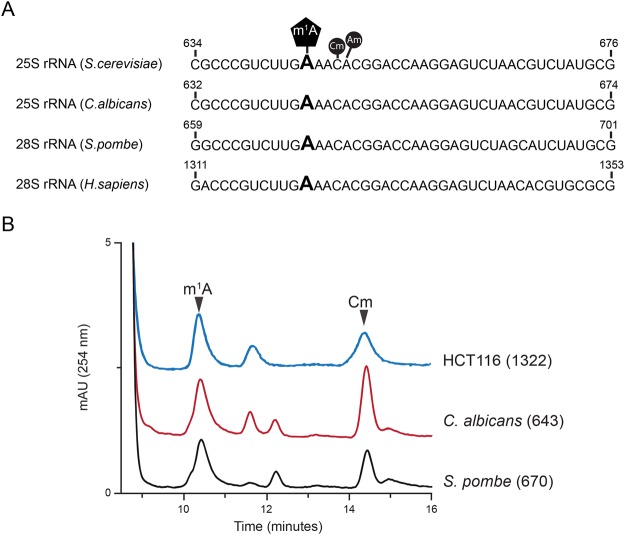
Figure 1 Conservation of the N1-methyladenosine in helix 25.1 of 25/28S rRNA. (A) rRNA sequence alignment of helix 25.1 from S. cerevisiae, C. albicans, S. pombe and H. sapiens. The m1A modified residue is highly conserved within helix 25.1 (B) Overlaid RP-HPLC chromatograms of nucleosides derived from fragments isolated using mung bean nuclease assay, containing m1A643 (Ca-Oligo643) isolated from wild-type C. albicans strain RM1000, m1A670 (Sp-Oligo643) isolated from wild-type S. pombe strain CBS356 and m1A1322 (Hs-Oligo1322) isolated from HCT116 cells. Cm (2′-O-ribose methylated cytidine) and Am (2′-O-ribose methylated adenosine) peaks are also labelled.
Figure 1 Conservation of the N1-methyladenosine in helix 25.1 of 25/28S rRNA. (A) rRNA sequence alignment of helix 25.1 from S. cerevisiae, C. albicans, S. pombe and H. sapiens. The m1A modified residue is highly conserved within helix 25.1 (B) Overlaid RP-HPLC chromatograms of nucleosides derived from fragments isolated using mung bean nuclease assay, containing m1A643 (Ca-Oligo643) isolated from wild-type C. albicans strain RM1000, m1A670 (Sp-Oligo643) isolated from wild-type S. pombe strain CBS356 and m1A1322 (Hs-Oligo1322) isolated from HCT116 cells. Cm (2′-O-ribose methylated cytidine) and Am (2′-O-ribose methylated adenosine) peaks are also labelled.CC BY · PubMed Central
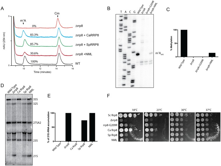
Figure 2 Heterologous expression of RRP8 homologs and biochemical and phenotypic analysis. (A) Overlaid RP-HPLC chromatograms of the nucleosides derived from fragments isolated using mung bean nuclease assay, containing m1A645 (Sc-Oligo645) isolated from wild-type (WT), deletion mutant (Δrrp8) or deletion mutant strains expressing homologs of RRP8 (CaRRP8 for C. albicans Rrp8, SpRRP8 for S. pombe Rrp8 and NML for H. sapiens Rrp8/NML). (B) Primer extension analysis of Δrrp8, the loss of methylation mutant rrp8G209R and the heterologous expressed human homolog NML. The presence of m1A at position 645 in the 25S rRNA from the wild-type cells led to a strong stop. The expression of the human homolog NML in the deletion mutant of RRP8 of S. cerevisiae also led to a stop at this position to an extent of about 40% compared with S. cerevisiae wild-type. (C) Percent methylation was quantified from the intensity of primer extension stop (panel B) using ImageJ software (http://imagej.nih.gov/ij/). (D) Northern blot analysis of RRP8 homologs expressed in S. cerevisiae. 5 µg of isolated total RNA was separated on an 1.2% agarose-gel and subsequently blotted onto a Hybond H+ nylon membrane. The membrane was hybridized with radioactive (32P)-labeled probes against the internal transcribed spacer 1 (ITS1) (region between cleavage sites A2-A3). (E) Percent of 21S rRNA accumulation in panel D was quantified using ImageJ software. (F) Ten-fold dilutions of strains expressing RRP8, RRP8 homologs or catalytically-defective mutants. Cells spotted onto solid YEPD plates were incubated at four different temperatures illustrating the cold temperature sensitive phenotype of Δrrp8. The differences in the methylation and complementation of Rrp8 homologs of other fungal species and human cells are likely due to sequence specificities that may influence how efficiently a heterologous protein is assembled within the ribosome assembly machinery of the host cell. For example, the S. pombe protein may be incorporated in maturing subunits sufficiently well to restore methylation but not well enough to suppress the RNA cleavage defect, while the C. albicans protein may be recruited to pre-ribosomes more efficiently, allowing it to suppress both defects.
Figure 2 Heterologous expression of RRP8 homologs and biochemical and phenotypic analysis. (A) Overlaid RP-HPLC chromatograms of the nucleosides derived from fragments isolated using mung bean nuclease assay, containing m1A645 (Sc-Oligo645) isolated from wild-type (WT), deletion mutant (Δrrp8) or deletion mutant strains expressing homologs of RRP8 (CaRRP8 for C. albicans Rrp8, SpRRP8 for S. pombe Rrp8 and NML for H. sapiens Rrp8/NML). (B) Primer extension analysis of Δrrp8, the loss of methylation mutant rrp8G209R and the heterologous expressed human homolog NML. The presence of m1A at position 645 in the 25S rRNA from the wild-type cells led to a strong stop. The expression of the human homolog NML in the deletion mutant of RRP8 of S. cerevisiae also led to a stop at this position to an extent of about 40% compared with S. cerevisiae wild-type. (C) Percent methylation was quantified from the intensity of primer extension stop (panel B) using ImageJ software (http://imagej.nih.gov/ij/). (D) Northern blot analysis of RRP8 homologs expressed in S. cerevisiae. 5 µg of isolated total RNA was separated on an 1.2% agarose-gel and subsequently blotted onto a Hybond H+ nylon membrane. The membrane was hybridized with radioactive (32P)-labeled probes against the internal transcribed spacer 1 (ITS1) (region between cleavage sites A2-A3). (E) Percent of 21S rRNA accumulation in panel D was quantified using ImageJ software. (F) Ten-fold dilutions of strains expressing RRP8, RRP8 homologs or catalytically-defective mutants. Cells spotted onto solid YEPD plates were incubated at four different temperatures illustrating the cold temperature sensitive phenotype of Δrrp8. The differences in the methylation and complementation of Rrp8 homologs of other fungal species and human cells are likely due to sequence specificities that may influence how efficiently a heterologous protein is assembled within the ribosome assembly machinery of the host cell. For example, the S. pombe protein may be incorporated in maturing subunits sufficiently well to restore methylation but not well enough to suppress the RNA cleavage defect, while the C. albicans protein may be recruited to pre-ribosomes more efficiently, allowing it to suppress both defects.CC BY · PubMed Central
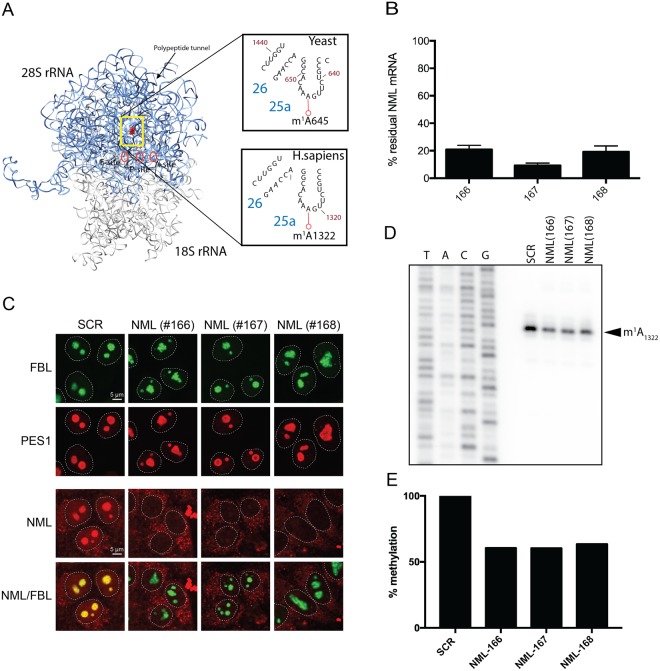
Figure 3 Validation of nucleomethylin (NML) as the human m1A large subunit rRNA methyltransferase. (A) 3-D structure model of the human rRNA. 4UG0 pdb file together with UCSF-Chimera were used to create the rRNA surface model. The N1-methylated residue A1322 in the 28S rRNA is highlighted as a red sphere. For comparison, the corresponding region of the yeast 25S rRNA is shown in an inset. (B) siRNA-mediated depletion of NML is highly efficient at mRNA level. Total RNA extracted from HCT116 cells transfected with a siRNA specific for NML (siRNA #166 to #168) and incubated for 72 h, was tested by RT-qPCR. (C) NML is a nucleolar protein. Immunofluorescence with an antibody against NML was performed in HCT116 cells stably expressing a green fluorescent fibrillarin construct, which labels the dense fibrillar component of the nucleolus. The granular component protein PES1 was detected with a specific antibody (Bethyl Lab Inc). (D) Primer extension analysis of m1A methylation after siRNA-mediated depletion of NML in HCT116 cells. 32P-labeled primer complementary to nucleotides 1362 to 1381 of human 28S rRNA were used for methylation analysis of m1A1322. (E) The reduction of stop intensity at the m1A1322 in siRNA-mediated depleted cells was quantified using ImageJ software and estimated to be of ~60%.
Figure 3 Validation of nucleomethylin (NML) as the human m1A large subunit rRNA methyltransferase. (A) 3-D structure model of the human rRNA. 4UG0 pdb file together with UCSF-Chimera were used to create the rRNA surface model. The N1-methylated residue A1322 in the 28S rRNA is highlighted as a red sphere. For comparison, the corresponding region of the yeast 25S rRNA is shown in an inset. (B) siRNA-mediated depletion of NML is highly efficient at mRNA level. Total RNA extracted from HCT116 cells transfected with a siRNA specific for NML (siRNA #166 to #168) and incubated for 72 h, was tested by RT-qPCR. (C) NML is a nucleolar protein. Immunofluorescence with an antibody against NML was performed in HCT116 cells stably expressing a green fluorescent fibrillarin construct, which labels the dense fibrillar component of the nucleolus. The granular component protein PES1 was detected with a specific antibody (Bethyl Lab Inc). (D) Primer extension analysis of m1A methylation after siRNA-mediated depletion of NML in HCT116 cells. 32P-labeled primer complementary to nucleotides 1362 to 1381 of human 28S rRNA were used for methylation analysis of m1A1322. (E) The reduction of stop intensity at the m1A1322 in siRNA-mediated depleted cells was quantified using ImageJ software and estimated to be of ~60%.CC BY · PubMed Central
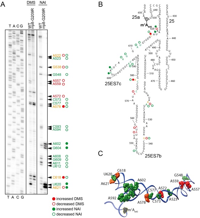
Figure 4 Loss of m1A645 leads to structural changes within the LSU. (A) Representative gel showing structure probing with DMS or SHAPE analysis with NAI in the rrp8G209R loss of methylation mutant. 32P-labeled primer (helix25_StrPrb) complementary to nucleotides 2428 to 2448 of human 28S rRNA were used for the analysis. Bands corresponding to modified residues are marked in red for DMS, green for NAI and orange for both. (B) 2-D structure model with mapped modified residues as described in (A). (C) The residues sensitive to chemical modification are represented as spheres on a 3-D structure model of helix 25 (color scheme as in panel A). The model was made using Chimera and PDB file 4V88.
Figure 4 Loss of m1A645 leads to structural changes within the LSU. (A) Representative gel showing structure probing with DMS or SHAPE analysis with NAI in the rrp8G209R loss of methylation mutant. 32P-labeled primer (helix25_StrPrb) complementary to nucleotides 2428 to 2448 of human 28S rRNA were used for the analysis. Bands corresponding to modified residues are marked in red for DMS, green for NAI and orange for both. (B) 2-D structure model with mapped modified residues as described in (A). (C) The residues sensitive to chemical modification are represented as spheres on a 3-D structure model of helix 25 (color scheme as in panel A). The model was made using Chimera and PDB file 4V88.CC BY · PubMed Central
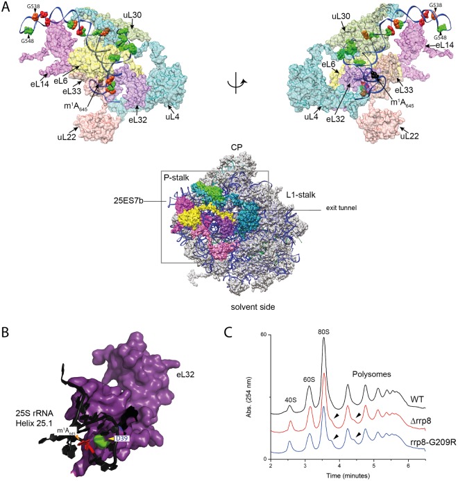
Figure 5 Loss of m1A645 leads to topological changes within the rRNA in the area of eL32 association (A) eL32 ‘neuron-like’ interaction network with five neighboring ribosomal proteins (uL22/L17, eL33/L33, eL6/L6, eL14/L14, uL30/L7 and uL4/L4) of the LSU around expansion segment ES7b and 7c. The models were made using Chimera and PDB files 4V88 (B) Possible electrostatic interaction of the D39 residue (green) of eL32 (colored violet) with the m1A645 modification (in red). 25S rRNA is colored black (C) Overlayed polysome profile analysis of S. cerevisiae wild-type (WT), Δrrp8 and rrp8G209R mutant strains. Polysome profiles of Δrrp8 and rrp8G209R mutants were performed to detect the translational status in comparison with its isogenic wild-type. Half-mer formation in Δrrp8 and rrp8G209R mutants are indicated by arrows.
Figure 5 Loss of m1A645 leads to topological changes within the rRNA in the area of eL32 association (A) eL32 ‘neuron-like’ interaction network with five neighboring ribosomal proteins (uL22/L17, eL33/L33, eL6/L6, eL14/L14, uL30/L7 and uL4/L4) of the LSU around expansion segment ES7b and 7c. The models were made using Chimera and PDB files 4V88 (B) Possible electrostatic interaction of the D39 residue (green) of eL32 (colored violet) with the m1A645 modification (in red). 25S rRNA is colored black (C) Overlayed polysome profile analysis of S. cerevisiae wild-type (WT), Δrrp8 and rrp8G209R mutant strains. Polysome profiles of Δrrp8 and rrp8G209R mutants were performed to detect the translational status in comparison with its isogenic wild-type. Half-mer formation in Δrrp8 and rrp8G209R mutants are indicated by arrows.CC BY · PubMed Central
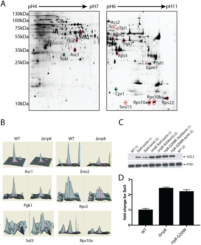
Figure 6 2D proteome analysis of rrp8 mutant. (A) Scanned images of a typical 2-D DIGE gel of 50 µg protein from the Cy3 labeled protein pool used for the rrp8 mutant strain (left gel: pH 4–7, right gel: pH 6–11). Differentially expressed proteins are annotated and were identified using ESI-MS/MS. Differentially expressed protein spots are annotated in red (down-regulated) or green (up-regulated). (B) DeCyder output showing the representative differentially expressed protein spots of Asc1, Eno2, Pgk1, Rps5, Sol3 and Rps10A along with their 3-D fluorescence intensity profiles. (C and D) Sol3 up-regulation is related to loss of m1A645 methylation. Western blot analysis using anti-HA antibodies to analyze the expression of 6xHA tagged SOL3 in wild-type, Δrrp8 and rrp8G209R mutant cells. As loading control, PYK1 was detected with a specific antibody. The represented fold changes in SOL3 expression were quantified using ImageJ software (http://imagej.nih.gov/ij/).
Figure 6 2D proteome analysis of rrp8 mutant. (A) Scanned images of a typical 2-D DIGE gel of 50 µg protein from the Cy3 labeled protein pool used for the rrp8 mutant strain (left gel: pH 4–7, right gel: pH 6–11). Differentially expressed proteins are annotated and were identified using ESI-MS/MS. Differentially expressed protein spots are annotated in red (down-regulated) or green (up-regulated). (B) DeCyder output showing the representative differentially expressed protein spots of Asc1, Eno2, Pgk1, Rps5, Sol3 and Rps10A along with their 3-D fluorescence intensity profiles. (C and D) Sol3 up-regulation is related to loss of m1A645 methylation. Western blot analysis using anti-HA antibodies to analyze the expression of 6xHA tagged SOL3 in wild-type, Δrrp8 and rrp8G209R mutant cells. As loading control, PYK1 was detected with a specific antibody. The represented fold changes in SOL3 expression were quantified using ImageJ software (http://imagej.nih.gov/ij/).CC BY · PubMed Central
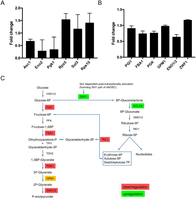
Figure 7 Quantitative PCR and biochemical activities of differentially regulated proteins in Δrrp8. (A) RTqPCR analysis of mRNAs from proteins that were differentially regulated in the rrp8 mutant strain are represented as fold change in mRNA expression. Expression of Asc1, Rps5, Eno2 and Pgk1 is regulated at the transcriptional level, whereas mRNA levels of Sol3 and Rps10A are not different from their wild-type level of expression. Data analyzed with the software REST®.41. (B) Regulation of specific activities of enzymes of the carbohydrate metabolism within Δrrp8. Changes in the specific enzyme activities of phosphoglucose isomerase (PGI1), fructose-1,6-bisphosphate aldolase (FBA1), phosphoglycerate kinase (PGK1), phosphoglycerate mutase (GPM1), enolase (ENO1/ENO2) and glucose-6-phosphate dehydrogenase (ZWF1) are represented as fold change. Activities of enzymes involved in the glycolytic pathway are downregulated due to deletion of RRP8 as compared to wild-type. Only activity of ZFW1, first enzyme of the pentose-phosphate pathway, is slightly upregulated. (C) Schematic overview of regulations in the carbohydrate metabolism in cells with inactive RRP8.
Figure 7 Quantitative PCR and biochemical activities of differentially regulated proteins in Δrrp8. (A) RTqPCR analysis of mRNAs from proteins that were differentially regulated in the rrp8 mutant strain are represented as fold change in mRNA expression. Expression of Asc1, Rps5, Eno2 and Pgk1 is regulated at the transcriptional level, whereas mRNA levels of Sol3 and Rps10A are not different from their wild-type level of expression. Data analyzed with the software REST®.41. (B) Regulation of specific activities of enzymes of the carbohydrate metabolism within Δrrp8. Changes in the specific enzyme activities of phosphoglucose isomerase (PGI1), fructose-1,6-bisphosphate aldolase (FBA1), phosphoglycerate kinase (PGK1), phosphoglycerate mutase (GPM1), enolase (ENO1/ENO2) and glucose-6-phosphate dehydrogenase (ZWF1) are represented as fold change. Activities of enzymes involved in the glycolytic pathway are downregulated due to deletion of RRP8 as compared to wild-type. Only activity of ZFW1, first enzyme of the pentose-phosphate pathway, is slightly upregulated. (C) Schematic overview of regulations in the carbohydrate metabolism in cells with inactive RRP8.CC BY · PubMed Central

Verification

Claims the paper makes

The claims and statistics of this paper haven’t been checked yet.

Run the check — free, no account needed. It reads the open-access text and checks the sources it cites.

A few free checks a day · papers already verified open instantly · results are public

Self-check

Percentages, sums and reported differences re-computed from the paper’s own numbers.

The self-check needs the paper’s full text.

Citations

Cites

Research works printed but not linked: 55

  1. Vogel, C. & Marcotte, E. M. Insights into the regulation of protein abundance from proteomic and transcriptomic analyses. Nat Rev Genet 5, 1512 (2012).
  2. Gebauer, F. & Hentze, M. W. Molecular mechanisms of translational control. Nat. Rev. Mol. Cell Biol. 5, 827–835 (2004). · DOI · Paper page
  3. Tian, Q. et al. Integrated Genomic and Proteomic Analyses of Gene Expression in Mammalian Cells. Mol. Cell Proteomics 3, 960–969 (2004). · DOI
  4. Vogel, C. et al. Sequence signatures and mRNA concentration can explain two-thirds of protein abundance variation in a human cell line. Molecular Systems Biology 6, 404 (2010). · DOI · Paper page
  5. Ramakrishnan, V. Ribosome structure and the mechanism of translation. Cell 108, 557–572 (2002). · DOI
  6. Woolford, J. L. & Baserga, S. J. Ribosome biogenesis in the yeast Saccharomyces cerevisiae. Genetics 195, 643–681 (2013). · DOI
  7. Henras, A. K., Plisson-Chastang, C., O’Donohue, M.-F., Chakraborty, A. & Gleizes, P.-E. An overview of pre-ribosomal RNA processing in eukaryotes. WIREs RNA 6, 225–242 (2015). · DOI
  8. Nissen, P., Hansen, J., Ban, N., Moore, P. B. & Steitz, T. A. The structural basis of ribosome activity in peptide bond synthesis. Science 289, 920–930 (2000). · DOI · Paper page
  9. Polacek, N. & Mankin, A. S. The Ribosomal Peptidyl Transferase Center: Structure, Function, Evolution, Inhibition. Crit. Rev. Biochem. Mol. Biol. 40, 285–311 (2005). · DOI
  10. Deusser, E. & Wittmann, H.-G. Biological Sciences: Ribosomal Proteins: Variation of the Protein Composition in Escherichia coli Ribosomes as Function of Growth Rate. Nature 238, 269–270 (1972). · DOI
  11. Byrgazov, K., Vesper, O. & Moll, I. Ribosome heterogeneity: another level of complexity in bacterial translation regulation. Curr Opin Microbiol 16, 133–139 (2013). · DOI
  12. Xue, S. & Barna, M. Specialized ribosomes: a new frontier in gene regulation and organismal biology. Nat. Rev. Mol. Cell Biol. 13, 355–369 (2012). · DOI
  13. Larsson, O. & Nadon, R. Gene expression - time to change point of view? Biotechnol. Genet. Eng. Rev. 25, 77–92 (2008). · DOI
  14. Vesper, O. et al. Selective Translation of Leaderless mRNAs by Specialized Ribosomes Generated by MazF in Escherichia coli. Cell 147, 147–157 (2011). · DOI
  15. Kondrashov, N. et al. Ribosome-Mediated Specificity in Hox mRNA Translation and Vertebrate Tissue Patterning. Cell 145, 383–397 (2011). · DOI
  16. Schosserer, M. et al. Methylation of ribosomal RNA by NSUN5 is a conserved mechanism modulating organismal lifespan. Nat Commun 6, 6158 (2015). · DOI
  17. Shi, Z. et al. Heterogeneous Ribosomes Preferentially Translate Distinct Subpools of mRNAs Genome-wide. Mol. Cell, 1–21, https://doi.org/10.1016/j.molcel.2017.05.021 (2017). · DOI
  18. Simsek, D. & Barna, M. An emerging role for the ribosome as a nexus for post-translational modifications. Curr Opin Cell Biol 45, 92–101 (2017). · DOI
  19. Decatur, W. A. & Fournier, M. J. rRNA modifications and ribosome function. Trends Biochem Sci 27, 344–351 (2002). · DOI
  20. Sharma, S. & Lafontaine, D. L. J. ‘View From A Bridge’: A New Perspective on Eukaryotic rRNA Base Modification. Trends Biochem Sci 40, 560–575 (2015). · DOI
  21. Lafontaine, D., Delcour, J., Glasser, A. L., Desgrès, J. & Vandenhaute, J. The DIM1 gene responsible for the conservedm6(2)Am6(2)A dimethylation in the 3′-terminal loop of 18 S rRNA is essential in yeast. J Mol Biol 241, 492–497 (1994). · DOI
  22. White, J. et al. Bud23 methylates G1575 of 18S rRNA and is required for efficient nuclear export of pre-40S subunits. Mol. Cell. Biol. 28, 3151–3161 (2008). · DOI
  23. Sharma, S. et al. Yeast Kre33 and human NAT10 are conserved 18S rRNA cytosine acetyltransferases that modify tRNAs assisted by the adaptor Tan1/THUMPD1. NAR 43, 2242–2258 (2015). · DOI
  24. Meyer, B. et al. Ribosome biogenesis factor Tsr3 is the aminocarboxypropyl transferase responsible for 18S rRNA hypermodification in yeast and humans. NAR 44, 4304–4316 (2016). · DOI
  25. Sharma, S. et al. Specialized box C/D snoRNPs act as antisense guides to target RNA base acetylation. Plos Genet 13, e1006804 (2017). · DOI
  26. Sharma, S., Watzinger, P., Kötter, P. & Entian, K.-D. Identification of a novel methyltransferase, Bmt2, responsible for the N-1-methyl-adenosine base modification of 25S rRNA in Saccharomyces cerevisiae. NAR 41, 5428–5443 (2013). · DOI
  27. Sharma, S. et al. Identification of novel methyltransferases, Bmt5 and Bmt6, responsible for the m3U methylations of 25S rRNA in Saccharomyces cerevisiae. NAR 42, 3246–3260 (2014). · DOI
  28. Sharma, S., Yang, J., Watzinger, P., Kötter, P. & Entian, K.-D. Yeast Nop2 and Rcm1 methylate C2870 and C2278 of the 25S rRNA, respectively. NAR 41, 9062–9076 (2013). · DOI
  29. Yang, J. et al. Mapping of Complete Set of Ribose and Base Modifications of Yeast rRNA by RP-HPLC and Mung Bean Nuclease Assay. Plos One 11, e0168873 (2016). · DOI
  30. Peifer, C. et al. Yeast Rrp8p, a novel methyltransferase responsible for m1A 645 base modification of 25S rRNA. NAR 41, 1151–1163 (2013). · DOI
  31. Waku, T. et al. NML-mediated rRNA base methylation links ribosomal subunit formation to cell proliferation in a p53-dependent manner. J Cell Sci 129, 2382–2393 (2016). · DOI
  32. Yokoyama, W. et al. rRNA adenine methylation requires T07A9.8 gene as rram-1 in Caenorhabditis elegans. The Journal of Biochemistry 163, 465–474 (2018). · DOI
  33. Bousquet-Antonelli, C., Vanrobays, E., Gélugne, J. P., Caizergues-Ferrer, M. & Henry, Y. Rrp8p is a yeast nucleolar protein functionally linked to Gar1p and involved in pre-rRNA cleavage at site A2. RNA 6, 826–843 (2000). · DOI
  34. Agris, P. F., Sierzputowska-Gracz, H. & Smith, C. Transfer RNA contains sites of localized positive charge: carbon NMR studies of [13C]methyl-enriched Escherichia coli and yeast tRNAPhe. Biochemistry 25, 5126–5131 (1986). · DOI
  35. Macon, J. B. & Wolfenden, R. 1-Methyladenosine. Dimroth rearrangement and reversible reduction. Biochemistry 7, 3453–3458 (1968). · DOI
  36. Mikhailov, S. N. et al. Chemical incorporation of 1-methyladenosine into oligonucleotides. NAR 30, 1124–1131 (2002). · DOI
  37. Peattie, D. A. & Gilbert, W. Chemical probes for higher-order structure in RNA. Proc. Natl. Acad. Sci. USA 77, 4679–4682 (1980). · DOI
  38. Spitale, R. C. et al. RNA SHAPE analysis in living cells. Nat Chem Biol 9, 18–20 (2012). · DOI
  39. Tijerina, P., Mohr, S. & Russell, R. DMS footprinting of structured RNAs and RNA–protein complexes. Nat Protoc 2, 2608–2623 (2007). · DOI
  40. Poirot, O. & Timsit, Y. Neuron-Like Networks Between Ribosomal Proteins Within the Ribosome. Sci. Rep. 6, 26485 (2016). · DOI
  41. Helser, T. L., Baan, R. A. & Dahlberg, A. E. Characterization of a 40S ribosomal subunit complex in polyribosomes of Saccharomyces cerevisiae treated with cycloheximide. 1, 51–57 (1981). · DOI
  42. Stanford, D. R. Division of Labor Among the Yeast Sol Proteins Implicated in tRNA Nuclear Export and Carbohydrate Metabolism. Genetics 168, 117–127 (2004). · DOI
  43. Duffieux, F. Molecular characterisation of the first two enzymes of the pentose-phosphate pathway of trypanosoma brucei. J Biol Chem, https://doi.org/10.1074/jbc.M004266200 (2000). · DOI
  44. Maitra, P. K. & Lobo, Z. A kinetic study of glycolytic enzyme synthesis in yeast. J. Biol. Chem. 246, 475–488 (1971). · DOI
  45. Zeller, C. E., Parnell, S. C. & Dohlman, H. G. The RACK1 Ortholog Asc1 Functions as a G-protein beta Subunit Coupled to Glucose Responsiveness in Yeast. J Biol Chem 282, 25168–25176 (2007). · DOI
  46. Lafontaine, D. L. J. Noncoding RNAs in eukaryotic ribosome biogenesis and function. Nature Structural & Molecular Biology 22, 11–19 (2015). · DOI
  47. De Keersmaecker, K., Sulima, S. O. & Dinman, J. D. Ribosomopathies and the paradox of cellular hypo-to hyperproliferation. Blood, https://doi.org/10.1182/blood-2014-10 (2015). · DOI
  48. Danilova, N. & Gazda, H. T. Ribosomopathies: how a common root can cause a tree of pathologies. Dis. Model. Mech. 8, 1013–1026 (2015). · DOI
  49. Di Tommaso, P. et al. T-Coffee: a web server for the multiple sequence alignment of protein and RNA sequences using structural information and homology extension. NAR 39, W13–7 (2011). · DOI
  50. Gouet, P. ESPript/ENDscript: extracting and rendering sequence and 3D information from atomic structures of proteins. NAR 31, 3320–3323 (2003). · DOI
  51. Edelheit, O., Hanukoglu, A. & Hanukoglu, I. Simple and efficient site-directed mutagenesis using two single-primer reactions in parallel to generate mutants for protein structure-function studies. BMC Biotechnol. 9, 61 (2009). · DOI
  52. Yang, J., Sharma, S., Kötter, P. & Entian, K.-D. Identification of a new ribose methylation in the 18S rRNA of S. cerevisiae. NAR 43, 2342–2352 (2015). · DOI
  53. Sharma, S., Marchand, V., Motorin, Y. & Lafontaine, D. L. J. Identification of sites of 2′-O-methylation vulnerability in human ribosomal RNAs by systematic mapping. Sci. Rep. 7, 11490 (2017). · DOI
  54. Pfaffl, M. W., Horgan, G. W. & Dempfle, L. Relative expression software tool (REST) for group-wise comparison and statistical analysis of relative expression results in real-time PCR. NAR 30, e36 (2002). · DOI
  55. Itzhaki, R. F. & Gill, D. M. A Micro-Biuret Method For Estimating Proteins. Analytical Biochemistry 9, 401–410 (1964). · DOI

Metadata

Authors
Sharma, S., Hartmann, J. D., Watzinger, P., Klepper, A., Peifer, C., Kötter, P., Lafontaine, D. L. J., & Entian, K.-D.
Affiliations
  • Goethe University Frankfurt (DE)
  • Université Libre de Bruxelles (BE)
Corresponding author
  • Sharma, Sunny
  • Entian, Karl-Dieter
Venue
Scientific Reports
Published
2018-08-09
Volume
8
Issue
1
Pages
11904-11904
ISSN
2045-2322 (ISSN-L)
OpenAlex
W2950745853
Cited by
60
References
2
Citations by year
2026: 4 · 2025: 8 · 2024: 9 · 2023: 7 · 2022: 14 · 2021: 11 · 2020: 2 · 2019: 5
FWCI
1.60
Type
journal article
Language
en
Open access
Locations
  • Scientific Reports (publishedVersion, cc-by)
  • bioRxiv (Cold Spring Harbor Laboratory) (acceptedVersion, cc-by-nc-nd)
  • Europe PMC (PubMed Central) (publishedVersion, cc-by)
  • PubMed Central (publishedVersion, cc-by)
  • PubMed (publishedVersion)
  • Dépôt institutionnel de l'Université libre de Bruxelles (Université Libre de Bruxelles) (submittedVersion, other-oa)
  • DOAJ (DOAJ: Directory of Open Access Journals) (submittedVersion, cc-by-sa)
  • Publication Server of Goethe University Frankfurt am Main (Goethe University Frankfurt) (submittedVersion, cc-by-nc-nd)
  • Publication Server of Goethe University Frankfurt am Main (Goethe University Frankfurt) (submittedVersion, cc-by)
Funders
  • Waalse Gewest
  • Deutsche Forschungsgemeinschaft
  • Fonds De La Recherche Scientifique - FNRS
  • Fédération Wallonie-Bruxelles
  • European Regional Development Fund
Topics
  • Life Sciences › Biochemistry, Genetics and Molecular Biology › Molecular Biology › RNA modifications and cancer
  • Life Sciences › Biochemistry, Genetics and Molecular Biology › Molecular Biology › RNA and protein synthesis mechanisms
  • Life Sciences › Biochemistry, Genetics and Molecular Biology › Molecular Biology › RNA regulation and disease
Keywords
N1-methyladenosine, 25S rRNA, 28S rRNA, rRNA methylation, translation initiation, 43S preinitiation complex, carbohydrate metabolism, metabolic enzymes, Schizosaccharomyces pombe, Candida albicans, human ribosome, obesity-related genes
MeSH terms
Protein Domains, Adenosine, Base Sequence, Humans, Methylation, Methyltransferases, Mutation, Nuclear Proteins, Nucleic Acid Conformation, Protein O-Methyltransferase, Ribosomal Proteins, RNA, Ribosomal, Electrophoresis, Gel, Two-Dimensional, RNA-Binding Proteins, Saccharomyces cerevisiae Proteins, Proteomics, HCT116 Cells, Ribosome Subunits, Large

About these checks

Registry, retraction, Cites and statistics results are computed, not judged, with one exception: a printed reference that no index lists is read once by a model before it counts as a citation error. Claims are extracted by a model and kept only when their quote appears verbatim in the text; each cited source is then checked, with a model’s help, for whether it says what the claim relies on. Those verdicts can be wrong — read the claim’s quote and the cited source before relying on one.

Statistics and arithmetic are recomputed, not judged.

Confidence bands are shown only for fields calibrated on our annotated test set. None is calibrated yet, so no band is shown.

What counts as a claim ›How we measure confidence ›How we check this ›

Are you an author, or do you see a problem with this page? Request a review