

The DOI matches its Crossref record; 64 of the 64 references it cites were found in a registry or an index.
Scribble co-operatively binds multiple α1D-adrenergic receptor C-terminal PDZ ligands
Open accessAbstract
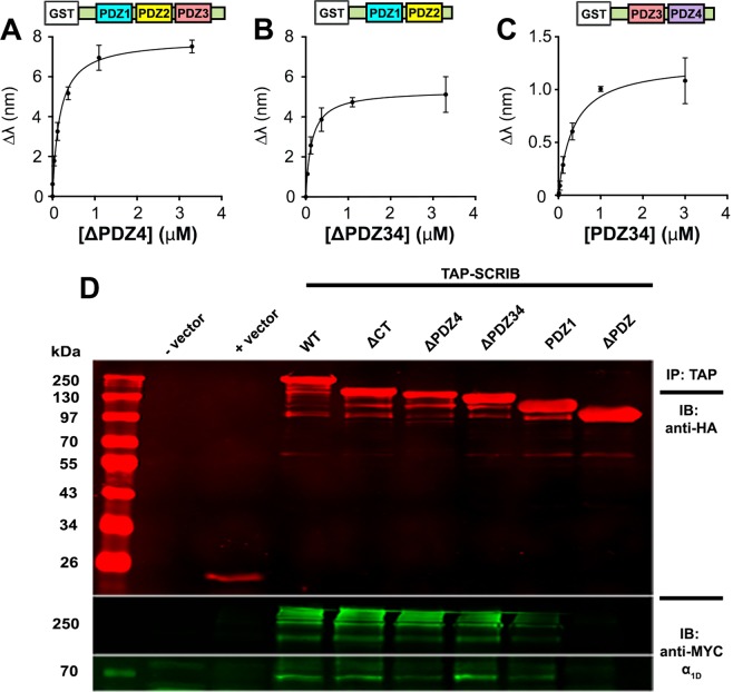
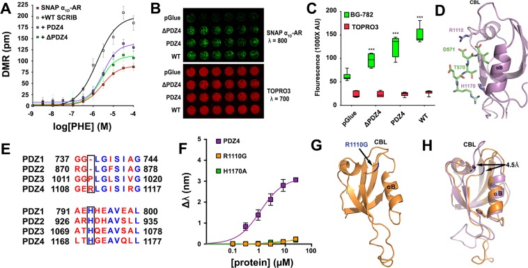
Many G protein-coupled receptors (GPCRs) are organized as dynamic macromolecular complexes in human cells. Unraveling the structural determinants of unique GPCR complexes may identify unique protein:protein interfaces to be exploited for drug development. We previously reported α 1D-adrenergic receptors (α 1D-ARs) – key regulators of cardiovascular and central nervous system function – form homodimeric, modular PDZ protein complexes with cell-type specificity. Towards mapping α 1D-AR complex architecture, biolayer interferometry (BLI) revealed the α 1D-AR C-terminal PDZ ligand selectively binds the PDZ protein scribble (SCRIB) with >8x higher affinity than known interactors syntrophin, CASK and DLG1. Complementary in situ and in vitro assays revealed SCRIB PDZ domains 1 and 4 to be high affinity α 1D-AR PDZ ligand interaction sites. SNAP-GST pull-down assays demonstrate SCRIB binds multiple α 1D-AR PDZ ligands via a co-operative mechanism. Structure-function analyses pinpoint R1110 PDZ4 as a unique, critical residue dictating SCRIB:α 1D-AR binding specificity. The crystal structure of SCRIB PDZ4 R1110G predicts spatial shifts in the SCRIB PDZ4 carboxylate binding loop dictate α 1D-AR binding specificity. Thus, the findings herein identify SCRIB PDZ domains 1 and 4 as high affinity α 1D-AR interaction sites, and potential drug targets to treat diseases associated with aberrant α 1D-AR signaling.
Overview
The claims and statistics of this paper haven’t been checked yet.
Free: it reads the open-access text and checks the first 5 claims that cite a source we can check. Create a free account to check every claim.
Integrity signals
Registry
Cites
Statistics
Figures




Verification
Claims the paper makes
The claims and statistics of this paper haven’t been checked yet.
Free: it reads the open-access text and checks the first 5 claims that cite a source we can check. Create a free account to check every claim.
Claims other papers make about this one
Each is checked against this paper’s own text: does it say what the citing paper says it does?
The source backs part of the claim, not all of it — or backs it for a different population, condition or comparison.
In the citing paper
“The finding that the isolated PDZ2 does not bind to TMIGD1 whereas the PDZ2 mutant in the context of the Scrib/PDZ domain construct impairs binding to TMIGD1 opens the possibility that regions adjacent to PDZ2 may influence ligand interactions with PDZ243,44.”
From Membrane recruitment of the polarity protein Scribble by the cell adhesion receptor TMIGD1 (2023) · cites this paper as [43]
Experimental — accuracy not yet measured
Self-check
Percentages, sums and reported differences re-computed from the paper’s own numbers.
The self-check needs the paper’s full text.
Citations
Cites
Coot : model-building tools for molecular graphics
Emsley, P., & Cowtan, K. (2004) · Acta Crystallographica Section D Biological Crystallography · doi:10.1107/s0907444904019158 · Cited by 30,571
PHENIX : a comprehensive Python-based system for macromolecular structure solution
Adams, P. D., Afonine, P. V., Bunkóczi, G., Chen, V. B., Davis, I. W., Echols, N., Headd, J. J., Hung, L.-W., Kapral, G. J., Grosse-Kunstleve, R. W., et al. (2010) · Acta Crystallographica Section D Biological Crystallography · doi:10.1107/s0907444909052925 · Cited by 23,716
Phaser crystallographic software
McCoy, A. J., Grosse-Kunstleve, R. W., Adams, P. D., Winn, M. D., Storoni, L. C., & Read, R. J. (2007) · Journal of Applied Crystallography · doi:10.1107/s0021889807021206 · Cited by 20,612
Kabsch, W. (2010) · Acta Crystallographica Section D Biological Crystallography · doi:10.1107/s0907444909047337 · Cited by 16,463
Overview of the CCP 4 suite and current developments
Winn, M. D., Ballard, C. C., Cowtan, K. D., Dodson, E. J., Emsley, P., Evans, P. R., Keegan, R. M., Krissinel, E. B., Leslie, A. G. W., McCoy, A., et al. (2011) · Acta Crystallographica Section D Biological Crystallography · doi:10.1107/s0907444910045749 · Cited by 12,353
PHENIX: a comprehensive Python-based system for macromolecular structure solution
Adams, P. D., Afonine, P. V., Bunkóczi, G., Chen, V. B., Davis, I. W., Echols, N., Headd, J. J., Hung, L.-W., Kapral, G. J., Grosse-Kunstleve, R. W., et al. (2012) · International Tables for Crystallography · doi:10.1107/97809553602060000865 · Cited by 7,334
Scaling and assessment of data quality
Evans, P. (2006) · Acta Crystallographica Section D Biological Crystallography · doi:10.1107/s0907444905036693 · Cited by 4,374
How many drug targets are there?
Overington, J. P., Al-Lazikani, B., & Hopkins, A. L. (2006) · Nature Reviews Drug Discovery · doi:10.1038/nrd2199 · Cited by 3,505
Recognition of Unique Carboxyl-Terminal Motifs by Distinct PDZ Domains
Songyang, Z., Fanning, A. S., Fu, C., Xu, J., Marfatia, S. M., Chishti, A. H., Crompton, A., Chan, A. C., Anderson, J. M., & Cantley, L. C. (1997) · Science · doi:10.1126/science.275.5296.73 · Cited by 1,385
Doyle, D. A., Lee, A., Lewis, J., Kim, E., Sheng, M., & MacKinnon, R. (1996) · Cell · doi:10.1016/s0092-8674(00)81307-0 · Cited by 1,142
Cited by
Li, Q., Yin, G., Wang, J., Li, L., Liang, Q., Zhao, X., Chen, Y., Zheng, X., & Zhao, X. (2022) · TrAC Trends in Analytical Chemistry · doi:10.1016/j.trac.2022.116728 · Cited by 29
Predicting Drug-Target Interaction Via Self-Supervised Learning
Chen, J., Zhang, L., Cheng, K., Jin, B., Lu, X., & Che, C. (2022) · IEEE Transactions on Computational Biology and Bioinformatics · doi:10.1109/tcbb.2022.3153963 · Cited by 27
“structure-based (or molecular docking-based) strategy [4], the ligand-based strategy [5] and machine learning-based approaches [1], [2], [6], [7], [8], [9].”
MethodUnraveling the Functional Significance of Unstructured Regions in G Protein-Coupled Receptors
Maggio, R., Fasciani, I., Petragnano, F., Coppolino, M. F., Scarselli, M., & Rossi, M. (2023) · Biomolecules · doi:10.3390/biom13101431 · Cited by 19
“As mentioned earlier, the free COOH-terminal peptide of the C-terminus of several GPCRs have the ability to bind PDZ domains [57,58].”
BackgroundChristensen, N. R., Pedersen, C. P., Sereikaité, V., Pedersen, J. N., Vistrup-Parry, M., Sørensen, A. T., Otzen, D. E., Teilum, K., Madsen, K. L., & Strømgaard, K. (2022) · iScience · doi:10.1016/j.isci.2022.103808 · Cited by 16
Janezic, E. M., Lauer, S. M., Williams, R. G., Chungyoun, M., Lee, K.-S., Navaluna, E. D., Lau, H.-T., Ong, S., & Hague, C. (2020) · Scientific Reports · doi:10.1038/s41598-020-64102-4 · Cited by 15
Self-citation
Metadata
- Authors
- Janezic, E. M., Harris, D.-A., Dinh, D., Lee, K.-S., Stewart, A., Hinds, T. R., Hsu, P. L., Zheng, N., & Hague, C.
- Affiliations
- University of Washington (US)
- Howard Hughes Medical Institute (US)
- Corresponding author
- Hague, Chris
- Venue
- Scientific Reports
- Published
- 2019-10-01
- Volume
- 9
- Issue
- 1
- Pages
- 14073-14073
- ISSN
- 2045-2322 (ISSN-L)
- PMC
- PMC6773690
- OpenAlex
- W2977907496
- Cited by
- 15
- References
- 64
- Citations by year
- 2026: 1 · 2025: 1 · 2024: 1 · 2023: 4 · 2022: 4 · 2021: 2 · 2020: 2
- FWCI
- 0.66
- Type
- journal article
- Language
- en
- Open access
- gold
- doi.org
- Licence
- cc-by
- creativecommons.org/licenses/by/4.0 (tdm)
- creativecommons.org/licenses/by/4.0 (vor)
- Locations
- Scientific Reports (publishedVersion, cc-by)
- PubMed Central (publishedVersion, cc-by)
- PubMed (publishedVersion)
- DOAJ (DOAJ: Directory of Open Access Journals) (submittedVersion)
- Funders
- U.S. Department of Health & Human Services | NIH | National Institute of General Medical Sciences (T32GM007750, R01GM100893)
- Howard Hughes Medical Institute
- Topics
- Life Sciences › Biochemistry, Genetics and Molecular Biology › Molecular Biology › Receptor Mechanisms and Signaling
- Life Sciences › Neuroscience › Cellular and Molecular Neuroscience › Neuroscience and Neuropharmacology Research
- Life Sciences › Biochemistry, Genetics and Molecular Biology › Molecular Biology › Protein Kinase Regulation and GTPase Signaling
- Keywords
- alpha1D-adrenergic receptor, Scribble, PDZ ligand, receptor homodimerization, syntrophin, CASK, DLG1, binding specificity, protein-protein interaction, therapeutic target
- MeSH terms
- Binding Sites, Humans, Interferometry, Membrane Proteins, Structure-Activity Relationship, Receptors, Adrenergic, alpha-1, Crystallography, X-Ray, Tumor Suppressor Proteins, Immunoprecipitation, PDZ Domains, HEK293 Cells, Molecular Docking Simulation
About these checks
Registry, retraction, Cites and statistics results are computed, not judged, with one exception: a printed reference that no index lists is read once by a model before it counts as a citation error. Claims are extracted by a model and kept only when their quote appears verbatim in the text; each cited source is then checked, with a model’s help, for whether it says what the claim relies on. Those verdicts can be wrong — read the claim’s quote and the cited source before relying on one.
Statistics and arithmetic are recomputed, not judged.
Confidence bands are shown only for fields calibrated on our annotated test set. None is calibrated yet, so no band is shown.
What counts as a claim ›How we measure confidence ›How we check this ›
Are you an author, or do you see a problem with this page? Request a review
How to cite
Cite CoChat for the checks on this page, and the paper for its own findings.
- This page
- CoChat. (2026). Citation check: Scribble co-operatively binds multiple α1D-adrenergic receptor C-terminal PDZ ligands. https://app.cochat.ai/papers/10.1038/s41598-019-50671-6
- The paper
- Janezic, E. M., Harris, D.-A., Dinh, D., Lee, K.-S., Stewart, A., Hinds, T. R., Hsu, P. L., Zheng, N., & Hague, C. (2019). Scribble co-operatively binds multiple α1D-adrenergic receptor C-terminal PDZ ligands. Scientific Reports. https://doi.org/10.1038/s41598-019-50671-6
Data sources
Bibliographic data: Semantic Scholar, OpenAlex, Crossref.
Contains information from the Semantic Scholar Academic Graph Datasets, made available under the ODC Attribution License (ODC-BY 1.0).
Semantic Scholar’s data is described in: Kinney, R., et al. (2023). The Semantic Scholar Open Data Platform. arXiv:2301.10140.
