

Effects of codon optimization on coagulation factor IX translation and structure: Implications for protein and gene therapies
Open accessThe DOI matches its Crossref record; 72 of the 72 references it cites were found in a registry or an index.
Abstract
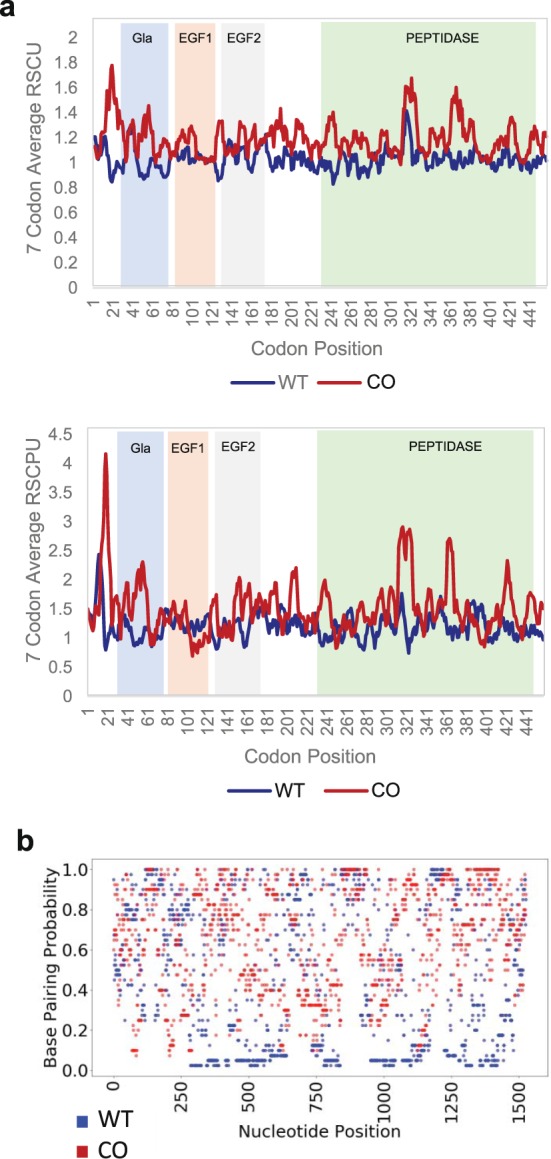
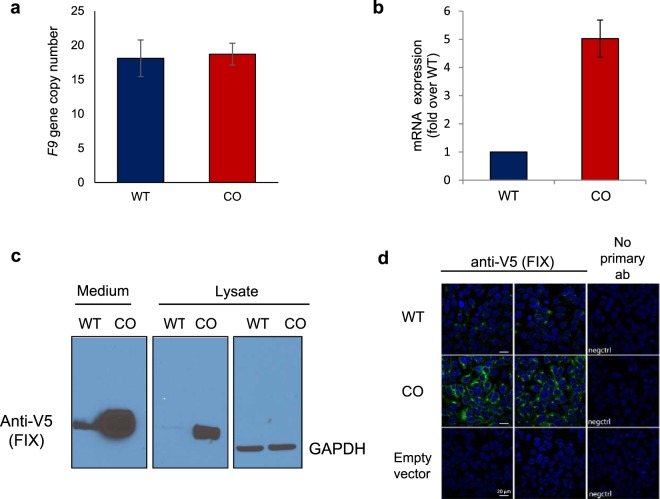
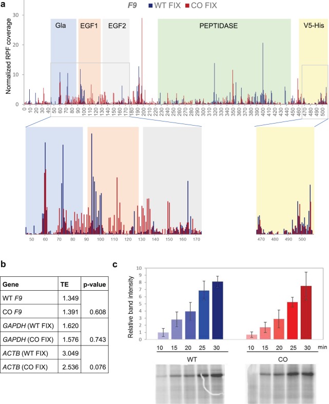
Synonymous codons occur with different frequencies in different organisms, a phenomenon termed codon usage bias. Codon optimization, a common term for a variety of approaches used widely by the biopharmaceutical industry, involves synonymous substitutions to increase protein expression. It had long been presumed that synonymous variants, which, by definition, do not alter the primary amino acid sequence, have no effect on protein structure and function. However, a critical mass of reports suggests that synonymous codon variations may impact protein conformation. To investigate the impact of synonymous codons usage on protein expression and function, we designed an optimized coagulation factor IX (FIX) variant and used multiple methods to compare its properties to the wild-type FIX upon expression in HEK293T cells. We found that the two variants differ in their conformation, even when controlling for the difference in expression levels. Using ribosome profiling, we identified robust changes in the translational kinetics of the two variants and were able to identify a region in the gene that may have a role in altering the conformation of the protein. Our data have direct implications for codon optimization strategies, for production of recombinant proteins and gene therapies.
Overview
The claims and statistics of this paper haven’t been checked yet.
Free: it reads the open-access text and checks the first 5 claims that cite a source we can check. Create a free account to check every claim.
Integrity signals
Registry
Cites
Statistics
Figures



Verification
Claims the paper makes
The claims and statistics of this paper haven’t been checked yet.
Free: it reads the open-access text and checks the first 5 claims that cite a source we can check. Create a free account to check every claim.
Claims other papers make about this one
Each is checked against this paper’s own text: does it say what the citing paper says it does?
In the citing paper
“Since CpG removal is usually performed by synonymous codon substitution, one concern is misfolding of transgene products due to species differences in codon usage (154).”
From A versatile toolkit for overcoming AAV immunity (2022) · cites this paper as [154]
Self-check
Percentages, sums and reported differences re-computed from the paper’s own numbers.
The self-check needs the paper’s full text.
Citations
Cites
Principles that Govern the Folding of Protein Chains
Anfinsen, C. B. (1973) · Science · doi:10.1126/science.181.4096.223 · Cited by 7,096
Sharp, P. M., & Li, W.-H. (1987) · Nucleic Acids Research · doi:10.1093/nar/15.3.1281 · Cited by 3,992
Genome-Wide Analysis in Vivo of Translation with Nucleotide Resolution Using Ribosome Profiling
Ingolia, N. T., Ghaemmaghami, S., Newman, J. R. S., & Weissman, J. S. (2009) · Science · doi:10.1126/science.1168978 · Cited by 3,853
A "Silent" Polymorphism in the MDR 1 Gene Changes Substrate Specificity
Kimchi-Sarfaty, C., Oh, J. M., Kim, I.-W., Sauna, Z. E., Calcagno, A. M., Ambudkar, S. V., & Gottesman, M. M. (2007) · Science · doi:10.1126/science.1135308 · Cited by 2,416
Self-citationIngolia, N. T., Lareau, L. F., & Weissman, J. S. (2011) · Cell · doi:10.1016/j.cell.2011.10.002 · Cited by 2,187
Aptamer-Based Multiplexed Proteomic Technology for Biomarker Discovery
Gold, L., Ayers, D., Bertino, J., Bock, C., Bock, A., Brody, E. N., Carter, J., Dalby, A. B., Eaton, B. E., Fitzwater, T., et al. (2010) · PLoS ONE · doi:10.1371/journal.pone.0015004 · Cited by 1,754
NUPACK: Analysis and design of nucleic acid systems
Zadeh, J. N., Steenberg, C. D., Bois, J. S., Wolfe, B. R., Pierce, M. B., Khan, A. R., Dirks, R. M., & Pierce, N. A. (2011) · Journal of Computational Chemistry · doi:10.1002/jcc.21596 · Cited by 1,670
Codon Optimality Is a Major Determinant of mRNA Stability
Presnyak, V., Alhusaini, N., Chen, Y.-H., Martin, S., Morris, N., Kline, N., Olson, S., Weinberg, D., Baker, K. E., Graveley, B. R., et al. (2015) · Cell · doi:10.1016/j.cell.2015.02.029 · Cited by 1,078
Ikemura, T. (1981) · Journal of Molecular Biology · doi:10.1016/0022-2836(81)90363-6 · Cited by 875 · cited 1×
“Arrow (2) points to a band that is absent from the WT (cathepsin 0.”
BackgroundSolving the riddle of codon usage preferences: a test for translational selection
Reis, M. D. (2004) · Nucleic Acids Research · doi:10.1093/nar/gkh834 · Cited by 755
Cited by
mRNA-based therapeutics: powerful and versatile tools to combat diseases
Qin, S., Tang, X., Chen, Y., Chen, K., Fan, N., Xiao, W., Zheng, Q., Li, G., Teng, Y., Wu, M., et al. (2022) · Signal Transduction and Targeted Therapy · doi:10.1038/s41392-022-01007-w · Cited by 771
mRNA-based cancer therapeutics
Liu, C., Shi, Q., Huang, X., Koo, S., Kong, N., & Tao, W. (2023) · Nature Reviews Cancer · doi:10.1038/s41568-023-00586-2 · Cited by 500
A code within the genetic code: codon usage regulates co-translational protein folding
Liu, Y. (2020) · Cell Communication and Signaling · doi:10.1186/s12964-020-00642-6 · Cited by 252
“More recently, codon usage optimization of the coagulation factor IX and a single SNP of the ADAMTS13 has been shown to affect protein folding or activity [82, 83].”
Synonymous but Not Silent: The Codon Usage Code for Gene Expression and Protein Folding
Liu, Y., Yang, Q., & Zhao, F. (2021) · Annual Review of Biochemistry · doi:10.1146/annurev-biochem-071320-112701 · Cited by 241
Nanomedicines to Deliver mRNA: State of the Art and Future Perspectives
Gómez-Aguado, I., Rodríguez-Castejón, J., Vicente-Pascual, M., Rodríguez-Gascón, A., Solinís, M. Á., & del Pozo-Rodríguez, A. (2020) · Nanomaterials · doi:10.3390/nano10020364 · Cited by 223
“Additionally, since most amino acids are encoded by different synonymous codons, codon optimization strategies have been developed to improve the cost efficiency of recombinant protein production [39].”
Background
Metadata
- Authors
- Alexaki, A., Hettiarachchi, G. K., Athey, J. C., Katneni, U. K., Simhadri, V., Hamasaki-Katagiri, N., Nanavaty, P., Lin, B., Takeda, K., Freedberg, D., Monroe, D., McGill, J. R., Peters, R., Kames, J. M., Holcomb, D. D., Hunt, R. C., Sauna, Z. E., Gelinas, A., Janjic, N., DiCuccio, M., Bar, H., Komar, A. A., & Kimchi-Sarfaty, C.
- Affiliations
- Center for Biologics Evaluation and Research (US)
- Cleveland State University (US)
- University of North Carolina at Chapel Hill (US)
- SomaLogic (United States) (US)
- National Institutes of Health (US)
- University of Connecticut (US)
- ORCID
- Venue
- Scientific Reports
- Published
- 2019-10-29
- Volume
- 9
- Issue
- 1
- Pages
- 15449-15449
- ISSN
- 2045-2322 (ISSN-L)
- PMC
- PMC6820528
- OpenAlex
- W2982639824
- Cited by
- 78
- References
- 72
- Citations by year
- 2026: 8 · 2025: 13 · 2024: 9 · 2023: 15 · 2022: 11 · 2021: 12 · 2020: 9 · 2019: 1
- FWCI
- 2.73
- Type
- journal article
- Language
- en
- Open access
- gold
- doi.org
- Licence
- cc-by
- creativecommons.org/licenses/by/4.0 (tdm)
- creativecommons.org/licenses/by/4.0 (vor)
- Locations
- Scientific Reports (publishedVersion, cc-by)
- Europe PMC (PubMed Central) (publishedVersion, cc-by)
- PubMed Central (publishedVersion, cc-by)
- PubMed (publishedVersion)
- OSTI OAI (U.S. Department of Energy Office of Scientific and Technical Information) (submittedVersion)
- DOAJ (DOAJ: Directory of Open Access Journals) (submittedVersion)
- Funders
- U.S. Department of Health & Human Services | NIH | Office of Extramural Research, National Institutes of Health (HL121779)
- U.S. Department of Health & Human Services | U.S. Food and Drug Administration
- Topics
- Life Sciences › Biochemistry, Genetics and Molecular Biology › Molecular Biology › RNA and protein synthesis mechanisms
- Health Sciences › Medicine › Hematology › Hemophilia Treatment and Research
- Life Sciences › Biochemistry, Genetics and Molecular Biology › Molecular Biology › Viral Infectious Diseases and Gene Expression in Insects
- Keywords
- codon optimization, codon usage bias, coagulation factor IX, synonymous codons, protein conformation, ribosome profiling, recombinant protein expression, gene therapy, HEK293T cells
- MeSH terms
- Codon, Factor IX, Genetic Code, Humans, Protein Conformation, Protein Biosynthesis, Genetic Therapy, HEK293 Cells
About these checks
Registry, retraction, Cites and statistics results are computed, not judged, with one exception: a printed reference that no index lists is read once by a model before it counts as a citation error. Claims are extracted by a model and kept only when their quote appears verbatim in the text; each cited source is then checked, with a model’s help, for whether it says what the claim relies on. Those verdicts can be wrong — read the claim’s quote and the cited source before relying on one.
Statistics and arithmetic are recomputed, not judged.
Confidence bands are shown only for fields calibrated on our annotated test set. None is calibrated yet, so no band is shown.
What counts as a claim ›How we measure confidence ›How we check this ›
Are you an author, or do you see a problem with this page? Request a review
How to cite
Cite CoChat for the checks on this page, and the paper for its own findings.
- This page
- CoChat. (2026). Citation check: Effects of codon optimization on coagulation factor IX translation and structure: Implications for protein and gene therapies. https://app.cochat.ai/papers/10.1038/s41598-019-51984-2
- The paper
- Alexaki, A., Hettiarachchi, G. K., Athey, J. C., Katneni, U. K., Simhadri, V., Hamasaki-Katagiri, N., Nanavaty, P., Lin, B., Takeda, K., Freedberg, D., Monroe, D., McGill, J. R., Peters, R., Kames, J. M., Holcomb, D. D., Hunt, R. C., Sauna, Z. E., Gelinas, A., Janjic, N., . . . Kimchi-Sarfaty, C. (2019). Effects of codon optimization on coagulation factor IX translation and structure: Implications for protein and gene therapies. Scientific Reports. https://doi.org/10.1038/s41598-019-51984-2
Data sources
Bibliographic data: Semantic Scholar, OpenAlex, Crossref.
Contains information from the Semantic Scholar Academic Graph Datasets, made available under the ODC Attribution License (ODC-BY 1.0).
Semantic Scholar’s data is described in: Kinney, R., et al. (2023). The Semantic Scholar Open Data Platform. arXiv:2301.10140.
