

The DOI matches its Crossref record; 42 of the 42 references it cites were found in a registry or an index.
Prostate Cancer Detection using Deep Convolutional Neural Networks
Open accessAbstract
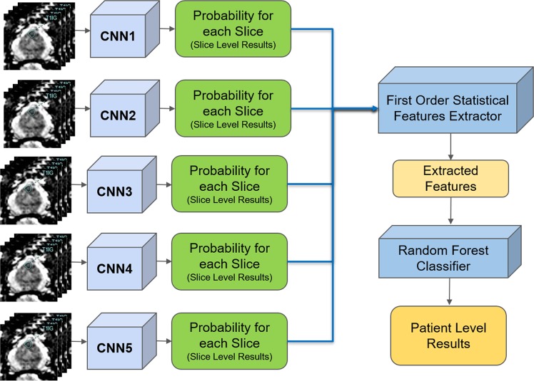
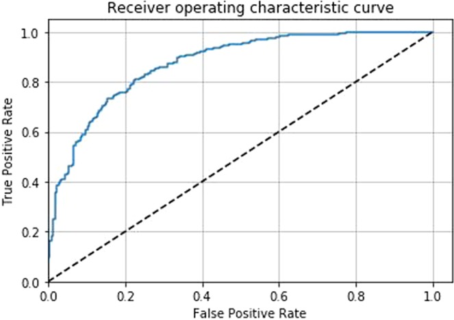
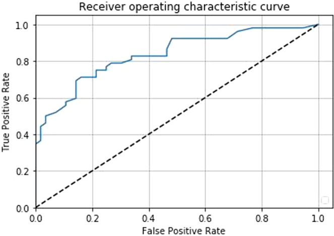
Prostate cancer is one of the most common forms of cancer and the third leading cause of cancer death in North America. As an integrated part of computer-aided detection (CAD) tools, diffusion-weighted magnetic resonance imaging (DWI) has been intensively studied for accurate detection of prostate cancer. With deep convolutional neural networks (CNNs) significant success in computer vision tasks such as object detection and segmentation, different CNN architectures are increasingly investigated in medical imaging research community as promising solutions for designing more accurate CAD tools for cancer detection. In this work, we developed and implemented an automated CNN-based pipeline for detection of clinically significant prostate cancer (PCa) for a given axial DWI image and for each patient. DWI images of 427 patients were used as the dataset, which contained 175 patients with PCa and 252 patients without PCa. To measure the performance of the proposed pipeline, a test set of 108 (out of 427) patients were set aside and not used in the training phase. The proposed pipeline achieved area under the receiver operating characteristic curve (AUC) of 0.87 (95[Formula: see text] Confidence Interval (CI): 0.84-0.90) and 0.84 (95[Formula: see text] CI: 0.76-0.91) at slice level and patient level, respectively.
Overview
The claims and statistics of this paper haven’t been checked yet.
Run the check — free, no account needed. It reads the open-access text and checks the sources it cites.
Integrity signals
Registry
Cites
Statistics
Figures
Verification
Claims the paper makes
The claims and statistics of this paper haven’t been checked yet.
Run the check — free, no account needed. It reads the open-access text and checks the sources it cites.
Claims other papers make about this one
Each is checked against this paper’s own text: does it say what the citing paper says it does?
The source backs part of the claim, not all of it — or backs it for a different population, condition or comparison.
In the citing paper
“Motivated by the dominant performance of CNNs, it is desirable to develop an end-to-end network for automatic placental maturity grading (PMG) from ultrasound images.”
From DilatedFormer: dilated granularity transformer network for placental maturity grading in ultrasound (2023) · cites this paper as [17]
Experimental — accuracy not yet measuredThe cited source says what the claim attributes to it.
In the citing paper
“They have been used successfully in a variety of medical imaging applications, including mammography [15], brain imaging [16], and prostate cancer detection [17].”
From DilatedFormer: dilated granularity transformer network for placental maturity grading in ultrasound (2023) · cites this paper as [17]
Self-check
Percentages, sums and reported differences re-computed from the paper’s own numbers.
The self-check needs the paper’s full text.
Citations
Cites
Deep Residual Learning for Image Recognition
He, K., Zhang, X., Ren, S., & Sun, J. (2016) · 2016 IEEE Conference on Computer Vision and Pattern Recognition (CVPR) · doi:10.1109/cvpr.2016.90 · Cited by 235,474
Breiman, L. (2001) · Machine Learning · doi:10.1023/a:1010933404324 · Cited by 133,538
Very Deep Convolutional Networks for Large-Scale Image Recognition
Simonyan, K., & Zisserman, A. (2014) · arXiv · doi:10.48550/arxiv.1409.1556 · Cited by 114,148
U-Net: Convolutional Networks for Biomedical Image Segmentation
Ronneberger, O., Fischer, P., & Brox, T. (2015) · Lecture notes in computer science · doi:10.1007/978-3-319-24574-4_28 · Cited by 99,404
LeCun, Y., Bengio, Y., & Hinton, G. (2015) · Nature · doi:10.1038/nature14539 · Cited by 84,126
Fully convolutional networks for semantic segmentation
Long, J., Shelhamer, E., & Darrell, T. (2015) · 2015 IEEE Conference on Computer Vision and Pattern Recognition (CVPR) · doi:10.1109/cvpr.2015.7298965 · Cited by 38,834
Batch Normalization: Accelerating Deep Network Training by Reducing Internal Covariate Shift
Ioffe, S., & Szegedy, C. (2015) · arXiv · doi:10.48550/arxiv.1502.03167 · Cited by 27,818
Advances in neural information processing systems 7
Cios, K. J., & Shields, M. E. (1997) · Neurocomputing · doi:10.1016/s0925-2312(97)90018-7 · Cited by 22,093
Book Review: Pattern Recognition and Machine Learning
Nasrabadi, N. M. (2007) · Journal of Electronic Imaging · doi:10.1117/1.2819119 · Cited by 22,066
The meaning and use of the area under a receiver operating characteristic (ROC) curve.
Hanley, J. A., & McNeil, B. J. (1982) · Radiology · doi:10.1148/radiology.143.1.7063747 · Cited by 21,856
Research works printed but not linked: 4
- Michalski, R. S., Carbonell, J. G. & Mitchell, T. M. Machine learning: An artificial intelligence approach (Springer Science & Business Media, 2013).
- LeCun, Y., Bengio, Y. & Hinton, G. Deep learning. nature 521, 436 (2015). · DOI · Paper page
- Vapnik, V.The nature of statistical learning theory (Springer science and business media, 2013).
- Brinkmann, R.The art and science of digital compositing: Techniques for visual effects, animation and motion graphics (Morgan Kaufmann, 2008).
Cited by
Sorted by citations
Oren, O., Gersh, B. J., & Bhatt, D. L. (2020) · The Lancet Digital Health · doi:10.1016/s2589-7500(20)30160-6 · Cited by 335
A systematic review of artificial intelligence techniques in cancer prediction and diagnosis
Kumar, Y., Gupta, S., Singla, R., & Hu, Y.-C. (2022) · Archives of Computational Methods in Engineering · doi:10.1007/s11831-021-09648-w · Cited by 223
COVID-19 Detection from Chest X-ray Images Using Feature Fusion and Deep Learning
Alam, N.-A.-A., Ahsan, M., Based, M. A., Haider, J., & Kowalski, M. (2021) · Sensors · doi:10.3390/s21041480 · Cited by 191
Saha, A., Hosseinzadeh, M., & Huisman, H. J. (2021) · Medical Image Analysis · doi:10.1016/j.media.2021.102155 · Cited by 175
Prostate Cancer Detection Using Deep Learning and Traditional Techniques
Iqbal, S., Siddiqui, G. F., Rehman, A. U., Hussain, L., Saba, T., Tariq, U., & Abbasi, A. A. (2021) · IEEE Access · doi:10.1109/access.2021.3057654 · Cited by 131
Radiomics in prostate cancer: an up-to-date review
Ferro, M., COBELLI, O. D., Musi, G., Giudice, F. D., Carrieri, G., Busetto, G. M., Falagario, U. G., Sciarra, A., Maggi, M., Crocetto, F., et al. (2022) · Therapeutic Advances in Urology · doi:10.1177/17562872221109020 · Cited by 129
Hosseinzadeh, M., Saha, A., Brand, P., Slootweg, I., de Rooij, M., & Huisman, H. J. (2021) · European Radiology · doi:10.1007/s00330-021-08320-y · Cited by 128
Pellicer-Valero, Ó. J., Jiménez, J. L. M., González‐Pérez, V., Ramón-Borja, J. L. C., García, I. M., Benito, M. B., Gómez, P. P., Rubio‐Briones, J., Rupérez, M. J., & Martín‐Guerrero, J. D. (2022) · Scientific Reports · doi:10.1038/s41598-022-06730-6 · Cited by 116
Design of Accurate Classification of COVID-19 Disease in X-Ray Images Using Deep Learning Approach
Chen, J. I. (2021) · Journal of ISMAC · doi:10.36548/jismac.2021.2.006 · Cited by 104
A deep learning study on osteosarcoma detection from histological images
Anisuzzaman, D., Barzekar, H., Tong, L., Luo, J., & Yu, Z. (2021) · Biomedical Signal Processing and Control · doi:10.1016/j.bspc.2021.102931 · Cited by 107
Javaheri, T., Homayounfar, M., Amoozgar, Z., Reiazi, R., Homayounieh, F., Abbas, E., Laali, A., Radmard, A. R., Gharib, M. H., Mousavi, S. A., et al. (2021) · npj Digital Medicine · doi:10.1038/s41746-021-00399-3 · Cited by 129
Twilt, J. J., van Leeuwen, K. G., Huisman, H. J., Fütterer, J. J., & de Rooij, M. (2021) · Diagnostics · doi:10.3390/diagnostics11060959 · Cited by 105
Sharma, P., Nayak, D. R., Balabantaray, B. K., Tanveer, M., & Nayak, R. (2023) · Neural Networks · doi:10.1016/j.neunet.2023.11.006 · Cited by 104
Hao, R., Namdar, K., Liu, L., Haider, M. A., & Khalvati, F. (2021) · Journal of Imaging Informatics in Medicine · doi:10.1007/s10278-021-00478-7 · Cited by 96
Self-citationGopatoti, A., & Vijayalakshmi, P. (2022) · Biomedical Signal Processing and Control · doi:10.1016/j.bspc.2022.103860 · Cited by 86
Cancer detection and segmentation using machine learning and deep learning techniques: a review
Rai, H. M. (2023) · Multimedia Tools and Applications · doi:10.1007/s11042-023-16520-5 · Cited by 76
Deep Learning Reconstruction Enables Highly Accelerated Biparametric MR Imaging of the Prostate
Johnson, P. M., Tong, A. Y., Donthireddy, A., Melamud, K., Petrocelli, R. D., Smereka, P. N., Qian, K., Keerthivasan, M., Chandarana, H., & Knöll, F. (2021) · Journal of Magnetic Resonance Imaging · doi:10.1002/jmri.28024 · Cited by 73
Machine and Deep Learning Prediction Of Prostate Cancer Aggressiveness Using Multiparametric MRI
Bertelli, E., Mercatelli, L., Marzi, C., Pachetti, E., Baccini, M., Barucci, A., Colantonio, S., Gherardini, L., Lattavo, L., Pascali, M. A., et al. (2022) · Frontiers in Oncology · doi:10.3389/fonc.2021.802964 · Cited by 69
Deep learning-based artificial intelligence applications in prostate MRI: brief summary
Türkbey, B., & Haider, M. A. (2021) · British Journal of Radiology · doi:10.1259/bjr.20210563 · Cited by 66
Self-citationProstate lesion segmentation in MR images using radiomics based deeply supervised U-Net
Hambarde, P., Talbar, S. N., Mahajan, A., Chavan, S., Thakur, M., & Sable, N. P. (2020) · Journal of Applied Biomedicine · doi:10.1016/j.bbe.2020.07.011 · Cited by 61
Hashemzadeh, H., Shojaeilangari, S., Allahverdi, A., Rothbauer, M., Ertl, P., & Naderi‐Manesh, H. (2021) · Scientific Reports · doi:10.1038/s41598-021-89352-8 · Cited by 58
RetractedShin, S., Lee, Y., Kim, S.-C., Choi, S., Kim, J. G., & Lee, K. (2021) · Food Chemistry · doi:10.1016/j.foodchem.2021.129329 · Cited by 56
Machine learning analyses of antibody somatic mutations predict immunoglobulin light chain toxicity
Garofalo, M., Piccoli, L., Romeo, M., Barzago, M. M., Ravasio, S., Foglierini, M., Matkovic, M., Sgrignani, J., Gasparo, R. D., Prunotto, M., et al. (2021) · Nature Communications · doi:10.1038/s41467-021-23880-9 · Cited by 59
Pandiyan, S., & Wang, L. (2022) · Computers in Biology and Medicine · doi:10.1016/j.compbiomed.2022.106140 · Cited by 63
Sushentsev, N., Silva, N. M. D., Yeung, M., Barrett, T., Sala, E., Roberts, M. T., & Rundo, L. (2022) · Insights into Imaging · doi:10.1186/s13244-022-01199-3 · Cited by 53
Shirzad, M., Salahvarzi, A., Razzaq, S., Javid‐Naderi, M. J., Rahdar, A., Fathi‐karkan, S., Ghadami, A., Kharaba, Z. J., & Ferreira, L. F. R. (2025) · Critical Reviews in Oncology/Hematology · doi:10.1016/j.critrevonc.2025.104653 · Cited by 59
Mokoatle, M., Marivate, V. N., Mapiye, D. S., Bornman, R. M. S., & Hayes, V. M. (2023) · BMC Bioinformatics · doi:10.1186/s12859-023-05235-x · Cited by 63
Zhang, T., Hasib, M. M., Chiu, Y., Han, Z.-F., Jin, Y.-F., Flores, M. A., Chen, Y., & Huang, Y. (2022) · Cancers · doi:10.3390/cancers14194763 · Cited by 59
Rai, H. M., Yoo, J., & Razaque, A. (2024) · Expert Systems with Applications · doi:10.1016/j.eswa.2024.124838 · Cited by 55
Fully Automatic Deep Learning in Bi-institutional Prostate Magnetic Resonance Imaging
Netzer, N., Weißer, C., Schelb, P., Wang, X., Qin, X., Görtz, M., Schütz, V., Radtke, J. P., Hielscher, T., Schwab, C., et al. (2021) · Investigative Radiology · doi:10.1097/rli.0000000000000791 · Cited by 51
Perera, M., Mirchandani, R., Papa, N. P., Breemer, G., Effeindzourou, A., Smith, L., Swindle, P., & Smith, E. (2020) · World Journal of Urology · doi:10.1007/s00345-020-03392-9 · Cited by 50
A Modified AUC for Training Convolutional Neural Networks: Taking Confidence Into Account
Namdar, K., Haider, M. A., & Khalvati, F. (2021) · Frontiers in Artificial Intelligence · doi:10.3389/frai.2021.582928 · Cited by 47
Self-citationRiaz, I. B., Harmon, S. A., Chen, Z., Naqvi, S. A. A., & Liang Cheng, C. (2024) · American Society of Clinical Oncology Educational Book · doi:10.1200/edbk_438516 · Cited by 49
Rethinking Assumptions in Deep Anomaly DetectionPreprint
Ruff, L., Vandermeulen, R. A., Franks, B. J., Müller, K., & Kloft, M. (2020) · arXiv (Cornell University) · doi:10.48550/arxiv.2006.00339 · Cited by 81
Harder, F. N., Weiss, K., Amiel, T., Peeters, J., Tauber, R., Ziegelmayer, S., Burian, E., Makowski, M. R., Sauter, A. P., Gschwend, J. E., et al. (2022) · Cancers · doi:10.3390/cancers14235741 · Cited by 45
Youn, S. Y., Choi, M. H., Kim, D. H., Lee, Y. J., Huisman, H. J., Johnson, E. J., Penzkofer, T., Shabunin, I., Winkel, D. J., Xing, P., et al. (2021) · European Journal of Radiology · doi:10.1016/j.ejrad.2021.109894 · Cited by 44
Afshar, P., Mohammadi, A., Tyrrell, P. N., Cheung, P., Sigiuk, A., Plataniotis, K. N., Nguyen, E. T., & Oikonomou, A. (2020) · Scientific Reports · doi:10.1038/s41598-020-69106-8 · Cited by 44
A comprehensive data‐level investigation of cancer diagnosis on imbalanced data
Gupta, S., & Gupta, M. K. (2021) · Computational Intelligence · doi:10.1111/coin.12452 · Cited by 44
Pneumonia Detection and Classification Using Deep Learning on Chest X-Ray Images
Darıcı, M. B., Dokur, Z., & Ölmez, T. (2020) · International Journal of Intelligent Systems and Applications in Engineering · doi:10.18201/ijisae.2020466310 · Cited by 43
Ibrahim, J., de Beeck, K. O., Fransén, E., Peeters, M., & Camp, G. V. (2022) · Molecular Oncology · doi:10.1002/1878-0261.13176 · Cited by 42
The Reconstruction of Magnetic Particle Imaging: Current Approaches Based on the System Matrix
Chen, X., Jiang, Z., Han, X. H., Wang, X., & Tang, X. (2021) · Diagnostics · doi:10.3390/diagnostics11050773 · Cited by 41
Exploration of Interpretability Techniques for Deep COVID-19 Classification Using Chest X-ray Images
Chatterjee, S., Saad, F., Sarasaen, C., Ghosh, S., Krug, V., Khatun, R., Mishra, R., Desai, N., Radeva, P. I., Rose, G., et al. (2024) · Journal of Imaging · doi:10.3390/jimaging10020045 · Cited by 46
Hesse, R., Krull, F., & Antonyuk, S. I. (2021) · Powder Technology · doi:10.1016/j.powtec.2021.07.056 · Cited by 41
Kim, J., Maranna, S., Watson, C. A., & Parange, N. (2025) · The American Journal of Emergency Medicine · doi:10.1016/j.ajem.2025.03.029 · Cited by 43
Rehman, A., Xing, H., Li, F., Hussain, M., Gulzar, N., Khan, M. A., Hussain, A., & Saeed, D. (2024) · Biomedical Signal Processing and Control · doi:10.1016/j.bspc.2023.105893 · Cited by 41
Artificial intelligence in oncology: From bench to clinic
Elkhader, J. A., & Elemento, O. (2021) · Seminars in Cancer Biology · doi:10.1016/j.semcancer.2021.04.013 · Cited by 39
Li, D., Han, X., Gao, J., Zhang, Q., Yang, H., Liao, S., Guo, H., & Zhang, B. (2022) · Frontiers in Medicine · doi:10.3389/fmed.2021.810995 · Cited by 37
Warrier, G. S., Amirthalakshmi, T. M., Nimala, K., Delsy, T. T. M., Malar, P. S. R., Ramkumar, G., & Raju, R. (2022) · Contrast Media & Molecular Imaging · doi:10.1155/2022/4356744 · Cited by 35
CovidCTNet: An Open-Source Deep Learning Approach to Identify Covid-19 Using CT ImagePreprint
Javaheri, T., Homayounfar, M., Amoozgar, Z., Reiazi, R., Homayounieh, F., Abbas, E., Laali, A., Radmard, A. R., Gharib, M. H., Mousavi, S. A., et al. (2020) · arXiv (Cornell University) · doi:10.48550/arxiv.2005.03059 · Cited by 36
The role of AI in prostate MRI quality and interpretation: Opportunities and challenges
Kim, H., Kang, S. W., Kim, J., Nagar, H., Sabuncu, M. R., Margolis, D., & Kim, C. K. (2023) · European Journal of Radiology · doi:10.1016/j.ejrad.2023.110887 · Cited by 35
Deniffel, D., Abraham, N., Namdar, K., Dong, X., Salinas, E., Milot, L. M., Khalvati, F., & Haider, M. A. (2020) · European Radiology · doi:10.1007/s00330-020-07030-1 · Cited by 33
Self-citationLai, C.-C., Wang, H.-K., Wang, F., Peng, Y., Lin, T., Peng, H., & Shen, S. (2021) · Sensors · doi:10.3390/s21082709 · Cited by 31
A review on deep learning applications in highly multiplexed tissue imaging data analysis
Zidane, M., Makky, A., Bruhns, M., Rochwarger, A., Babaei, S., Claassen, M., & Schürch, C. M. (2023) · Frontiers in Bioinformatics · doi:10.3389/fbinf.2023.1159381 · Cited by 32
Flap failure prediction in microvascular tissue reconstruction using machine learning algorithms
Shi, Y.-C., Li, J., Li, S.-J., Li, Z., Zhang, H., Wu, Z., & Wu, Z. (2022) · World Journal of Clinical Cases · doi:10.12998/wjcc.v10.i12.3729 · Cited by 30
Ramkumar, G., Bhuvaneswari, P., Radhika, R., Saranya, S., Vijayalakshmi, S., Karpagam, M., & Wilfred, F. (2022) · Contrast Media & Molecular Imaging · doi:10.1155/2022/6862083 · Cited by 29
Cross-Organ, Cross-Modality Transfer Learning: Feasibility Study for Segmentation and Classification
Lee, J., & Nishikawa, R. M. (2020) · IEEE Access · doi:10.1109/access.2020.3038909 · Cited by 29
Roest, C., Fransen, S. J., Kwee, T. C., & Yakar, D. (2022) · Life · doi:10.3390/life12101490 · Cited by 30
Belue, M. J., Harmon, S. A., Lay, N. S., Daryanani, A., Phelps, T. E., Choyke, P. L., & Turkbey, B. (2023) · Journal of the American College of Radiology · doi:10.1016/j.jacr.2022.05.022 · Cited by 27
Rai, H. M., Yoo, J., Moqurrab, S. A., & Dashkevych, S. (2023) · Measurement · doi:10.1016/j.measurement.2023.114059 · Cited by 26
Schelb, P., Tavakoli, A. A., Tubtawee, T., Hielscher, T., Radtke, J.-P., Görtz, M., Schütz, V., Kuder, T. A., Schimmöller, L., Stenzinger, A., et al. (2020) · RöFo - Fortschritte auf dem Gebiet der Röntgenstrahlen und der bildgebenden Verfahren · doi:10.1055/a-1290-8070 · Cited by 27
Automatic Diagnosis of COVID-19 Using a tailored Transformer-Like Network
Liu, C., & Yin, Q. (2021) · Journal of Physics Conference Series · doi:10.1088/1742-6596/2010/1/012175 · Cited by 27
A Novel Multi-Neural Ensemble Approach for Cancer Diagnosis
Gupta, S., Gupta, M. K., & Kumar, R. (2021) · Applied Artificial Intelligence · doi:10.1080/08839514.2021.2018182 · Cited by 26
Accurate prediction of immunoglobulin proteins using machine learning model
Ghulam, A., Sikander, R., Ali, F., Swati, Z. N. K., Unar, A., & Talpur, D. B. (2022) · Informatics in Medicine Unlocked · doi:10.1016/j.imu.2022.100885 · Cited by 26
Neural Networks for the Detection of COVID-19 and Other Diseases: Prospects and Challenges
Azeem, M., Javaid, S., Khalil, R. A., Fahim, H., Althobaiti, T., Al-Sharif, N., & Saeed, N. (2023) · Bioengineering · doi:10.3390/bioengineering10070850 · Cited by 29
Bhattacharjee, S., Kim, C., Prakash, D., Park, H., Cho, N.-H., & Choi, H. (2020) · Applied Sciences · doi:10.3390/app10228013 · Cited by 29
Wu, X., Yin, C., Chen, X., Zhang, Y., Su, Y., Shi, J., Weng, D., Jiang, X., Zhang, A., Zhang, W., et al. (2022) · Frontiers in Pharmacology · doi:10.3389/fphar.2022.878764 · Cited by 25
The performance of artificial intelligence in prostate magnetic resonance imaging screening
Owida, H. A., Hassan, M. R., Ali, A. M., Alnaimat, F., Sharah, A. A., Abuowaida, S. F. A., & Alshdaifat, N. (2024) · International Journal of Power Electronics and Drive Systems/International Journal of Electrical and Computer Engineering · doi:10.11591/ijece.v14i2.pp2234-2241 · Cited by 30
Unet based Xception Model for Prostate Cancer Segmentation from MRI Images
Chahal, E. S., Patel, A., Gupta, A. K., Purwar, A., & Gopinathan, D. (2021) · Multimedia Tools and Applications · doi:10.1007/s11042-021-11334-9 · Cited by 24
Song, E., Long, J., Ma, G., Liu, H., Hung, C., Jin, R., Wang, P., & Wang, W. (2023) · Magnetic Resonance Imaging · doi:10.1016/j.mri.2023.01.015 · Cited by 23
Context aware machine learning techniques for brain tumor classification and detection – A review
Amjad, U., Raza, A., Fahad, M., Farid, D., Akhunzada, A., Abubakar, M., & Beenish, H. (2025) · Heliyon · doi:10.1016/j.heliyon.2025.e41835 · Cited by 24
Mitigating Data Imbalance Issues in Medical Image Analysis
Banik, D., & Bhattacharjee, D. (2021) · Advances in data mining and database management book series · doi:10.4018/978-1-7998-7371-6.ch004 · Cited by 22
A Survey on Human Cancer Categorization Based on Deep Learning
Ibrahim, A., Mohamed, H. K., Maher, A., & Zhang, B. (2022) · Frontiers in Artificial Intelligence · doi:10.3389/frai.2022.884749 · Cited by 21
Wang, K., Karalis, J. D., Elamir, A. M., Bifolco, A., Wachsmann, M., Capretti, G. L., Spaggiari, P., Enrico, S., Balasubramanian, K., Fatimah, N., et al. (2023) · Annals of Surgical Oncology · doi:10.1245/s10434-023-14805-5 · Cited by 21
Prostate Cancer Prognosis Using Multi-Layer Perceptron and Class Balancing Techniques
Gupta, S., & Kumar, M. (2021) · International Conference on Contemporary Computing · doi:10.1145/3474124.3474125 · Cited by 21
Zhou, X., Huang, N., Chen, W., Tang, X., Mahdavi, B., Raoofi, A., Mahdian, D., & Atabati, H. (2020) · AMB Express · doi:10.1186/s13568-020-01138-9 · Cited by 20
Incidental detection of prostate cancer with computed tomography scans
Korevaar, S., Tennakoon, R., Page, M., Brotchie, P., Thangarajah, J., Florescu, C., Sutherland, T., Kam, N. M., & Bab‐Hadiashar, A. (2021) · Scientific Reports · doi:10.1038/s41598-021-86972-y · Cited by 20
Rashmi, R., Umapathy, S., Krishnan, P. T., & Dhanraj, V. (2022) · Soft Computing · doi:10.1007/s00500-021-06668-3 · Cited by 21
RetractedAboutalebi, H., Pavlova, M., Gunraj, H., Shafiee, M. J., Sabri, A., Alaref, A. A., & Wong, A. M. (2022) · Frontiers in Medicine · doi:10.3389/fmed.2021.821120 · Cited by 25
Zhu, L., Gao, G., Zhu, Y. C., Han, C., Liu, X., Li, D., Liu, W., Wang, X., Zhang, J., Zhang, X., et al. (2022) · Frontiers in Oncology · doi:10.3389/fonc.2022.958065 · Cited by 19
Mehmood, M., Abbasi, S. H., Aurangzeb, K., Majeed, M. F., Anwar, M. S., & Alhussein, M. A. (2023) · Frontiers in Oncology · doi:10.3389/fonc.2023.1225490 · Cited by 22
Mohseni, N., Moodi, M., Kefayat, A., Shokati, F., & Molaabasi, F. (2024) · ACS Omega · doi:10.1021/acsomega.3c06884 · Cited by 19
Koç, M., Sut, S. K., Serhatlioğlu, İ., Baygin, M., & Tuncer, T. (2022) · Multimedia Tools and Applications · doi:10.1007/s11042-022-11906-3 · Cited by 19
Ragab, M., Kateb, F., El-Sawy, E.-S. K., Binyamin, S. S., Al-Rabia, M. W., & Mansouri, R. A. (2023) · Healthcare · doi:10.3390/healthcare11040590 · Cited by 18
Talyshinskii, A., Hameed, B. M. Z., Ravinder, P. P., Naik, N., Randhawa, P., Shah, M., Rai, B. P., Tokas, T., & Somani, B. K. (2024) · Cancers · doi:10.3390/cancers16101809 · Cited by 18
Salama, W. M., & Aly, M. H. (2021) · Multimedia Tools and Applications · doi:10.1007/s11042-021-10849-5 · Cited by 18
Olabanjo, O. A., Ashiribo, W. U. S. U., Asokere, M., Afisi, O. T., Okugbesan, B., Olabanjo, O., Folorunso, O. O., & Mazzara, M. (2023) · Analytics · doi:10.3390/analytics2030039 · Cited by 21
Lin, J., Ma, L., & Cui, J. (2020) · IEEE Access · doi:10.1109/access.2020.2996250 · Cited by 18
Yıldırım, K., Yıldırım, M., Eryeşil, H., Talo, M., Yıldırım, Ö., Karabatak, M., Oğraş, M. S., Artaş, H., & Acharya, U. R. (2022) · Computers & Electrical Engineering · doi:10.1016/j.compeleceng.2022.108275 · Cited by 16
Real-time Prostate Cancer Detection via YOLO-Tiny Variants
Aishwarya, N., & S, Y. K. G. (2023) · International Conference on I-SMAC (IoT in Social, Mobile, Analytics and Cloud) (I-SMAC) · doi:10.1109/i-smac58438.2023.10290712 · Cited by 16
Multistage Segmentation of Prostate Cancer Tissues Using Sample Entropy Texture Analysis
Ali, T. M., Masood, K., Irfan, M., Draz, U., Nagra, A. A., Asif, M., Alshehri, B. M., Głowacz, A., Tadeusiewicz, R., Mahnashi, M. H., et al. (2020) · Entropy · doi:10.3390/e22121370 · Cited by 16
Artificial Intelligence Assessment of Renal Scarring (AIRS Study)
Chantaduly, C., Troutt, H. R., Reyes, K. A. P., Zuckerman, J. E., Chang, P., & Lau, W. L. (2021) · Kidney360 · doi:10.34067/kid.0003662021 · Cited by 16
Zhang, K. S., Schelb, P., Netzer, N., Tavakoli, A. A., Keymling, M., Wehrse, E., Hog, R., Rotkopf, L. T., Wennmann, M., Glemser, P. A., et al. (2022) · Investigative Radiology · doi:10.1097/rli.0000000000000878 · Cited by 16
Prasad, J. R., Prasad, R. S., Dhumane, A., Ranjan, N. M., & Tamboli, M. S. (2023) · Biomedical Signal Processing and Control · doi:10.1016/j.bspc.2023.105474 · Cited by 14
Prostate Cancer Detection from MRI Using Efficient Feature Extraction with Transfer Learning
Islam, M. R., Imran, A., & Rabbi, M. F. (2024) · Prostate Cancer · doi:10.1155/2024/1588891 · Cited by 14
Artificial intelligence: a new era in prostate cancer diagnosis and treatment
Vidiyala, N., Parupathi, P. R., Sunkishala, P., Muppavarapu, C. S., Gujja, A., Kanagala, P., Meduri, S. K., & Nyavanandi, D. (2025) · International Journal of Pharmaceutics · doi:10.1016/j.ijpharm.2025.126024 · Cited by 13
Kaandorp, M., Zijlstra, F., Karimi, D., Gholipour, A., & While, P. T. (2024) · Medical Image Analysis · doi:10.1016/j.media.2024.103414 · Cited by 13
Ali, A. M., & Mohammed, A. A. (2022) · Multimedia Tools and Applications · doi:10.1007/s11042-022-12102-z · Cited by 13
A review of deep learning methods for the prediction of prostate cancer
Al-Gburi, R. M., Gaib, R., Al-Sabti, S. M. B., Tharwat, M. E. A. A., Mustafa, A., & Elhenidy, A. M. (2025) · Neural Computing and Applications · doi:10.1007/s00521-025-11563-1 · Cited by 12
On the Effectiveness of 3D Vision Transformers for the Prediction of Prostate Cancer Aggressiveness
Pachetti, E., Colantonio, S., & Pascali, M. A. (2022) · Lecture notes in computer science · doi:10.1007/978-3-031-13324-4_27 · Cited by 12
Zhou, C., Zhang, Y., Guo, S., Wang, D., Lv, H.-X., Qiao, X.-N., Wang, R., Chang, D., Zhao, L.-M., & Zhou, F. (2023) · Discover Oncology · doi:10.1007/s12672-023-00752-w · Cited by 12
Metadata
- Authors
- Yoo, S., Gujrathi, I., Haider, M. A., & Khalvati, F.
- Affiliations
- Lunenfeld-Tanenbaum Research Institute (CA)
- University of Toronto (CA)
- Corresponding author
- Khalvati, Farzad
- ORCID
- Haider, Masoom A.
- Venue
- Scientific Reports
- Published
- 2019-12-20
- Volume
- 9
- Issue
- 1
- Pages
- 19518-19518
- ISSN
- 2045-2322 (ISSN-L)
- PMC
- PMC6925141
- OpenAlex
- W2995218128
- Cited by
- 230
- References
- 39
- Citations by year
- 2026: 14 · 2025: 31 · 2024: 37 · 2023: 36 · 2022: 42 · 2021: 50 · 2020: 17 · 2019: 3
- FWCI
- 12.77
- Type
- journal article
- Language
- en
- Open access
- gold
- doi.org
- Locations
- Scientific Reports (publishedVersion, cc-by)
- Europe PMC (PubMed Central) (publishedVersion, cc-by)
- PubMed Central (publishedVersion, cc-by)
- PubMed (publishedVersion)
- arXiv (Cornell University) (submittedVersion)
- DOAJ (DOAJ: Directory of Open Access Journals) (submittedVersion)
- arXiv (Cornell University)
- Funders
- Ontario Institute for Cancer Research
- Government of Ontario
- Topics
- Physical Sciences › Computer Science › Artificial Intelligence › AI in cancer detection
- Health Sciences › Medicine › Radiology, Nuclear Medicine and Imaging › Radiomics and Machine Learning in Medical Imaging
- Health Sciences › Medicine › Radiology, Nuclear Medicine and Imaging › MRI in cancer diagnosis
- Keywords
- prostate cancer detection, clinically significant prostate cancer, diffusion-weighted imaging, computer-aided detection, deep convolutional neural networks, medical image classification
- MeSH terms
- Deep Learning, Humans, Male, Prostatic Neoplasms, ROC Curve, Neural Networks, Computer, Diffusion Magnetic Resonance Imaging
About these checks
Registry, retraction, Cites and statistics results are computed, not judged, with one exception: a printed reference that no index lists is read once by a model before it counts as a citation error. Claims are extracted by a model and kept only when their quote appears verbatim in the text; each cited source is then checked, with a model’s help, for whether it says what the claim relies on. Those verdicts can be wrong — read the claim’s quote and the cited source before relying on one.
Statistics and arithmetic are recomputed, not judged.
Confidence bands are shown only for fields calibrated on our annotated test set. None is calibrated yet, so no band is shown.
What counts as a claim ›How we measure confidence ›How we check this ›
Are you an author, or do you see a problem with this page? Request a review
