

The DOI matches its Crossref record; 42 of the 42 references it cites were found in a registry or an index.
Prostate Cancer Detection using Deep Convolutional Neural Networks
Open accessAbstract
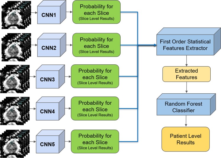
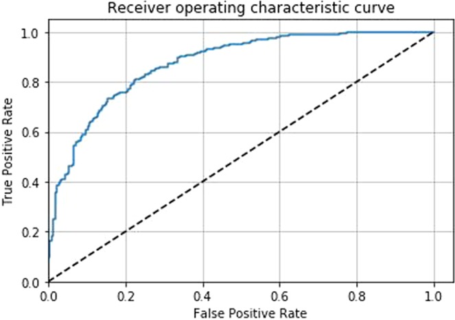
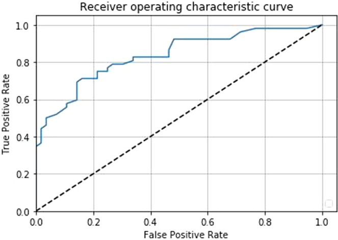
Prostate cancer is one of the most common forms of cancer and the third leading cause of cancer death in North America. As an integrated part of computer-aided detection (CAD) tools, diffusion-weighted magnetic resonance imaging (DWI) has been intensively studied for accurate detection of prostate cancer. With deep convolutional neural networks (CNNs) significant success in computer vision tasks such as object detection and segmentation, different CNN architectures are increasingly investigated in medical imaging research community as promising solutions for designing more accurate CAD tools for cancer detection. In this work, we developed and implemented an automated CNN-based pipeline for detection of clinically significant prostate cancer (PCa) for a given axial DWI image and for each patient. DWI images of 427 patients were used as the dataset, which contained 175 patients with PCa and 252 patients without PCa. To measure the performance of the proposed pipeline, a test set of 108 (out of 427) patients were set aside and not used in the training phase. The proposed pipeline achieved area under the receiver operating characteristic curve (AUC) of 0.87 (95[Formula: see text] Confidence Interval (CI): 0.84-0.90) and 0.84 (95[Formula: see text] CI: 0.76-0.91) at slice level and patient level, respectively.
Overview
The claims and statistics of this paper haven’t been checked yet.
Run the check — free, no account needed. It reads the open-access text and checks the sources it cites.
Integrity signals
Registry
Cites
Statistics
Figures
Verification
Claims the paper makes
The claims and statistics of this paper haven’t been checked yet.
Run the check — free, no account needed. It reads the open-access text and checks the sources it cites.
Claims other papers make about this one
Each is checked against this paper’s own text: does it say what the citing paper says it does?
The source backs part of the claim, not all of it — or backs it for a different population, condition or comparison.
In the citing paper
“Motivated by the dominant performance of CNNs, it is desirable to develop an end-to-end network for automatic placental maturity grading (PMG) from ultrasound images.”
From DilatedFormer: dilated granularity transformer network for placental maturity grading in ultrasound (2023) · cites this paper as [17]
Experimental — accuracy not yet measuredThe cited source says what the claim attributes to it.
In the citing paper
“They have been used successfully in a variety of medical imaging applications, including mammography [15], brain imaging [16], and prostate cancer detection [17].”
From DilatedFormer: dilated granularity transformer network for placental maturity grading in ultrasound (2023) · cites this paper as [17]
Self-check
Percentages, sums and reported differences re-computed from the paper’s own numbers.
The self-check needs the paper’s full text.
Citations
Cites
Deep Residual Learning for Image Recognition
He, K., Zhang, X., Ren, S., & Sun, J. (2016) · 2016 IEEE Conference on Computer Vision and Pattern Recognition (CVPR) · doi:10.1109/cvpr.2016.90 · Cited by 235,474
Breiman, L. (2001) · Machine Learning · doi:10.1023/a:1010933404324 · Cited by 133,538
Very Deep Convolutional Networks for Large-Scale Image Recognition
Simonyan, K., & Zisserman, A. (2014) · arXiv · doi:10.48550/arxiv.1409.1556 · Cited by 114,148
U-Net: Convolutional Networks for Biomedical Image Segmentation
Ronneberger, O., Fischer, P., & Brox, T. (2015) · Lecture notes in computer science · doi:10.1007/978-3-319-24574-4_28 · Cited by 99,404
LeCun, Y., Bengio, Y., & Hinton, G. (2015) · Nature · doi:10.1038/nature14539 · Cited by 84,126
Fully convolutional networks for semantic segmentation
Long, J., Shelhamer, E., & Darrell, T. (2015) · 2015 IEEE Conference on Computer Vision and Pattern Recognition (CVPR) · doi:10.1109/cvpr.2015.7298965 · Cited by 38,834
Batch Normalization: Accelerating Deep Network Training by Reducing Internal Covariate Shift
Ioffe, S., & Szegedy, C. (2015) · arXiv · doi:10.48550/arxiv.1502.03167 · Cited by 27,818
Advances in neural information processing systems 7
Cios, K. J., & Shields, M. E. (1997) · Neurocomputing · doi:10.1016/s0925-2312(97)90018-7 · Cited by 22,093
Book Review: Pattern Recognition and Machine Learning
Nasrabadi, N. M. (2007) · Journal of Electronic Imaging · doi:10.1117/1.2819119 · Cited by 22,066
The meaning and use of the area under a receiver operating characteristic (ROC) curve.
Hanley, J. A., & McNeil, B. J. (1982) · Radiology · doi:10.1148/radiology.143.1.7063747 · Cited by 21,856
Siegel, R. L., Miller, K. D., & Jemal, A. (2017) · CA: A Cancer Journal for Clinicians · doi:10.3322/caac.21387 · Cited by 14,712
Fully convolutional networks for semantic segmentation
Shelhamer, E., Long, J., & Darrell, T. (2017) · IEEE Transactions on Pattern Analysis and Machine Intelligence · doi:10.1109/tpami.2016.2572683 · Cited by 11,898
Identity mappings in deep residual networks
He, K., Zhang, X., Ren, S., & Sun, J. (2016) · Lecture Notes in Computer Science · doi:10.1007/978-3-319-46493-0_38 · Cited by 10,741
Wolpert, D. H. (1992) · Neural Networks · doi:10.1016/s0893-6080(05)80023-1 · Cited by 7,951
Large-Scale Machine Learning with Stochastic Gradient Descent
Bottou, L. (2010) · Proceedings of COMPSTAT'2010 · doi:10.1007/978-3-7908-2604-3_16 · Cited by 5,795
Recurrent neural network based language model
Mikolov, T., Karafiát, M., Burget, L., Černocký, J., & Khudanpur, S. (2010) · Interspeech 2010 · doi:10.21437/interspeech.2010-343 · Cited by 5,390
A review of feature selection techniques in bioinformatics
Saeys, Y., Inza, I., & Larrañaga, P. (2007) · Bioinformatics · doi:10.1093/bioinformatics/btm344 · Cited by 5,389
Kim, H.-Y. (2013) · Restorative Dentistry & Endodontics · doi:10.5395/rde.2013.38.1.52 · Cited by 4,035
Genetic Algorithms and Machine Learning
Goldberg, D. E., & Holland, J. H. (1988) · Machine Learning · doi:10.1023/a:1022602019183 · Cited by 3,193
A Tutorial on the Cross-Entropy Method
de Boer, P.-T., Kroese, D. P., Mannor, S., & Rubinstein, R. Y. (2005) · Annals of Operations Research · doi:10.1007/s10479-005-5724-z · Cited by 3,110
Diffusion-Weighted Magnetic Resonance Imaging as a Cancer Biomarker: Consensus and Recommendations
Padhani, A. R., Liu, G., Koh, D., Chenevert, T. L., Thoeny, H. C., Takahara, T., Dzik-Jurasz, A. S. K., Ross, B. D., CAUTEREN, M. V., Collins, D., et al. (2009) · Neoplasia · doi:10.1593/neo.81328 · Cited by 1,992
Machine learning for medical imaging
Erickson, B. J., Korfiatis, P., Akkus, Z., & Kline, T. L. (2017) · RadioGraphics · doi:10.1148/rg.2017160130 · Cited by 1,672
Orrù, G., Pettersson-Yeo, W., Marquand, A. F., Sartori, G., & Mechelli, A. (2012) · Neuroscience & Biobehavioral Reviews · doi:10.1016/j.neubiorev.2012.01.004 · Cited by 1,012
Rosenkrantz, A. B., Ginocchio, L. A., Cornfeld, D., Froemming, A. T., Gupta, R. T., Türkbey, B., Westphalen, A. C., Babb, J. S., & Margolis, D. (2016) · Radiology · doi:10.1148/radiol.2016152542 · Cited by 501
Computer-Aided Detection of Prostate Cancer in MRI
Litjens, G. J. S., Debats, O. A., Barentsz, J. O., Karssemeijer, N., & Huisman, H. J. (2014) · IEEE Transactions on Medical Imaging · doi:10.1109/tmi.2014.2303821 · Cited by 453
Fehr, D., Veeraraghavan, H., Wibmer, A. G., Gondo, T., Matsumoto, K., Vargas, H. A., Sala, E., Hricak, H., & Deasy, J. O. (2015) · Proceedings of the National Academy of Sciences · doi:10.1073/pnas.1505935112 · Cited by 391
Random forest classifier combined with feature selection for breast cancer diagnosis and prognostic
Nguyen, C., Wang, Y., & Nguyen, H. N. (2013) · Journal of Biomedical Science and Engineering · doi:10.4236/jbise.2013.65070 · Cited by 310
Prostate Magnetic Resonance Imaging Interpretation Varies Substantially Across Radiologists
Sonn, G. A., Fan, R. E., Ghanouni, P., Wang, N. N., Brooks, J. D., Loening, A. M., Daniel, B. L., To’o, K. J., Thong, A. E. C., & Leppert, J. T. (2017) · European Urology Focus · doi:10.1016/j.euf.2017.11.010 · Cited by 279
Wang, X., Yang, W. H., Weinreb, J. C., Han, J., Li, Q., Kong, X., Yan, Y., Ke, Z., Luo, B., Liu, T., et al. (2017) · Scientific Reports · doi:10.1038/s41598-017-15720-y · Cited by 200
MAPS: A Quantitative Radiomics Approach for Prostate Cancer Detection
Cameron, A., Khalvati, F., Haider, M. A., & Wong, A. K. C. (2015) · IEEE Transactions on Biomedical Engineering · doi:10.1109/tbme.2015.2485779 · Cited by 185
Self-citationLe, M. H., Chen, J., Wang, L., Wang, Z., Liu, W., Cheng, K.-T. T., & Yang, X. Y. (2017) · Physics in Medicine and Biology · doi:10.1088/1361-6560/aa7731 · Cited by 161
Prostate cancer diagnosis using deep learning with 3D multiparametric MRI
Liu, S., Zheng, H., Feng, Y., & Li, W. (2017) · Proceedings of SPIE, the International Society for Optical Engineering/Proceedings of SPIE · doi:10.1117/12.2277121 · Cited by 160
Khalvati, F., Wong, A. M., & Haider, M. A. (2015) · BMC Medical Imaging · doi:10.1186/s12880-015-0069-9 · Cited by 158
Self-citationWang, S., Burtt, K. E., Türkbey, B., Choyke, P. L., & Summers, R. M. (2014) · BioMed Research International · doi:10.1155/2014/789561 · Cited by 121
Overdiagnosis of Prostate Cancer
Sandhu, G. S., & Andriole, G. L. (2012) · JNCI Monographs · doi:10.1093/jncimonographs/lgs031 · Cited by 97
The meninges: new therapeutic targets for multiple sclerosis
Russi, A. E., & Brown, M. A. (2014) · Translational research · doi:10.1016/j.trsl.2014.08.005 · Cited by 90
Mehrtash, A., Sedghi, A., Ghafoorian, M., Taghipour, M., Tempany, C. M., Wells, W. M., Kapur, T., Mousavi, P., Abolmaesumi, P., & Fedorov, A. (2017) · Proceedings of SPIE, the International Society for Optical Engineering/Proceedings of SPIE · doi:10.1117/12.2277123 · Cited by 85
Khalvati, F., Zhang, J., Chung, A. G., Shafiee, M. J., Wong, A. M., & Haider, M. A. (2018) · BMC Medical Imaging · doi:10.1186/s12880-018-0258-4 · Cited by 52
Self-citationTsehay, Y. K., Lay, N. S., Wang, X., Kwak, J. T., Türkbey, B., Choyke, P. L., Pinto, P. A., Wood, B. J., & Summers, R. M. (2017) · IEEE International Symposium on Biomedical Imaging (ISBI) · doi:10.1109/isbi.2017.7950602 · Cited by 45
Glaister, J., Cameron, A., Wong, A., & Haider, M. (2012) · Annual International Conference of the IEEE Engineering in Medicine and Biology Society (EMBC) · doi:10.1109/embc.2012.6345957 · Cited by 19
Self-citationPROSTATEx: Prostate MR Classification Challenge (Conference Presentation)
Armato, S. G., Petrick, N., & Drukker, K. (2017) · SPIE Medical Imaging: Computer-Aided Diagnosis · doi:10.1117/12.2280374 · Cited by 6
Ishioka, J., Matsuoka, Y., Uehara, S., Yasuda, Y., Kijima, T., Yoshida, S., Yokoyama, M., Saito, K., Kimura, T., Kudo, K., et al. (2018) · The Journal of Urology · doi:10.1016/j.juro.2018.02.680 · Cited by 5
Research works printed but not linked: 4
- Michalski, R. S., Carbonell, J. G. & Mitchell, T. M. Machine learning: An artificial intelligence approach (Springer Science & Business Media, 2013).
- LeCun, Y., Bengio, Y. & Hinton, G. Deep learning. nature 521, 436 (2015). · DOI · Paper page
- Vapnik, V.The nature of statistical learning theory (Springer science and business media, 2013).
- Brinkmann, R.The art and science of digital compositing: Techniques for visual effects, animation and motion graphics (Morgan Kaufmann, 2008).
Cited by
Sorted by citations
Oren, O., Gersh, B. J., & Bhatt, D. L. (2020) · The Lancet Digital Health · doi:10.1016/s2589-7500(20)30160-6 · Cited by 335
A systematic review of artificial intelligence techniques in cancer prediction and diagnosis
Kumar, Y., Gupta, S., Singla, R., & Hu, Y.-C. (2022) · Archives of Computational Methods in Engineering · doi:10.1007/s11831-021-09648-w · Cited by 223
COVID-19 Detection from Chest X-ray Images Using Feature Fusion and Deep Learning
Alam, N.-A.-A., Ahsan, M., Based, M. A., Haider, J., & Kowalski, M. (2021) · Sensors · doi:10.3390/s21041480 · Cited by 191
Saha, A., Hosseinzadeh, M., & Huisman, H. J. (2021) · Medical Image Analysis · doi:10.1016/j.media.2021.102155 · Cited by 175
Prostate Cancer Detection Using Deep Learning and Traditional Techniques
Iqbal, S., Siddiqui, G. F., Rehman, A. U., Hussain, L., Saba, T., Tariq, U., & Abbasi, A. A. (2021) · IEEE Access · doi:10.1109/access.2021.3057654 · Cited by 131
Metadata
- Authors
- Yoo, S., Gujrathi, I., Haider, M. A., & Khalvati, F.
- Affiliations
- Lunenfeld-Tanenbaum Research Institute (CA)
- University of Toronto (CA)
- Corresponding author
- Khalvati, Farzad
- ORCID
- Haider, Masoom A.
- Venue
- Scientific Reports
- Published
- 2019-12-20
- Volume
- 9
- Issue
- 1
- Pages
- 19518-19518
- ISSN
- 2045-2322 (ISSN-L)
- PMC
- PMC6925141
- OpenAlex
- W2995218128
- Cited by
- 230
- References
- 39
- Citations by year
- 2026: 14 · 2025: 31 · 2024: 37 · 2023: 36 · 2022: 42 · 2021: 50 · 2020: 17 · 2019: 3
- FWCI
- 12.77
- Type
- journal article
- Language
- en
- Open access
- gold
- doi.org
- Locations
- Scientific Reports (publishedVersion, cc-by)
- Europe PMC (PubMed Central) (publishedVersion, cc-by)
- PubMed Central (publishedVersion, cc-by)
- PubMed (publishedVersion)
- arXiv (Cornell University) (submittedVersion)
- DOAJ (DOAJ: Directory of Open Access Journals) (submittedVersion)
- arXiv (Cornell University)
- Funders
- Ontario Institute for Cancer Research
- Government of Ontario
- Topics
- Physical Sciences › Computer Science › Artificial Intelligence › AI in cancer detection
- Health Sciences › Medicine › Radiology, Nuclear Medicine and Imaging › Radiomics and Machine Learning in Medical Imaging
- Health Sciences › Medicine › Radiology, Nuclear Medicine and Imaging › MRI in cancer diagnosis
- Keywords
- prostate cancer detection, clinically significant prostate cancer, diffusion-weighted imaging, computer-aided detection, deep convolutional neural networks, medical image classification
- MeSH terms
- Deep Learning, Humans, Male, Prostatic Neoplasms, ROC Curve, Neural Networks, Computer, Diffusion Magnetic Resonance Imaging
About these checks
Registry, retraction, Cites and statistics results are computed, not judged, with one exception: a printed reference that no index lists is read once by a model before it counts as a citation error. Claims are extracted by a model and kept only when their quote appears verbatim in the text; each cited source is then checked, with a model’s help, for whether it says what the claim relies on. Those verdicts can be wrong — read the claim’s quote and the cited source before relying on one.
Statistics and arithmetic are recomputed, not judged.
Confidence bands are shown only for fields calibrated on our annotated test set. None is calibrated yet, so no band is shown.
What counts as a claim ›How we measure confidence ›How we check this ›
Are you an author, or do you see a problem with this page? Request a review
