

The DOI matches its Crossref record; 38 of the 38 references it cites were found in a registry or an index.
Radiomics feature reproducibility under inter-rater variability in segmentations of CT images
Open accessAbstract
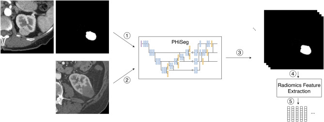
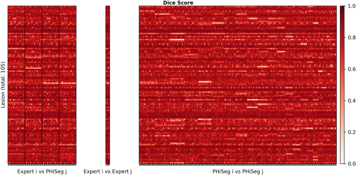
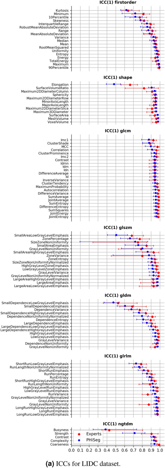
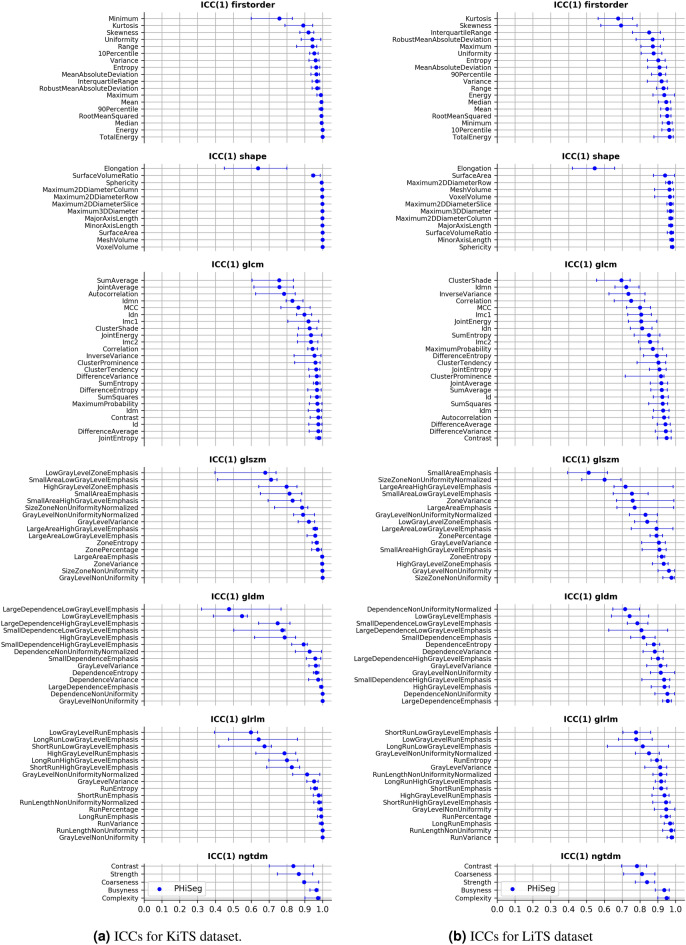
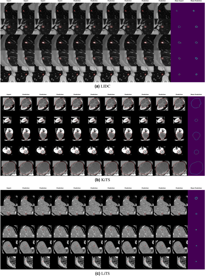
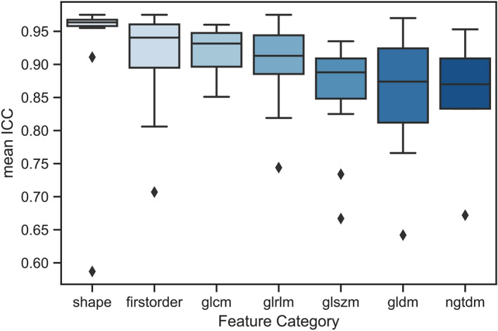
Identifying image features that are robust with respect to segmentation variability is a tough challenge in radiomics. So far, this problem has mainly been tackled in test–retest analyses. In this work we analyse radiomics feature reproducibility in two phases: first with manual segmentations provided by four expert readers and second with probabilistic automated segmentations using a recently developed neural network (PHiseg). We test feature reproducibility on three publicly available datasets of lung, kidney and liver lesions. We find consistent results both over manual and automated segmentations in all three datasets and show that there are subsets of radiomic features which are robust against segmentation variability and other radiomic features which are prone to poor reproducibility under differing segmentations. By providing a detailed analysis of robustness of the most common radiomics features across several datasets, we envision that more reliable and reproducible radiomic models can be built in the future based on this work.
Overview
The claims and statistics of this paper haven’t been checked yet.
Run the check — free, no account needed. It reads the open-access text and checks the sources it cites.
Integrity signals
Registry
Cites
Statistics
Figures
Verification
Claims the paper makes
The claims and statistics of this paper haven’t been checked yet.
Run the check — free, no account needed. It reads the open-access text and checks the sources it cites.
Claims other papers make about this one
Each is checked against this paper’s own text: does it say what the citing paper says it does?
In the citing paper
“Moreover, these statistical features are affected by several confounders, such as inter-reader variability in segmentations26 and acquisition settings of the scanners27, limiting their applicability in diverse settings.”
From Foundation Model for Cancer Imaging Biomarkers (2024) · cites this paper as [26]
Self-check
Percentages, sums and reported differences re-computed from the paper’s own numbers.
The self-check needs the paper’s full text.
Citations
Cites
U-Net: Convolutional Networks for Biomedical Image Segmentation
Ronneberger, O., Fischer, P., & Brox, T. (2015) · Lecture notes in computer science · doi:10.1007/978-3-319-24574-4_28 · Cited by 99,404
A guideline of selecting and reporting intraclass correlation coefficients for reliability research
Koo, T. K., & Li, M. Y. (2016) · Journal of Chiropractic Medicine · doi:10.1016/j.jcm.2016.02.012 · Cited by 31,709
Intraclass correlations: Uses in assessing rater reliability.
Shrout, P. E., & Fleiss, J. L. (1979) · Psychological Bulletin · doi:10.1037/0033-2909.86.2.420 · Cited by 24,532
Advances in neural information processing systems 7
Cios, K. J., & Shields, M. E. (1997) · Neurocomputing · doi:10.1016/s0925-2312(97)90018-7 · Cited by 22,093
Cicchetti, D. V. (1994) · Psychological Assessment · doi:10.1037/1040-3590.6.4.284 · Cited by 9,349 · cited 1×
“Possible choices are the often-used interpretation of defining excellent agreement as ICC > 0.75 36 or the interpretation proposed by Koo and Li 37 , stating that ICC > 0.9 corresponds to excellent agreement.”
MethodInfluentialComputational Radiomics System to Decode the Radiographic Phenotype
(2017) · doi:10.1158/0008-5472.can-17-0339 · Cited by 6,593
Decoding tumour phenotype by noninvasive imaging using a quantitative radiomics approach
Aerts, H. J. W. L., Velazquez, E. R., Leijenaar, R. T. H., Parmar, C., Grossmann, P., Carvalho, S., Bussink, J., Monshouwer, R., Haibe-Kains, B., Rietveld, D., et al. (2014) · Nature Communications · doi:10.1038/ncomms5006 · Cited by 5,132
The Cancer Imaging Archive (TCIA): Maintaining and Operating a Public Information Repository
Clark, K., Vendt, B., Smith, K., Freymann, J., Kirby, J., Koppel, P., Moore, S., Phillips, S., Maffitt, D., Pringle, M., et al. (2013) · Journal of Digital Imaging · doi:10.1007/s10278-013-9622-7 · Cited by 4,599
Zwanenburg, A., Vallières, M., Abdalah, M. A., Aerts, H. J. W. L., Andrearczyk, V., Apte, A., Ashrafinia, S., Bakas, S., Beukinga, R. J., Boellaard, R., et al. (2020) · Radiology · doi:10.1148/radiol.2020191145 · Cited by 4,177
Armato, S. G., McLennan, G., Bidaut, L., McNitt‐Gray, M. F., Meyer, C. R., Reeves, A. P., Zhao, B., Aberle, D. R., Henschke, C. I., Hoffman, E. A., et al. (2011) · Medical Physics · doi:10.1118/1.3528204 · Cited by 2,887
Cited by
Sorted by citations
Items 101–139 of 139
Saadh, M. J., Albadr, R. J., Sur, D. H., Yadav, A., Roopashree, R., Sangwan, G., Krithiga, T., Aminov, Z., Taher, W. M., Alwan, M., et al. (2025) · Neuroradiology · doi:10.1007/s00234-025-03725-8 · Cited by 2
Jain, A., Amanian, A., Nagururu, N. V., Creighton, F. X., & Prisman, E. (2025) · Head & Neck · doi:10.1002/hed.28200 · Cited by 1
Chai, Y., Chai, Y., & Mao, D. (2025) · Journal of Radiation Research and Applied Sciences · doi:10.1016/j.jrras.2025.101832 · Cited by 1
Coşkun, B. N., Barbüroğlu, M., Aksop, C., Durmaz, S., Seyrek, S., Ünverengil, G., Yıldırım, A. Y., & Sencer, A. (2025) · Clinical Radiology · doi:10.1016/j.crad.2025.107146 · Cited by 1
Szekely-Kohn, A. C., Oliveira, D., Castellani, M., Douglas, M., Ahmed, Z., & Espino, D. M. (2026) · Computers in Biology and Medicine · doi:10.1016/j.compbiomed.2026.111519 · Cited by 1
Toward Robust and Generalizable Radio(gen)omics Predictive Models for Brain Tumor Characterization
Nadeem, M., Shaheen, A., Chaudhary, M. F. A., & Mohy‐ud‐Din, H. (2026) · American Journal of Neuroradiology · doi:10.3174/ajnr.a9229 · Cited by 1
Park, I., Song, H. J., & Seo, H.-J. (2026) · European Journal of Clinical Pharmacology · doi:10.1007/s00228-026-04022-w · Cited by 1
Huang, J., Su, Y., Chen, L., & Long, Y. (2026) · Frontiers in Medicine · doi:10.3389/fmed.2026.1802716 · Cited by 1
Hong, D.-S., Wu, X.-H., Ruan, C.-X., Dong-shan, P., Huang, X., En-ming, Y., Wang, J., Wang, F., Li, K., Lin, J.-B., et al. (2026) · Cancer Imaging · doi:10.1186/s40644-026-01034-8 · Cited by 1
Artificial Intelligence and Radiomics in Molecular Oncology Imaging
Truhn, D., & Nebelung, S. (2026) · Recent results in cancer research/Recent Results in Cancer Research · doi:10.1007/978-3-032-15314-2_12 · Cited by 1
Self-citationDong, N., Li, Z., Cong, Z., Ba, X., Qin, J., Lin, Z., Wang, X., & Liang, P. (2026) · Frontiers in Oncology · doi:10.3389/fonc.2026.1752652 · Cited by 3
Wu, M., Liu, L., Wang, X., Xiao, Y., Yang, S., & Dong, J. (2024) · iLiver · doi:10.1016/j.iliver.2024.100079 · Cited by 1
Felicetti, F., Costanzo, S., Carnì, D. L., & Lamonaca, F. (2025) · Measurement Sensors · doi:10.1016/j.measen.2024.101656 · Cited by 1
Using ensemble radiomic models to identify uncertainty in lung nodule classifications
Pfaehler, E. A. G., Krieger, L., Assent, I., Nebelung, S., Zwanenburg, A., & Truhn, D. (2025) · European Journal of Radiology Artificial Intelligence · doi:10.1016/j.ejrai.2025.100047 · Cited by 1
Self-citationGottlich, H. C., Korfiatis, P. D., Gregory, A., & Kline, T. L. (2023) · Frontiers in Radiology · doi:10.3389/fradi.2023.1223294 · Cited by 2
Priya, S., Aggarwal, T., Ward, C. E., Bathla, G., Jacob, M., Gerke, A. K., Hoffman, E. A., & Nagpal, P. (2021) · Research Square · doi:10.21203/rs.3.rs-242269/v1 · Cited by 1
Naseri, H., Skamene, S., Tolba, M., Faye, M. D., Ramia, P., Khriguian, J., Patrick, H. M., Andrade, A. X., David, M., & Kildea, J. (2022) · Research Square · doi:10.21203/rs.3.rs-1446196/v1 · Cited by 1
Weakly supervised Unet: an image classifier which learns to explain itselfPreprint
O’Shea, R. J., Horst, C., Manickavasagar, T., Hughes, D. J., Cusack, J., Tsoka, S., Cook, G., & Goh, V. (2022) · bioRxiv (Cold Spring Harbor Laboratory) · doi:10.1101/2022.09.09.507144 · Cited by 1
Aziz, D., Jamthikar, A. D., Bhattaru, A., Desingu, K., Rajiv, N., Maganti, K., Hamirani, Y. S., Bokhari, S., Yanamala, N. V. K., & Sengupta, P. P. (2024) · medRxiv · doi:10.1101/2024.03.28.24305048 · Cited by 1
Singh, A., Horng, H., Roshkovan, L., Hershman, M., Shinohara, R. T., Katz, S. I., & Despina Kontos, B. (2022) · SPIE Medical Imaging: Computer-Aided Diagnosis · doi:10.1117/12.2611467
AI applications in routine clinical imaging
Vaidyanathan, A. (2023) · Research Publications (Maastricht University) · doi:10.26481/dis.20230228av
Haniff, N. S. M., Ng, K. H., Kamal, I., Zain, N. M., & Karim, M. K. A. (2023) · SSRN Electronic Journal · doi:10.2139/ssrn.4599355
Cancer radiomic feature variations due to reconstruction kernel choice and integral tube current.Preprint
Salanon, E. M. B., Fu, A., Apte, A., Mahmood, U. A., Belkhatir, Z., Shukla‐Dave, A., & Deasy, J. O. (2024) · bioRxiv (Cold Spring Harbor Laboratory) · doi:10.1101/2024.06.04.596806
Beaumont, H., Iannessi, A., Thinnes, A., JACQUES, S., & Quintás‐Cardama, A. (2025) · Cancers · doi:10.3390/cancers17030463
A deep learning lung cancer segmentation pipeline to facilitate CT-based radiomics
So, A., Cheng, D., Aslani, S., Azimbagirad, M., Yamada, D., Dunn, R., Josephides, E., McDowall, E., Henry, A.-R., Billè, A., et al. (2026) · Clinical Radiology · doi:10.1016/j.crad.2026.107375
Multimodal transformer-based fusion for breast ultrasound: Segmentation and BI-RADS classification
Lu, W., Tang, Y., Luo, J., Zhang, C., & Zhu, L. (2026) · Journal of Radiation Research and Applied Sciences · doi:10.1016/j.jrras.2026.102260
Qin, L., Jiang, J., Ma, S., Liu, X., Lv, P., Wang, W., Takiff, H. E., Xie, Y. L., Liu, Q., & Li, W. (2026) · Diagnostics · doi:10.3390/diagnostics16060892
Zhang, Y., Wu, G., Sun, F., Wang, B., Guo, Y., & Wu, J. (2026) · Frontiers in Oncology · doi:10.3389/fonc.2026.1838755
O’Dwyer, M., Murray, C., Harley, M., John, A., & Matiullah, S. (2026) · Hearts · doi:10.3390/hearts7030023
Nieberle, F., Berger, M., Napodano, G., Strotzer, Q., Schuderer, J. G., Himmelstoß, K., Eichberger, J., Ettl, T., Reichert, T. E., Erber, R., et al. (2026) · Research Square · doi:10.21203/rs.3.rs-10503624/v1
Mulvany, T., Griffiths-King, D., Worthington, L., Crombie, K., Rose, H. E. L., Peet, A. C., Apps, J. R., & Novak, J. (2026) · PLoS ONE · doi:10.1371/journal.pone.0356382
Radiomics in spinal research: a narrative review
Tao, B. S., Tao, K. A., Keko, M., Haouchine, N., & Alkalay, R. N. (2026) · Frontiers in Bioengineering and Biotechnology · doi:10.3389/fbioe.2026.1769254
Guo, D., Wang, Y., Yang, C., Zhan, J., Han, X., Wang, Y., Huang, Z., & Zhang, P. (2026) · Frontiers in Medicine · doi:10.3389/fmed.2026.1929252
Šojo, R., Benčević, M., Galić, I., Košuta, M., & Kopačin, V. (2026) · International Symposium ELMAR (ELMAR) · doi:10.1109/elmar71231.2026.11712250
Haniff, N. S. M., Karim, M. K. A., Izham, M. ‘. S., Ibahim, M. J., & Zakaria, M. H. (2026) · Biomedical Signal Processing and Control · doi:10.1016/j.bspc.2026.111642
Tan, Y., Xu, S., 刘小方, Liao, W.-W., Zhu, R., Shen, M., Wang, W., Liu, M., & Liu, J. (2026) · Abdominal Radiology · doi:10.1007/s00261-026-05788-4
Yang, Q., Liao, W., Sun, J., Mu, D., Yang, M., Li, Q., Zhao, Y., & Zhang, S. (2026) · Frontiers in Oncology · doi:10.3389/fonc.2026.1926379
Jiang, J., Bi, S., Gui, S., Zheng, L., Li, Y., & Liu, Q. (2026) · Frontiers in Neurology · doi:10.3389/fneur.2026.1883223
Metadata
- Authors
- Haarburger, C., Müller-Franzes, G., Weninger, L., Kuhl, C., Truhn, D., & Merhof, D.
- Affiliations
- RWTH Aachen University (DE)
- Universitätsklinikum Aachen (DE)
- Fraunhofer Institute for Digital Medicine (DE)
- Corresponding author
- Haarburger, Christoph
- Venue
- Scientific Reports
- Published
- 2020-07-29
- Volume
- 10
- Issue
- 1
- Pages
- 12688-12688
- ISSN
- 2045-2322 (ISSN-L)
- PMC
- PMC7391354
- OpenAlex
- W3046015102
- Cited by
- 139
- References
- 38
- Citations by year
- 2026: 24 · 2025: 20 · 2024: 17 · 2023: 28 · 2022: 26 · 2021: 22 · 2020: 2
- FWCI
- 9.49
- Type
- journal article
- Language
- en
- Update notices
- correction (2021-11-16)
- Open access
- gold
- doi.org
- Locations
- Scientific Reports (publishedVersion, cc-by)
- PubMed Central (publishedVersion, cc-by)
- PubMed (publishedVersion)
- Publikationsdatenbank der Fraunhofer-Gesellschaft (Fraunhofer-Gesellschaft) (submittedVersion, other-oa)
- Fraunhofer-Publica (Fraunhofer-Gesellschaft) (submittedVersion)
- RWTH Publications (RWTH Aachen) (submittedVersion, other-oa)
- Funders
- RWTH Aachen University
- Interreg
- Topics
- Health Sciences › Medicine › Radiology, Nuclear Medicine and Imaging › Radiomics and Machine Learning in Medical Imaging
- Physical Sciences › Computer Science › Computer Vision and Pattern Recognition › Medical Image Segmentation Techniques
- Physical Sciences › Computer Science › Computer Vision and Pattern Recognition › Image Retrieval and Classification Techniques
- Keywords
- radiomics, feature reproducibility, interobserver variability, segmentation variability, CT images, manual segmentation, automatic segmentation, lung lesions, renal lesions, liver lesions, feature robustness
- MeSH terms
- Automation, Humans, Neoplasms, Radiographic Image Interpretation, Computer-Assisted, Tomography, X-Ray Computed, Observer Variation, Databases, Factual, Neural Networks, Computer
About these checks
Registry, retraction, Cites and statistics results are computed, not judged, with one exception: a printed reference that no index lists is read once by a model before it counts as a citation error. Claims are extracted by a model and kept only when their quote appears verbatim in the text; each cited source is then checked, with a model’s help, for whether it says what the claim relies on. Those verdicts can be wrong — read the claim’s quote and the cited source before relying on one.
Statistics and arithmetic are recomputed, not judged.
Confidence bands are shown only for fields calibrated on our annotated test set. None is calibrated yet, so no band is shown.
What counts as a claim ›How we measure confidence ›How we check this ›
Are you an author, or do you see a problem with this page? Request a review
