

The DOI matches its Crossref record; 38 of the 38 references it cites were found in a registry or an index.
Radiomics feature reproducibility under inter-rater variability in segmentations of CT images
Open accessAbstract
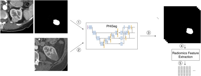
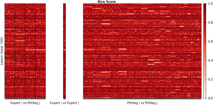
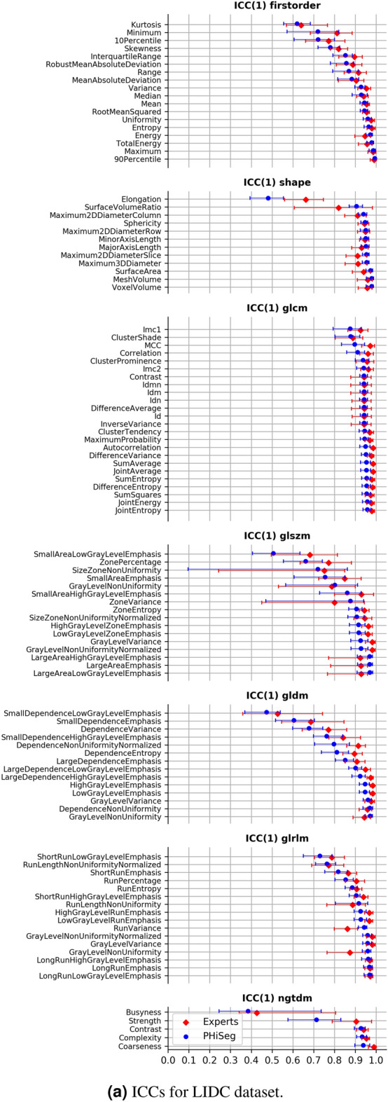
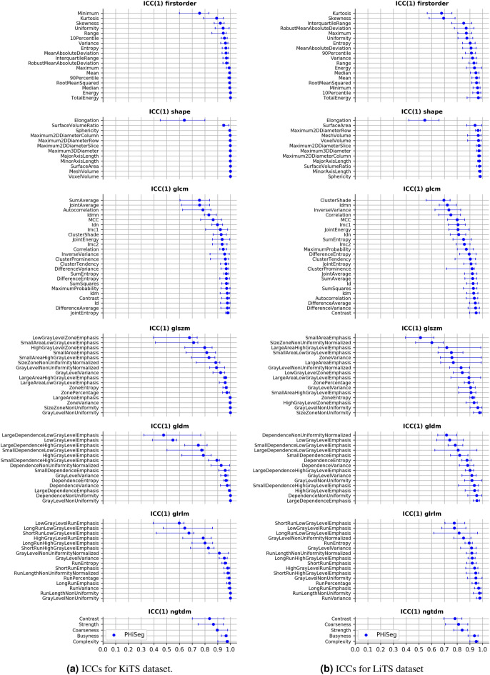
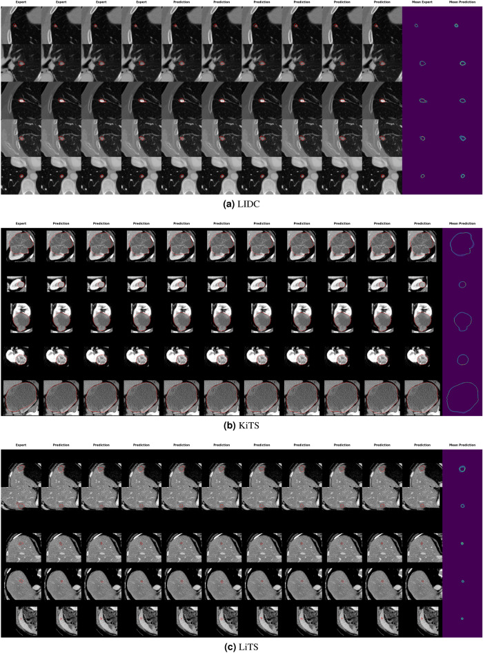
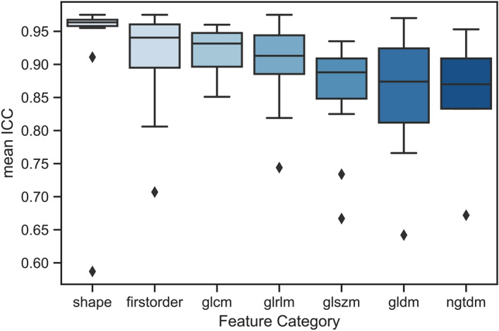
Identifying image features that are robust with respect to segmentation variability is a tough challenge in radiomics. So far, this problem has mainly been tackled in test–retest analyses. In this work we analyse radiomics feature reproducibility in two phases: first with manual segmentations provided by four expert readers and second with probabilistic automated segmentations using a recently developed neural network (PHiseg). We test feature reproducibility on three publicly available datasets of lung, kidney and liver lesions. We find consistent results both over manual and automated segmentations in all three datasets and show that there are subsets of radiomic features which are robust against segmentation variability and other radiomic features which are prone to poor reproducibility under differing segmentations. By providing a detailed analysis of robustness of the most common radiomics features across several datasets, we envision that more reliable and reproducible radiomic models can be built in the future based on this work.
Overview
The claims and statistics of this paper haven’t been checked yet.
Run the check — free, no account needed. It reads the open-access text and checks the sources it cites.
Integrity signals
Registry
Cites
Statistics
Figures
Verification
Claims the paper makes
The claims and statistics of this paper haven’t been checked yet.
Run the check — free, no account needed. It reads the open-access text and checks the sources it cites.
Claims other papers make about this one
Each is checked against this paper’s own text: does it say what the citing paper says it does?
In the citing paper
“Moreover, these statistical features are affected by several confounders, such as inter-reader variability in segmentations26 and acquisition settings of the scanners27, limiting their applicability in diverse settings.”
From Foundation Model for Cancer Imaging Biomarkers (2024) · cites this paper as [26]
Self-check
Percentages, sums and reported differences re-computed from the paper’s own numbers.
The self-check needs the paper’s full text.
Citations
Cites
U-Net: Convolutional Networks for Biomedical Image Segmentation
Ronneberger, O., Fischer, P., & Brox, T. (2015) · Lecture notes in computer science · doi:10.1007/978-3-319-24574-4_28 · Cited by 99,404
A guideline of selecting and reporting intraclass correlation coefficients for reliability research
Koo, T. K., & Li, M. Y. (2016) · Journal of Chiropractic Medicine · doi:10.1016/j.jcm.2016.02.012 · Cited by 31,709
Intraclass correlations: Uses in assessing rater reliability.
Shrout, P. E., & Fleiss, J. L. (1979) · Psychological Bulletin · doi:10.1037/0033-2909.86.2.420 · Cited by 24,532
Advances in neural information processing systems 7
Cios, K. J., & Shields, M. E. (1997) · Neurocomputing · doi:10.1016/s0925-2312(97)90018-7 · Cited by 22,093
Cicchetti, D. V. (1994) · Psychological Assessment · doi:10.1037/1040-3590.6.4.284 · Cited by 9,349 · cited 1×
“Possible choices are the often-used interpretation of defining excellent agreement as ICC > 0.75 36 or the interpretation proposed by Koo and Li 37 , stating that ICC > 0.9 corresponds to excellent agreement.”
MethodInfluentialComputational Radiomics System to Decode the Radiographic Phenotype
(2017) · doi:10.1158/0008-5472.can-17-0339 · Cited by 6,593
Decoding tumour phenotype by noninvasive imaging using a quantitative radiomics approach
Aerts, H. J. W. L., Velazquez, E. R., Leijenaar, R. T. H., Parmar, C., Grossmann, P., Carvalho, S., Bussink, J., Monshouwer, R., Haibe-Kains, B., Rietveld, D., et al. (2014) · Nature Communications · doi:10.1038/ncomms5006 · Cited by 5,132
The Cancer Imaging Archive (TCIA): Maintaining and Operating a Public Information Repository
Clark, K., Vendt, B., Smith, K., Freymann, J., Kirby, J., Koppel, P., Moore, S., Phillips, S., Maffitt, D., Pringle, M., et al. (2013) · Journal of Digital Imaging · doi:10.1007/s10278-013-9622-7 · Cited by 4,599
Zwanenburg, A., Vallières, M., Abdalah, M. A., Aerts, H. J. W. L., Andrearczyk, V., Apte, A., Ashrafinia, S., Bakas, S., Beukinga, R. J., Boellaard, R., et al. (2020) · Radiology · doi:10.1148/radiol.2020191145 · Cited by 4,177
Armato, S. G., McLennan, G., Bidaut, L., McNitt‐Gray, M. F., Meyer, C. R., Reeves, A. P., Zhao, B., Aberle, D. R., Henschke, C. I., Hoffman, E. A., et al. (2011) · Medical Physics · doi:10.1118/1.3528204 · Cited by 2,887
Cited by
Sorted by citations
A review in radiomics: Making personalized medicine a reality via routine imaging
Guiot, J., Vaidyanathan, A., Deprez, L., Zerka, F., Danthine, D., Frix, A.-N., Lambin, P., Bottari, F., Tsoutzidis, N., Miraglio, B., et al. (2021) · Medicinal Research Reviews · doi:10.1002/med.21846 · Cited by 356
Radiomics and artificial intelligence for precision medicine in lung cancer treatment
Chen, M., Copley, S. J., Viola, P., Lu, H., & Aboagye, E. O. (2023) · Seminars in Cancer Biology · doi:10.1016/j.semcancer.2023.05.004 · Cited by 213
Zhang, Y., Zhang, X., Cheng, Y., Li, B., Teng, X.-Z., Zhang, J., Lam, S., Zhou, T., Ma, Z., Sheng, J., et al. (2023) · Military Medical Research · doi:10.1186/s40779-023-00458-8 · Cited by 218
Foundation Model for Cancer Imaging Biomarkers
Pai, S., Bontempi, D., Hadzic, I., Prudente, V., Sokač, M., Chaunzwa, T. L., Bernatz, S., Hosny, A., Mak, R. H., Birkbak, N. J., et al. (2024) · Nature Machine Intelligence · doi:10.1038/s42256-024-00807-9 · Cited by 199
Radiomics feature reliability assessed by intraclass correlation coefficient: a systematic review
Xue, C., Yuan, J., Lo, G. G., Chang, A. T. Y., Poon, D. M. C., Wong, O. L., Zhou, Y., & Chu, C. W. (2021) · Quantitative Imaging in Medicine and Surgery · doi:10.21037/qims-21-86 · Cited by 194
Pfaehler, E. A. G., Zhovannik, I., Wei, L., Boellaard, R., Dekker, A. L. A. J., Monshouwer, R., Naqa, I. E., Bussink, J., Gillies, R. J., Wee, L., et al. (2021) · Physics and Imaging in Radiation Oncology · doi:10.1016/j.phro.2021.10.007 · Cited by 109
Radiomics: a primer on high-throughput image phenotyping
Lafata, K. J., Wang, Y., Konkel, B., Yin, F.-F., & Bashir, M. R. (2021) · Abdominal Radiology · doi:10.1007/s00261-021-03254-x · Cited by 92
Feature selection methods and predictive models in CT lung cancer radiomics
Ge, G. R., & Zhang, J. (2022) · Journal of Applied Clinical Medical Physics · doi:10.1002/acm2.13869 · Cited by 77
Setu, M. A. K., Horstmann, J., Schmidt, S., Stern, M. E., & Steven, P. (2021) · Scientific Reports · doi:10.1038/s41598-021-87314-8 · Cited by 71
Challenges for machine learning in clinical translation of big data imaging studies
Dinsdale, N. K., Bluemke, E., Sundaresan, V., Jenkinson, M., Smith, S. M., & Namburete, A. I. L. (2022) · Neuron · doi:10.1016/j.neuron.2022.09.012 · Cited by 74
Salmanpour, M. R., Hosseinzadeh, M., Rezaeijo, S. M., & Rahmim, A. (2023) · Computer Methods and Programs in Biomedicine · doi:10.1016/j.cmpb.2023.107714 · Cited by 69
Lambin, P., Woodruff, H. C., Mali, S. A., Zhong, X., Kuang, S., Lavrova, E., Khan, H., Lekadir, K., Zwanenburg, A., Deasy, J. O., et al. (2025) · Nature Reviews Clinical Oncology · doi:10.1038/s41571-025-01067-1 · Cited by 72
Oncologic Imaging and Radiomics: A Walkthrough Review of Methodological Challenges
Stanzione, A., Cuocolo, R., Ugga, L., Verde, F., Romeo, V., Brunetti, A., & Maurea, S. (2022) · Cancers · doi:10.3390/cancers14194871 · Cited by 64
Dunn, B., Pierobon, M., & Wei, Q. (2023) · Bioengineering · doi:10.3390/bioengineering10060690 · Cited by 64
Radiomics in Oncology, Part 1: Technical Principles and Gastrointestinal Application in CT and MRI
Caruso, D., Polici, M., Zerunian, M., Pucciarelli, F., Guido, G., Polidori, T., Landolfi, F., Nicolai, M., Lucertini, E., Tarallo, M., et al. (2021) · Cancers · doi:10.3390/cancers13112522 · Cited by 59
Wong, J., Baine, M. J., Wisnoskie, S., Bennion, N. R., Zheng, D., Lei, Y., Dalal, V., Hollingsworth, M. A., Lin, C. H., & Zheng, D. (2021) · Scientific Reports · doi:10.1038/s41598-021-95152-x · Cited by 55
Ferrante, M., Rinaldi, L. M., Botta, F., Hu, X., Dolp, A., Minotti, M., Piano, F. D., Funicelli, G., Volpe, S., Bellerba, F., et al. (2022) · Journal of Clinical Medicine · doi:10.3390/jcm11247334 · Cited by 43
Automated Tumor Segmentation in Radiotherapy
Savjani, R. R., Lauria, M., Bose, S., Deng, J., Yuan, Y., & Andrearczyk, V. (2022) · Seminars in Radiation Oncology · doi:10.1016/j.semradonc.2022.06.002 · Cited by 44
Re, M. D., Cucchiara, F., Rofi, E., Fontanelli, L., Petrini, I., Gri, N., Pasquini, G., Rizzo, M., Gabelloni, M., Belluomini, L., et al. (2020) · Cancer Immunology Immunotherapy · doi:10.1007/s00262-020-02810-6 · Cited by 38
Multiparametric MRI and Radiomics in Prostate Cancer: A Review of the Current Literature
Midiri, F., Vernuccio, F., Purpura, P., Alongi, P., & Bartolotta, T. V. (2021) · Diagnostics · doi:10.3390/diagnostics11101829 · Cited by 37
Teng, X., Zhang, J., Ma, Z., Zhang, Y., Lam, S., Li, W., Xiao, H., Li, T., Li, B., Zhou, T., et al. (2022) · Frontiers in Oncology · doi:10.3389/fonc.2022.974467 · Cited by 37
Majumder, S., Katz, S., Kontos, D., & Roshkovan, L. (2023) · BJR|Open · doi:10.1093/bjro/tzad004 · Cited by 42
Dutta, K., Roy, S. S., Whitehead, T., Luo, J. R., Jha, A. K., Li, S., Quirk, J. D., & Shoghi, K. I. (2021) · Cancers · doi:10.3390/cancers13153795 · Cited by 37
Staal, F. C., Taghavi, M., van der Reijd, D. J., Gómez, F. M., Imani, F., Klompenhouwer, E. G., Meek, D. B., Roberti, S., de Boer, M., Lambregts, D. M. J., et al. (2021) · European Journal of Radiology · doi:10.1016/j.ejrad.2021.109773 · Cited by 34
Granata, V., Fusco, R., Muzio, F. D., Brunese, M. C., Setola, S. V., Ottaiano, A., Cardone, C., Avallone, A., Patrone, R., Pradella, S., et al. (2023) · La radiologia medica · doi:10.1007/s11547-023-01710-w · Cited by 32
Haniff, N. S. M., Karim, M. K. A., Osman, N. H., Saripan, M. I., Isa, I. N. C., & Ibahim, M. J. (2021) · Diagnostics · doi:10.3390/diagnostics11091573 · Cited by 32
Lee, S. B., Cho, Y. J., Hong, Y., Jeong, D., Lee, J., Kim, S., Lee, S., & Choi, Y. H. (2021) · Investigative Radiology · doi:10.1097/rli.0000000000000839 · Cited by 32
Impact of image quality on radiomics applications
Cui, Y., & Yin, F.-F. (2022) · Physics in Medicine and Biology · doi:10.1088/1361-6560/ac7fd7 · Cited by 30
Endo, Y., Alaimo, L., Lima, H. A., Moazzam, Z., Ratti, F., Marques, H. P., Soubrane, O., Lam, V. W. T., Kitago, M., Poultsides, G. A., et al. (2022) · Annals of Surgical Oncology · doi:10.1245/s10434-022-12494-0 · Cited by 31
Teng, X., Wang, Y., Nicol, A. J., Ching, J. C. F., Wong, E. K. Y., Lam, K. T. C., Zhang, J., Lee, S. W. Y., & Cai, J. (2024) · Diagnostics · doi:10.3390/diagnostics14161835 · Cited by 30
Yousefi, B., Lariviere, M. J., Cohen, E. A., Buckingham, T. H., Yee, S. S., Black, T. A., Chien, A. L., Noël, P. B., Hwang, W., Katz, S. I., et al. (2021) · Scientific Reports · doi:10.1038/s41598-021-88239-y · Cited by 28
Park, T., Yoon, M. A., Cho, Y. C., Ham, S. J., Ko, Y., Kim, S., Jeong, H., & Lee, J. (2022) · Scientific Reports · doi:10.1038/s41598-022-10807-7 · Cited by 29
Comprehensive Analysis of Tumour Sub-Volumes for Radiomic Risk Modelling in Locally Advanced HNSCC
Leger, S., Zwanenburg, A., Leger, K., Lohaus, F., Linge, A., Schreiber, A., Kalinauskaitė, G., Tinhofer, I., Guberina, N., Guberina, M., et al. (2020) · Cancers · doi:10.3390/cancers12103047 · Cited by 29
Zaridis, D. I., Mylona, E., Tachos, N. S., Pezoulas, V. C., Grigoriadis, G., Tsiknakis, N., Kostas Marias, A., Tsiknakis, M., & Fotiadis, D. I. (2023) · Scientific Reports · doi:10.1038/s41598-023-27671-8 · Cited by 28
Shankar, L., Schöder, H., Sharon, E., Wolchok, J., Knopp, M., Wahl, R., Ellingson, B., Hall, N., Yaffe, M., Towbin, A., et al. (2023) · The Lancet Oncology · doi:10.1016/s1470-2045(22)00742-2 · Cited by 19
Naseri, H., Skamene, S., Tolba, M., Faye, M. D., Ramia, P., Khriguian, J., Patrick, H. M., Andrade, A. X., David, M., & Kildea, J. (2022) · Scientific Reports · doi:10.1038/s41598-022-13379-8 · Cited by 27
Spatial assessments in texture analysis: what the radiologist needs to know
Varghese, B. A., Fields, B. K. K., Hwang, D., Duddalwar, V. A., Matcuk, G. R., & Cen, S. (2023) · Frontiers in Radiology · doi:10.3389/fradi.2023.1240544 · Cited by 26
Ferrari, R., Trinci, M., Casinelli, A., Treballi, F., Leone, E., Caruso, D., Polici, M., Faggioni, L., Neri, E., & Galluzzo, M. (2024) · La radiologia medica · doi:10.1007/s11547-024-01904-w · Cited by 25
Challenges in Glioblastoma Radiomics and the Path to Clinical Implementation
Martin, P., Holloway, L. C., Metcalfe, P. E., Koh, E., & Brighi, C. (2022) · Cancers · doi:10.3390/cancers14163897 · Cited by 24
Kothari, G., Woon, B., Patrick, C. J., Korte, J. C., Wee, L., Hanna, G. G., Kron, T., Hardcastle, N., & Siva, S. (2022) · Scientific Reports · doi:10.1038/s41598-022-16520-9 · Cited by 27
Reiazi, R., Arrowsmith, C., Welch, M. L., Aghababazadeh, F. A., Eeles, C. B., Tadic, T., Hope, A. J., Bratman, S. V., & Haibe‐Kains, B. (2021) · Cancers · doi:10.3390/cancers13092269 · Cited by 22
Mottola, M., Ursprung, S., Rundo, L., Sánchez, L. E., Klatte, T., Mendichovszky, I. A., Stewart, G. D., Sala, E., & Bevilacqua, A. (2021) · Scientific Reports · doi:10.1038/s41598-021-90985-y · Cited by 23
Radiomics feature reproducibility: The elephant in the room
Klontzas, M. E. (2024) · European Journal of Radiology · doi:10.1016/j.ejrad.2024.111430 · Cited by 22
Singh, A., Horng, H., Roshkovan, L., Weeks, J. K., Hershman, M., Noël, P. B., Luna, J. M., Cohen, E. A., Pantalone, L., Shinohara, R. T., et al. (2022) · Scientific Reports · doi:10.1038/s41598-022-14160-7 · Cited by 21
Priya, S., Aggarwal, T., Ward, C. E., Bathla, G., Jacob, M., Gerke, A. K., Hoffman, E. A., & Nagpal, P. (2021) · Scientific Reports · doi:10.1038/s41598-021-92155-6 · Cited by 21
Siow, T. Y., Yeh, C., Lin, G., Lin, C., Wang, H., Liao, C., Toh, C., Chan, S.-C., Lin, C.-P., & Ng, S. (2022) · Cancers · doi:10.3390/cancers14246119 · Cited by 20
Pasini, G., Russo, G. I., Mantarro, C., Bini, F., Richiusa, S., Morgante, L., Comelli, A., Russo, G. I., SABINI, M. G., Cosentino, S., et al. (2023) · Diagnostics · doi:10.3390/diagnostics13243640 · Cited by 20
Mukherjee, S., Korfiatis, P. D., Patnam, N. G., Trivedi, K. H., Karbhari, A., Suman, G., Fletcher, J. G., & Goenka, A. H. (2024) · Abdominal Radiology · doi:10.1007/s00261-023-04127-1 · Cited by 20
Berta, L., Rizzetto, F., Mattia, C. D., Lizio, D., Felisi, M. M. J., Colombo, P. E., Carrazza, S., Gelmini, S., Bianchi, L. M. G., Artioli, D., et al. (2021) · Physica Medica · doi:10.1016/j.ejmp.2021.06.001 · Cited by 19
Granata, V., Fusco, R., Setola, S. V., Galdiero, R., Maggialetti, N., Patrone, R., Ottaiano, A., Nasti, G., Silvestro, L., Cassata, A., et al. (2023) · Infectious Agents and Cancer · doi:10.1186/s13027-023-00495-x · Cited by 19
Ren, Q.-Q., Zhu, P., Li, C.-D., Yan, M.-J., Liu, S., Zheng, C., & Xia, X. (2022) · Frontiers in Bioengineering and Biotechnology · doi:10.3389/fbioe.2022.872044 · Cited by 18
Radiomics in veterinary medicine: Overview, methods, and applications
Basran, P. S., & Porter, I. R. (2022) · Veterinary Radiology & Ultrasound · doi:10.1111/vru.13156 · Cited by 19
Gottlich, H. C., Gregory, A., Sharma, V., Khanna, A., Moustafa, A. U., Lohse, C. M., Potretzke, T. A., Korfiatis, P. D., Potretzke, A. M., Đenić, A., et al. (2023) · Journal of Imaging Informatics in Medicine · doi:10.1007/s10278-023-00804-1 · Cited by 17
Joo, L., Jung, S. C., Lee, H., Park, S. Y., Kim, M., Park, J. E., & Choi, K. M. (2021) · Scientific Reports · doi:10.1038/s41598-021-96621-z · Cited by 17
Du, D., Lv, W., Lv, J., Chen, X., Wu, H., Rahmim, A., & Lu, L. (2022) · European Radiology · doi:10.1007/s00330-022-09229-w · Cited by 16
Variability Matters: Evaluating Inter-Rater Variability in Histopathology for Robust Cell Detection
Kang, C. M., Lee, C., Song, H., Ma, M., & Pereira, S. (2023) · Lecture notes in computer science · doi:10.1007/978-3-031-25082-8_37 · Cited by 14
HeLLePhant: A phantom mimicking non-small cell lung cancer for texture analysis in CT images
Rinaldi, L. M., Pezzotta, F., Santaniello, T., Marco, P. D., Bianchini, L., Origgi, D., Cremonesi, M., Milani, P., Mariani, M., & Botta, F. (2022) · Physica Medica · doi:10.1016/j.ejmp.2022.03.010 · Cited by 14
Robust Radiomic Signatures of Intervertebral Disc Degeneration From MRI
McSweeney, T. P., Tiulpin, A., Kowlagi, N., Määttä, J. H., Karppinen, J. I., & Saarakkala, S. S. (2025) · Spine · doi:10.1097/brs.0000000000005435 · Cited by 13
Mohammadi-Sadr, M., Cheki, M., Moslehi, M., Zarasvandnia, M., & Salamat, M. R. (2024) · Academic Radiology · doi:10.1016/j.acra.2024.10.049 · Cited by 13
Translating radiomics into clinical practice: A step-by-step guide to study design and evaluation
Chen, M., Linton‐Reid, K., Aboagye, E. O., & Copley, S. J. (2025) · Clinical Radiology · doi:10.1016/j.crad.2025.107053 · Cited by 15
PRISM: A Promptable and Robust Interactive Segmentation Model with Visual Prompts
Li, H., Liu, H., Hu, D., Wang, J., & Oguz, I. (2024) · Lecture Notes in Computer Science · doi:10.1007/978-3-031-72384-1_37 · Cited by 12
Jahanshahi, A., Soleymani, Y., Ghaziyani, M. F., & Khezerloo, D. (2023) · The Egyptian Journal of Radiology and Nuclear Medicine · doi:10.1186/s43055-023-01029-6 · Cited by 12
Alsoof, D., McDonald, C. L., Durand, W. M., Diebo, B. G., Kuris, E. O., & Daniels, A. H. (2023) · The International Journal of Spine Surgery · doi:10.14444/8501 · Cited by 11
From pixels to prognosis: radiomics and AI in Alzheimer’s disease management
Peng, D., Huang, W., Liu, R. P., & Zhong, W. (2025) · Frontiers in Neurology · doi:10.3389/fneur.2025.1536463 · Cited by 12
Predictive performance of radiomic models based on features extracted from pretrained deep networks
Demircioğlu, A. (2022) · Insights into Imaging · doi:10.1186/s13244-022-01328-y · Cited by 10
Bianconi, F., Fravolini, M. L., Palumbo, I., Pascoletti, G., Nuvoli, S. M. F., Rondini, M., Spanu, A., & Palumbo, B. (2021) · Diagnostics · doi:10.3390/diagnostics11071224 · Cited by 10
Soleymani, Y., Jahanshahi, A., Pourfarshid, A., & Khezerloo, D. (2022) · Journal of medical imaging and radiation sciences · doi:10.1016/j.jmir.2022.09.018 · Cited by 10
Bakker, M. A. G., Orvalho, M. L., Matela, N., & Mota, A. M. (2024) · Journal of Imaging · doi:10.3390/jimaging10090218 · Cited by 11
Foundation Models for Quantitative Biomarker Discovery in Cancer ImagingPreprint
Pai, S., Bontempi, D., Hadžić, I., Prudente, V., Sokač, M., Chaunzwa, T. L., Bernatz, S., Hosny, A., Mak, R. H., Birkbak, N. J., et al. (2023) · medRxiv · doi:10.1101/2023.09.04.23294952 · Cited by 9
Zeng, D., Song, Z., Liu, Q., Huang, J., Wang, X. W., & Tang, Z. (2025) · BMC Medical Imaging · doi:10.1186/s12880-025-01664-7 · Cited by 8
Whybra, P., & Spezi, E. (2023) · Scientific Reports · doi:10.1038/s41598-023-41475-w · Cited by 8
Robustness and performance of radiomic features in diagnosing cystic renal masses
Könik, A., Miskin, N. P., Guo, Y., Shinagare, A. B., & Qin, L. (2021) · Abdominal Radiology · doi:10.1007/s00261-021-03241-2 · Cited by 8
Bartholomeus, G. A., van Amsterdam, W. A. C., den Harder, A. M., Willemink, M. J., van Hamersvelt, R. W., de Jong, P. A., & Leiner, T. (2023) · European Radiology · doi:10.1007/s00330-023-09643-8 · Cited by 8
Stability of Radiomic Models and Strategies to Enhance Reproducibility
Chaddad, A., & Liang, X. (2024) · IEEE Transactions on Radiation and Plasma Medical Sciences · doi:10.1109/trpms.2024.3365778 · Cited by 7
Ma, J., Yang, H., Chou, Y., Yoon, J. H., Allison, T., Komandur, R., McDunn, J., Tasneem, A., Do, R. K., Schwartz, L. H., et al. (2024) · Medical Physics · doi:10.1002/mp.17504 · Cited by 7
Dong, Y., Wang, P., Geng, H., Liu, Y., & Wang, E. (2025) · Journal of X-Ray Science and Technology · doi:10.1177/08953996241304988 · Cited by 6
Müller‐Franzes, G. A., Nebelung, S., Schock, J., Haarburger, C., Khader, F., Pedersoli, F., Schulze-Hagen, M. F., Kuhl, C. K., & Truhn, D. (2022) · Diagnostics · doi:10.3390/diagnostics12020247 · Cited by 6
Self-citationGe, G. R., Siddique, A., & Zhang, J. (2023) · Physics in Medicine and Biology · doi:10.1088/1361-6560/acce1c · Cited by 6
Widaatalla, Y., Wolswijk, T., Khan, M. D., Halilaj, I., Mosterd, K., Woodruff, H. C., & Lambin, P. (2025) · Cancers · doi:10.3390/cancers17050768 · Cited by 5
Haniff, N. S. M., Ng, K. H., Kamal, I., Zain, N. M., & Karim, M. K. A. (2024) · Heliyon · doi:10.1016/j.heliyon.2024.e36313 · Cited by 5
Chirra, P. V., Giriprakash, P. P., Rizk, A. G., Kurowski, J. A., Viswanath, S. E., & Gandhi, N. S. (2024) · Journal of Imaging Informatics in Medicine · doi:10.1007/s10278-024-01303-7 · Cited by 5
Xu, F., Zhang, Y., Ma, Q., Hu, L., Li, Y. H., Gao, C., Guo, P., Yang, X., Zhou, Y., Zhang, J., et al. (2025) · BMC Pregnancy and Childbirth · doi:10.1186/s12884-025-07508-0 · Cited by 5
Qian, W.-L., Chen, Q., Jb, Z., Xu, J.-M., & Hu, C.-H. (2023) · Clinical Radiology · doi:10.1016/j.crad.2023.03.001 · Cited by 5
Chen, Y., Zhao, B., Zhu, C., Bie, C., He, X., Zheng, Z., & Song, X. (2023) · Magnetic Resonance Imaging · doi:10.1016/j.mri.2023.07.003 · Cited by 5
The impact of Inter-observation variation on radiomic features of pulmonary nodules
Zhu, W., Xu, F., Lou, K., Qiu, X., Huang, D., Huang, S., Xie, D., & Hu, H. (2025) · Frontiers in Oncology · doi:10.3389/fonc.2025.1567028 · Cited by 4
Arisar, F. A. Q., Salinas-Miranda, E., Ali, H. A., Lajkosz, K., Chen, C., Azhie, A., Healy, G. M., Deniffel, D., Haider, M. A., & Bhat, M. (2023) · Transplant International · doi:10.3389/ti.2023.11149 · Cited by 4
Stability of Multi-Parametric Prostate MRI Radiomic Features to Variations in Segmentation
Seetha, S. T., Garanzini, E. M., Tenconi, C., Marenghi, C., Avuzzi, B., Catanzaro, M. A., Stagni, S., Villa, S., Chiorda, B. N., Badenchini, F., et al. (2023) · Journal of Personalized Medicine · doi:10.3390/jpm13071172 · Cited by 4
Artificial Intelligence Applications for Imaging Metabolic Bone Diseases
Isaac, A., Akdoğan, A. I., Dalili, D., Saber, N., Drobny, D., Guglielmi, G., Marc Modat, A., & Bazzocchi, A. (2024) · Seminars in Musculoskeletal Radiology · doi:10.1055/s-0044-1789218 · Cited by 4
Chest CT in COVID-19 Pneumonia: Potentials and Limitations of Radiomics and Artificial Intelligence
Vernuccio, F., Cutaia, G., Cannella, R., Vernuccio, L., Lagalla, R., & Midiri, M. (2021) · Studies in computational intelligence · doi:10.1007/978-3-030-74761-9_3 · Cited by 4
Ramlee, S., Delgado-Ortet, M., Sánchez, L. E., Aloj, L., & Manavaki, R. (2025) · PLoS ONE · doi:10.1371/journal.pone.0335219 · Cited by 3
Pediatric Personalized Deep Learning Models for Segmentation of Hepatoblastoma at CT and MRI
Modanwal, G., Kumar, S., Viswanathan, V. S., Morin, C. E., Rees, M. A., Squires, J. E., Tang, E., Katzenstein, H. M., Towbin, A. J., Madabhushi, A., et al. (2026) · Radiology Imaging Cancer · doi:10.1148/rycan.250041 · Cited by 3
Radiomics-Based Classification of Pathological Patterns in Common Carotid Artery Wall
Jadoon, M., Poli, F., Boutouyrie, P., Khettab, H., Joywin-Melitus, G., Sam, D.-O., Bianchini, E., Faita, F., Jouven, X., Empana, J. P., et al. (2026) · Ultrasound in Medicine & Biology · doi:10.1016/j.ultrasmedbio.2026.02.017 · Cited by 3
Soleymani, Y., Sheikhzadeh, P., Mohammadzadeh, M., & Khezerloo, D. (2023) · Journal of Biomedical Physics and Engineering · doi:10.31661/jbpe.v0i0.2208-1525 · Cited by 3
Mirza‐Aghazadeh‐Attari, M., Afyouni, S., Zandieh, G., Nia, I. Y., Mohseni, A., Borhani, A., Madani, S. P., Shahbazian, H., Ansari, G., Kim, A., et al. (2023) · Journal of Computer Assisted Tomography · doi:10.1097/rct.0000000000001557 · Cited by 3
Martin, P., Holloway, L. C., Metcalfe, P. E., Koh, E., Aly, F., Chan, E., & Brighi, C. (2025) · Physical and Engineering Sciences in Medicine · doi:10.1007/s13246-025-01613-2 · Cited by 2
From diagnosis to treatment: navigating the course for pancreatic neuroendocrine neoplasms
Mastnak, L., & Badovinac, D. (2025) · Annals of Medicine · doi:10.1080/07853890.2025.2569992 · Cited by 2
Chupetlovska, K., Lipman, K. B. W. G., Bodalal, Z., Aricò, F. M., Topff, L., Maas, M., & Trebeschi, S. (2025) · European Journal of Radiology Artificial Intelligence · doi:10.1016/j.ejrai.2025.100053 · Cited by 2
Sümer, E., Danyeli, D. A. E., Yakicier, M. C., Pamir, M. N., Özduman, K., Di̇nçer, A., & Öztürk-Işık, E. (2026) · Frontiers in Oncology · doi:10.3389/fonc.2026.1775958 · Cited by 2
Mori, M., Alborghetti, L., Palumbo, D., Broggi, S., Raspanti, D., Rovere, P. Q., Vecchio, A. D., COBELLI, F. D., & Fiorino, C. (2022) · Physica Medica · doi:10.1016/j.ejmp.2022.06.018 · Cited by 2
A Deep Learning Framework for Analysis of the Eustachian Tube and the Internal Carotid Artery
Amanian, A., Jain, A., Xiao, Y., Kim, C., Ding, A. S., Sahu, M., Taylor, R. H., Unberath, M., Ward, B. K., Galaiya, D. J., et al. (2024) · Otolaryngology · doi:10.1002/ohn.789 · Cited by 2
Metadata
- Authors
- Haarburger, C., Müller-Franzes, G., Weninger, L., Kuhl, C., Truhn, D., & Merhof, D.
- Affiliations
- RWTH Aachen University (DE)
- Universitätsklinikum Aachen (DE)
- Fraunhofer Institute for Digital Medicine (DE)
- Corresponding author
- Haarburger, Christoph
- Venue
- Scientific Reports
- Published
- 2020-07-29
- Volume
- 10
- Issue
- 1
- Pages
- 12688-12688
- ISSN
- 2045-2322 (ISSN-L)
- PMC
- PMC7391354
- OpenAlex
- W3046015102
- Cited by
- 139
- References
- 38
- Citations by year
- 2026: 24 · 2025: 20 · 2024: 17 · 2023: 28 · 2022: 26 · 2021: 22 · 2020: 2
- FWCI
- 9.49
- Type
- journal article
- Language
- en
- Update notices
- correction (2021-11-16)
- Open access
- gold
- doi.org
- Locations
- Scientific Reports (publishedVersion, cc-by)
- PubMed Central (publishedVersion, cc-by)
- PubMed (publishedVersion)
- Publikationsdatenbank der Fraunhofer-Gesellschaft (Fraunhofer-Gesellschaft) (submittedVersion, other-oa)
- Fraunhofer-Publica (Fraunhofer-Gesellschaft) (submittedVersion)
- RWTH Publications (RWTH Aachen) (submittedVersion, other-oa)
- Funders
- RWTH Aachen University
- Interreg
- Topics
- Health Sciences › Medicine › Radiology, Nuclear Medicine and Imaging › Radiomics and Machine Learning in Medical Imaging
- Physical Sciences › Computer Science › Computer Vision and Pattern Recognition › Medical Image Segmentation Techniques
- Physical Sciences › Computer Science › Computer Vision and Pattern Recognition › Image Retrieval and Classification Techniques
- Keywords
- radiomics, feature reproducibility, interobserver variability, segmentation variability, CT images, manual segmentation, automatic segmentation, lung lesions, renal lesions, liver lesions, feature robustness
- MeSH terms
- Automation, Humans, Neoplasms, Radiographic Image Interpretation, Computer-Assisted, Tomography, X-Ray Computed, Observer Variation, Databases, Factual, Neural Networks, Computer
About these checks
Registry, retraction, Cites and statistics results are computed, not judged, with one exception: a printed reference that no index lists is read once by a model before it counts as a citation error. Claims are extracted by a model and kept only when their quote appears verbatim in the text; each cited source is then checked, with a model’s help, for whether it says what the claim relies on. Those verdicts can be wrong — read the claim’s quote and the cited source before relying on one.
Statistics and arithmetic are recomputed, not judged.
Confidence bands are shown only for fields calibrated on our annotated test set. None is calibrated yet, so no band is shown.
What counts as a claim ›How we measure confidence ›How we check this ›
Are you an author, or do you see a problem with this page? Request a review
