

The DOI matches its Crossref record; the references it cites are not checked yet.
Magnetic domains oscillation in the brain with neurodegenerative disease
Open accessAbstract
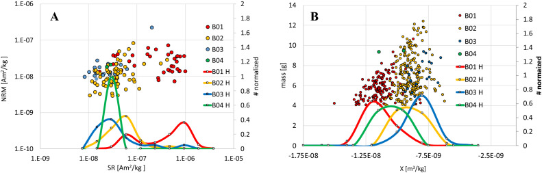
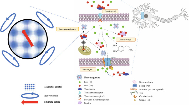
Geomagnetic fields interfere with the accumulation of iron in the human brain. Magnetic sensing of the human brain provides compelling evidence of new electric mechanisms in human brains and may interfere with the evolution of neurodegenerative diseases. We revealed that the human brain may have a unique susceptibility to conduct electric currents as feedback of magnetic dipole fluctuation in superparamagnetic grains. These grains accumulate and grow with brain aging. The electric feedback creates an electronic noise background that depends on geomagnetic field intensity and may compromise functional stability of the human brain, while induced currents are spontaneously generated near superparamagnetic grains. Grain growth due to an increase of iron mobility resulted in magnetic remanence enhancement during the final years of the studied brains.
Overview
The claims and statistics of this paper haven’t been checked yet.
Run the check — free, no account needed. It reads the open-access text and checks the sources it cites.
Integrity signals
Registry
Cites
Statistics
Figures
Verification
Claims the paper makes
The claims and statistics of this paper haven’t been checked yet.
Run the check — free, no account needed. It reads the open-access text and checks the sources it cites.
Claims other papers make about this one
Each is checked against this paper’s own text: does it say what the citing paper says it does?
The source backs part of the claim, not all of it — or backs it for a different population, condition or comparison.
In the citing paper
“In addition to this, magnetic fields at these frequencies have the potential to disturb intracellular ionic concentrations, which can indirectly affect neural activity and cellular homeostasis (Kletetschka et al., 2021; Panagopoulos et al., 2021).”
From Comparative analysis of energy transfer mechanisms for neural implants (2024) · cites this paper as Kletetschka et al., 2021
Experimental — accuracy not yet measured
Self-check
Percentages, sums and reported differences re-computed from the paper’s own numbers.
The self-check needs the paper’s full text.
Citations
The citation lists haven’t been fetched yet
References 67 · Cited by 25
Research works printed but not linked: 60
- Kirschvink, J. L., Kobayashi-Kirschvink, A. & Woodford, B. J. Magnetite biomineralization in the human brain. Proc. Natl. Acad. Sci. USA 89, 7683–7687. https://doi.org/10.1073/pnas.89.16.7683 (1992). · DOI
- Maher, B. A. et al. Magnetite pollution nanoparticles in the human brain. Proc. Natl. Acad. Sci. USA 113, 10797–10801. https://doi.org/10.1073/pnas.1605941113 (2016). · DOI
- Bilgic, B., Pfefferbaum, A., Rohlfing, T., Sullivan, E. V. & Adalsteinsson, E. MRI estimates of brain iron concentration in normal aging using quantitative susceptibility mapping. Neuroimage 59, 2625–2635. https://doi.org/10.1016/j.neuroimage.2011.08.077 (2012). · DOI · Paper page
- Langkammer, C. et al. Susceptibility induced gray-white matter MRI contrast in the human brain. Neuroimage 59, 1413–1419. https://doi.org/10.1016/j.neuroimage.2011.08.045 (2012). · DOI
- Langkammer, C. et al. Quantitative susceptibility mapping (QSM) as a means to measure brain iron? A post mortem validation study. Neuroimage 62, 1593–1599. https://doi.org/10.1016/j.neuroimage.2012.05.049 (2012). · DOI · Paper page
- Acosta-Cabronero, J. et al. In vivo quantitative susceptibility mapping (QSM) in Alzheimer’s disease. PLoS ONE 8, 15. https://doi.org/10.1371/journal.pone.0081093 (2013). · DOI
- Deh, K. et al. Reproducibility of quantitative susceptibility mapping in the brain at two field strengths from two vendors. J. Magn. Reson. Imaging 42, 1592–1600. https://doi.org/10.1002/jmri.24943 (2015). · DOI
- Hinoda, T. et al. Quantitative susceptibility mapping at 3 T and 1.5 T evaluation of consistency and reproducibility. Invest. Radiol. 50, 522–530. https://doi.org/10.1097/rli.0000000000000159 (2015). · DOI
- Dunlop, J. D. & Özdemir, Ö. Rock Magnetism: Fundamentals and Frontiers 595 (Cambridge University Press, Cambridge, 1997). · DOI · Paper page
- Kletetschka, G. & Wieczorek, M. A. Fundamental relations of mineral specific magnetic carriers for paleointensity determination. Phys. Earth Planet. Int. 272, 44–49. https://doi.org/10.1016/j.pepi.2017.09.008 (2017). · DOI
- Chang, C. H., Lane, H. Y. & Lin, C. H. Brain stimulation in Alzheimer’s disease. Front. Psychiatry 9, 13. https://doi.org/10.3389/fpsyt.2018.00201 (2018). · DOI
- Barker, A. T. & Jalinous, R. Non-invasive magnetic stimulation of human motor cortex. Lancet 1, 1106–1107 (1985). · DOI
- Hallett, M. Transcranial magnetic stimulation: a primer. Neuron 55, 187–199. https://doi.org/10.1016/j.neuron.2007.06.026 (2007). · DOI
- Tiepolt, S. et al. Quantitative susceptibility mapping of amyloid-beta aggregates in Alzheimer’s disease with 7T MR. J. Alzheimers Dis. 64, 393–404. https://doi.org/10.3233/jad-180118 (2018). · DOI
- Fernandez, T., Martinez-Serrano, A., Cusso, L., Desco, M. & Ramos-Gomez, M. Functionalization and characterization of magnetic nanoparticles for the detection of ferritin accumulation in Alzheimer’s disease. ACS Chem. Neurosci. 9, 912–924. https://doi.org/10.1021/acschemneuro.7b00260 (2018). · DOI
- Altamura, S. & Muckenthaler, M. U. Iron toxicity in diseases of aging: Alzheimer’s disease, Parkinson’s disease and atherosclerosis. J. Alzheimers Dis. 16, 879–895. https://doi.org/10.3233/jad-2009-1010 (2009). · DOI · Paper page
- Bartzokis, G. et al. MR evaluation of age-related increase of brain iron in young adult and older normal males. Magn. Reson. Imaging 15, 29–35. https://doi.org/10.1016/s0730-725x(96)00234-2 (1997). · DOI
- Acosta-Cabronero, J., Betts, M. J., Cardenas-Blanco, A., Yang, S. & Nestor, P. J. In vivo MRI mapping of brain iron deposition across the adult lifespan. J. Neurosci. 36, 364–374. https://doi.org/10.1523/jneurosci.1907-15.2016 (2016). · DOI
- Prousek, J. Fenton chemistry in biology and medicine. Pure Appl. Chem. 79, 2325–2338. https://doi.org/10.1351/pac200779122325 (2007). · DOI
- Abeyawardhane, D. L. & Lucas, H. R. Iron redox chemistry and implications in the Parkinson’s disease brain. Oxid. Med. Cell. Longev. 2019, 11. https://doi.org/10.1155/2019/4609702 (2019). · DOI · Paper page
- Zhao, Z. Iron and oxidizing species in oxidative stress and Alzheimer’s disease. Aging Med. 2, 82–87. https://doi.org/10.1002/agm2.12074 (2019). · DOI
- Youdim, M. B. H., Benshachar, D. & Riederer, P. The possible role of iron in the etiopathology of Parkinsons-disease. Mov. Disord. 8, 1–12. https://doi.org/10.1002/mds.870080102 (1993). · DOI
- Zecca, L., Youdim, M. B. H., Riederer, P., Connor, J. R. & Crichton, R. R. Iron, brain ageing and neurodegenerative disorders. Nat. Rev. Neurosci. 5, 863–873. https://doi.org/10.1038/nrn1537 (2004). · DOI · Paper page
- Youdim, M. B. H., Stephenson, G. & Ben Shachar, D. In Redox-Active Metals in Neurological Disorders Vol. 1012 Annals of the New York Academy of Sciences (eds LeVine, S. M. et al.) 306–325 (New York Acad Sciences, New York, 2004).
- Riederer, P. et al. Transition-metals, ferritin, glutathione, and ascorbic-acid in Parkinsonian brains. J. Neurochem. 52, 515–520. https://doi.org/10.1111/j.1471-4159.1989.tb09150.x (1989). · DOI · Paper page
- Jenner, P. & Olanow, C. W. Understanding cell death in Parkinson’s disease. Ann. Neurol. 44, S72–S84. https://doi.org/10.1002/ana.410440712 (1998). · DOI
- Masaldan, S., Bush, A. I., Devos, D., Rolland, A. S. & Moreau, C. Striking while the iron is hot: iron metabolism and ferroptosis in neurodegeneration. Free Radic. Biol. Med. 133, 221–233. https://doi.org/10.1016/j.freeradbiomed.2018.09.033 (2019). · DOI
- Cenini, G., Lloret, A. & Cascella, R. Oxidative stress in neurodegenerative diseases: from a mitochondrial point of view. Oxid. Med. Cell. Longev. 2019, 18. https://doi.org/10.1155/2019/2105607 (2019). · DOI
- Ayton, S. et al. Cerebral quantitative susceptibility mapping predicts amyloid-beta-related cognitive decline. Brain 140, 2112–2119. https://doi.org/10.1093/brain/awx137 (2017). · DOI
- Dunlop, J. D. & Özdemir, Ö. Rock Magnetism: Fundamental and Frontiers 58 (Cambridge University Press, Cambridge, 1997). · DOI · Paper page
- Duce, J. A. et al. Iron-export ferroxidase activity of beta-amyloid precursor protein is inhibited by zinc in Alzheimer’s disease. Cell 142, 857–867. https://doi.org/10.1016/j.cell.2010.08.014 (2010). · DOI
- Lee, D. W. & Andersen, J. K. Iron elevations in the aging Parkinsonian brain: a consequence of impaired iron homeostasis?. J. Neurochem. 112, 332–339. https://doi.org/10.1111/j.1471-4159.2009.06470.x (2010). · DOI
- Piccinelli, P. & Samuelsson, T. Evolution of the iron-responsive element. RNA 13, 952–966. https://doi.org/10.1261/rna.464807 (2007). · DOI
- Mastroberardino, P. G. et al. A novel transferrin/TfR2-mediated mitochondrial iron transport system is disrupted in Parkinson’s disease. Neurobiol. Dis. 34, 417–431. https://doi.org/10.1016/j.nbd.2009.02.009 (2009). · DOI · Paper page
- Febbraro, F., Giorgi, M., Caldarola, S., Loreni, F. & Romero-Ramos, M. alpha-Synuclein expression is modulated at the translational level by iron. NeuroReport 23, 576–580. https://doi.org/10.1097/WNR.0b013e328354a1f0 (2012). · DOI
- Zecca, L. et al. The absolute concentration of nigral neuromelanin, assayed by a new sensitive method, increases throughout the life and is dramatically decreased in Parkinson’s disease. FEBS Lett. 510, 216–220. https://doi.org/10.1016/s0014-5793(01)03269-0 (2002). · DOI
- Adlard, P. A. et al. Rapid restoration of cognition in Alzheimer’s transgenic mice with 8-hydroxy quinoline analogs is associated with decreased interstitial A beta. Neuron 59, 43–55. https://doi.org/10.1016/j.neuron.2008.06.018 (2008). · DOI · Paper page
- Rogers, J. T. et al. An iron-responsive element type II in the 5 ’-untranslated region of the Alzheimer’s amyloid precursor protein transcript. J. Biol. Chem. 277, 45518–45528. https://doi.org/10.1074/jbc.M207435200 (2002). · DOI · Paper page
- Klausner, R. D., Rouault, T. A. & Harford, J. B. Regulating the fate of messenger-RNA—the control of cellular iron-metabolism. Cell 72, 19–28. https://doi.org/10.1016/0092-8674(93)90046-s (1993). · DOI · Paper page
- Schwaller, B. Cytosolic Ca2+ buffers are inherently Ca2+ signal modulators. Cold Spring Harbor Perspect. Biol. 12, 24. https://doi.org/10.1101/cshperspect.a035543 (2020). · DOI
- Tong, B. C. K., Wu, A. J., Li, M. & Cheung, K. H. Calcium signaling in Alzheimer’s disease and therapies. Biochim. Biophys. Acta Mol. Cell Res. 1745–1760, 2018. https://doi.org/10.1016/j.bbamcr.2018.07.018 (1865). · DOI
- Ku, J. G. et al. Modelling external magnetic fields of magnetite particles: from micro- to macro-scale. Geosciences 9, 7. https://doi.org/10.3390/geosciences9030133 (2019). · DOI
- Kobayashi, K. et al. Significance of high-frequency electrical brain activity. Acta Med. Okayama 71, 191–200 (2017).
- Seoane, F. et al. in Proceedings of the 26th Annual International Conference of the Ieee Engineering in Medicine and Biology Society, Vols 1–7, Vol. 26 Proceedings of Annual International Conference of the IEEE Engineering in Medicine and Biology Society 2322–2325 (IEEE, 2004).
- Haas, J. S. A new measure for the strength of electrical synapses. Front. Cell. Neurosci. 9, 5. https://doi.org/10.3389/fncel.2015.00378 (2015). · DOI
- Kletetschka, G., Inoue, Y., Lindauer, J. & Hulka, Z. Magnetic tunneling with CNT-based metamaterial. Sci. Rep. 9, 6. https://doi.org/10.1038/s41598-019-39325-9 (2019). · DOI
- Nishida, K. et al. Differences in quantitative EEG between frontotemporal dementia and Alzheimer’s disease as revealed by LORETA. Clin. Neurophysiol. 122, 1718–1725. https://doi.org/10.1016/j.clinph.2011.02.011 (2011). · DOI
- Dlhan, L., Kopani, M. & Baca, R. Magnetic properties of iron oxides present in the human brain. Polyhedron 157, 505–510. https://doi.org/10.1016/j.poly.2018.10.032 (2019). · DOI
- Bulk, M. et al. Quantitative comparison of different iron forms in the temporal cortex of Alzheimer patients and control subjects. Sci. Rep. 8, 6898. https://doi.org/10.1038/s41598-018-25021-7 (2018). · DOI
- Peters, C. & Dekkers, M. J. Selected room temperature magnetic parameters as a function of mineralogy, concentration and grain size. Phys. Chem. Earth 28, 659–667. https://doi.org/10.1016/s1474-7065(03)00120-7 (2003). · DOI
- Hautot, D., Pankhurst, Q. A., Khan, N. & Dobson, J. Preliminary evaluation of nanoscale biogenic magnetite in Alzheimer’s disease brain tissue. Proc. R. Soc. B Biol. Sci. 270, S62–S64. https://doi.org/10.1098/rsbl.2003.0012 (2003). · DOI
- Langkammer, C., Ropele, S., Pirpamer, L., Fazekas, F. & Schmidt, R. MRI for iron mapping in Alzheimer’s disease. Neurodegener. Dis. 13, 189–191. https://doi.org/10.1159/000353756 (2014). · DOI
- Langkammer, C. et al. Fast quantitative susceptibility mapping using 3D EPI and total generalized variation. Neuroimage 111, 622–630. https://doi.org/10.1016/j.neuroimage.2015.02.041 (2015). · DOI
- Cogswell, P. M. et al. Associations of quantitative susceptibility mapping with Alzheimer’s disease clinical and imaging markers. Neuroimage 224, 117433. https://doi.org/10.1016/j.neuroimage.2020.117433 (2021). · DOI
- Fukunaga, M. et al. Layer-specific variation of iron content in cerebral cortex as a source of MRI contrast. Proc. Natl. Acad. Sci. USA 107, 3834–3839 (2011). · DOI · Paper page
- Zhang, Y. Y. et al. Longitudinal atlas for normative human brain development and aging over the lifespan using quantitative susceptibility mapping. Neuroimage 171, 176–189. https://doi.org/10.1016/j.neuroimage.2018.01.008 (2018). · DOI
- Zhang, S. et al. Clinical feasibility of brain quantitative susceptibility mapping. Magn. Reson. Imaging 60, 44–51. https://doi.org/10.1016/j.mri.2019.04.003 (2019). · DOI
- Kim, H. G. et al. Quantitative susceptibility mapping to evaluate the early stage of Alzheimer’s disease. NeuroImage Clin. 16, 429–438. https://doi.org/10.1016/j.nicl.2017.08.019 (2017). · DOI
- Schubert, D. & Chevion, M. The role of iron in beta amyloid toxicity. Biochem. Biophys. Res. Commun. 216, 702–707 (1995). · DOI
- Leskovjan, A. C. et al. Increased brain iron coincides with early plaque formation in a mouse model of Alzheimer’s disease. Neuroimage 55, 32–38 (2011). · DOI
Metadata
- Authors
- Kletetschka, G., Bazala, R., Takáč, M., & Svecova, E.
- Affiliations
- University of Alaska Fairbanks (US)
- Charles University (CZ)
- Corresponding author
- Kletetschka, Gunther
- Venue
- Scientific Reports
- Published
- 2021-01-12
- Volume
- 11
- Issue
- 1
- Pages
- 714-714
- ISSN
- 2045-2322 (ISSN-L)
- PMC
- PMC7804002
- OpenAlex
- W3120171020
- Cited by
- 25
- References
- 67
- Citations by year
- 2026: 2 · 2025: 5 · 2024: 2 · 2023: 6 · 2022: 6 · 2021: 4
- FWCI
- 1.41
- Type
- journal article
- Language
- en
- Open access
- gold
- doi.org
- Locations
- Scientific Reports (publishedVersion, cc-by)
- Europe PMC (PubMed Central) (publishedVersion, other-oa)
- PubMed Central (publishedVersion, cc-by)
- PubMed (publishedVersion)
- DOAJ (DOAJ: Directory of Open Access Journals) (submittedVersion, cc-by-sa)
- Funders
- Ministry of Education, Youth and Science (LTAUSA 19141)
- Grantová Agentura České Republiky (20-08294S)
- Topics
- Physical Sciences › Earth and Planetary Sciences › Geophysics › Geomagnetism and Paleomagnetism Studies
- Physical Sciences › Engineering › Biomedical Engineering › Characterization and Applications of Magnetic Nanoparticles
- Life Sciences › Biochemistry, Genetics and Molecular Biology › Biophysics › Electromagnetic Fields and Biological Effects
- Keywords
- magnetic domains, neurodegenerative diseases, brain aging, geomagnetic field, induced current, electronic noise, remanent magnetization, iron accumulation
- MeSH terms
- Aged, Aged, 80 and over, Brain, Female, Humans, Iron, Male, Middle Aged, Neurodegenerative Diseases, Magnetic Phenomena
About these checks
Registry, retraction, Cites and statistics results are computed, not judged, with one exception: a printed reference that no index lists is read once by a model before it counts as a citation error. Claims are extracted by a model and kept only when their quote appears verbatim in the text; each cited source is then checked, with a model’s help, for whether it says what the claim relies on. Those verdicts can be wrong — read the claim’s quote and the cited source before relying on one.
Statistics and arithmetic are recomputed, not judged.
Confidence bands are shown only for fields calibrated on our annotated test set. None is calibrated yet, so no band is shown.
What counts as a claim ›How we measure confidence ›How we check this ›
Are you an author, or do you see a problem with this page? Request a review
How to cite
Cite CoChat for the checks on this page, and the paper for its own findings.
- This page
- CoChat. (2026). Citation check: Magnetic domains oscillation in the brain with neurodegenerative disease. https://app.cochat.ai/papers/10.1038/s41598-020-80212-5
- The paper
- Kletetschka, G., Bazala, R., Takáč, M., & Svecova, E. (2021). Magnetic domains oscillation in the brain with neurodegenerative disease. Scientific Reports. https://doi.org/10.1038/s41598-020-80212-5
