

The DOI matches its Crossref record; 1 of the 1 references it cites were found in a registry or an index.
Molecular docking assisted biological functions and phytochemical screening of Amaranthus lividus L. extract
Abstract
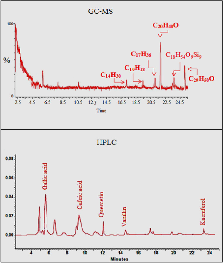
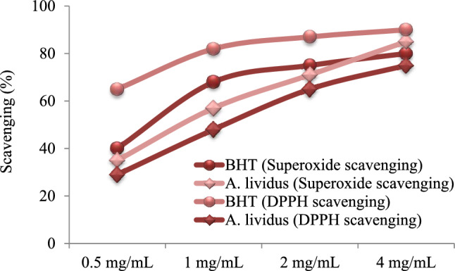
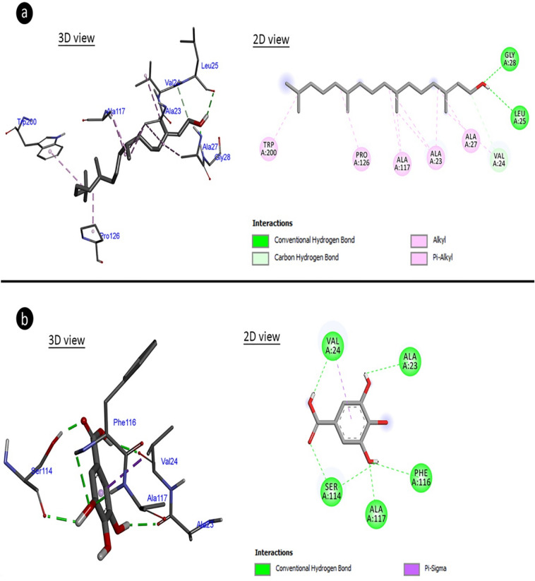
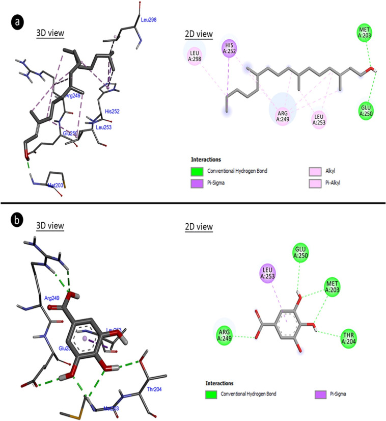
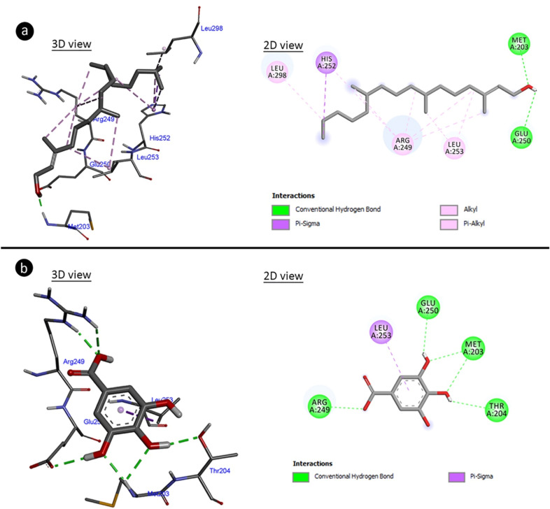
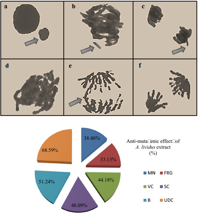
In this study, the phytochemical content of Amaranthus lividus extract and its multi-biological activities were investigated. Total protein, phenol, flavonoid, saponin and condensed tannin contents were determined for phytochemical analysis. In addition, GC–MS and HPLC analyzes were carried out for the determination of the active components of the extract. In determining the multi-biological activities, radical scavenging, anti-mutagenic, anti-proliferative and anti-microbial activities of the extract were investigated. GC–MS analysis revealed that the leaf extract of A. lividus contains phytol and β-sitosterol as major compounds and the presence of gallic acid, caffeic acid, quercetin, vanillin and kaemferol compounds were determined with HPLC analysis. The radical scavenging effect of A. lividus extract was determined as 75.6% against 2,2-diphenyl-1-picrylhydrazyl and 85.2% against superoxide. In anti-bacterial studies, it was determined that A.lividus extract formed different inhibition zones against all tested bacteria. The highest inhibition zone was 14.3 ± 0.7 mm against Bacillus subtilis . In addition, the anti-microbial activity of the extract was demonstrated by molecular docking studies of the binding of gallic acid and phytol to aquaporin and arginase enzyme of bacteria, and the mechanism of anti-microbial activity was explained. A. lividus extract, which provided a 68.59–33.13% reduction in the formation of chromosomal aberrations such as unequal distribution of chromatin, micronucleus formation, fragment, sticky chromosome, bridge and vagrant chromosome, exhibited a strong anti-mutagenic effect. A. lividus extract has a reducing effect on the number of dividing cells and exhibits an anti-proliferative effect of 25.7% compared to the control group. The antiproliferative mechanism of action was investigated by molecular docking and it was determined that the gallic acid and phytol in the extract decreased proliferation by interacting with telomerase. As a result, A.lividus extract consumed as food is a potential natural anti-microbial, anti-oxidant, anti-mutagenic and anti-proliferative source with its rich phytochemical content.
Overview
The claims and statistics of this paper haven’t been checked yet.
Run the check — free, no account needed. It reads the open-access text and checks the sources it cites.
Integrity signals
Registry
Cites
Statistics
Figures
Verification
Claims the paper makes
The claims and statistics of this paper haven’t been checked yet.
Run the check — free, no account needed. It reads the open-access text and checks the sources it cites.
Claims other papers make about this one
Each is checked against this paper’s own text: does it say what the citing paper says it does?
In the citing paper
“NaN3 is a potent mutagen that has been shown to induce high rates of chromosomal abnormalities19.”
From Synergistic and antagonistic contributions of main components to the bioactivity profile of Anethum graveolens extract (2025) · cites this paper as [19]
Self-check
Percentages, sums and reported differences re-computed from the paper’s own numbers.
The self-check needs the paper’s full text.
Citations
Cites
Lambert, A. J., & Brand, M. D. (2004) · Journal of Biological Chemistry · doi:10.1074/jbc.m406576200 · Cited by 480
Research works printed but not linked: 67
- Graça, V. C., Ferreira, I. C. & Santos, P. F. Phytochemical composition and biological activities of Geranium robertianum L.: A review. Ind. Crops Prod. 87, 363–437 (2016). · DOI
- Camejo-Rodrigues, J., Ascensao, L., Bonet, M. À. & Valles, J. An ethnobotanical study of medicinal and aromatic plants in the Natural Park of Serra de São Mamed” (Portugal). J. Ethnopharmacol. 89(2–3), 199–209 (2003). · DOI
- Rasul, M. G. Extraction, isolation and characterization of natural products from medicinal plants. Int. J. Basic Sci. Appl. Comput. 2, 1–6 (2018).
- Krogsgaard-Larsen, S. et al. (eds) Natural Products and Drug Development 34–45 (Munksgaard, 1994).
- Ozsoy, N., Yilmaz, T., Kurt, O., Can, A. & Yanardag, R. In vitro antioxidant activity of Amaranthus lividus L.. Food Chem. 116(4), 867–872 (2009). · DOI
- López, O. A., López, M. A. & Palou, E. Antioxidant capacity of extracts from amaranth seeds or leaves. Ind. Crops Prod. 53, 55–59 (2014). · DOI
- Tikekar, R. V., Ludescher, R. D. & Karwe, M. V. Processing stability of squalene in amaranth and antioxidant potential of amaranth extract. J. Agric. Food Chem. 56, 10675–10678 (2008). · DOI
- Shukla, S. et al. Mineral profile and variability in vegetable amaranth (Amaranthus tricolor). Plant Foods Hum. Nutr. 61, 23–28 (2006). · DOI
- Chaudhary, M. A. et al. Evaluation of gut modulatory and bronchodilator activities of Amaranthus spinosus Linn.. BMC Complement. Altern. Med. 12, 166 (2012). · DOI
- Nehal, N., Mann, S. & Gupta, R. K. Nutritional and phytochemical evaluation of A. lividus L. syn. Amaranthus blitum subsp. oleraceus (L.). IJTK 15(4), 669–674 (2016).
- Erdoğan, E. A. Using fields of plant essential oils and potential genetic effects. Mersin Uni. School Med. Lokman Hekim J. History Medicine Folk Med. 2(2), 21–24 (2012).
- Gündüz, A., Yalçın, E. & Çavuşoğlu, K. Combined toxic effects of aflatoxin B2 and the protective role of resveratrol in Swiss albino mice. Sci. Rep. 11(1), 1–14 (2021). · DOI
- Ayhan, B. S., Yalçın, E., Çavuşoğlu, K. & Acar, A. Antidiabetic potential and multi-biological activities of Trachystemon orientalis extracts. J. Food Meas. Charact. 13(4), 2887–2893 (2019). · DOI
- Aktumsek, A., Zengin, G., Guler, G. O., Cakmak, Y. S. & Duran, A. Antioxidant potentials and anticholinesterase activities of methanolic and aqueous extracts of three endemic Centaurea L. species. Food Chem. Toxicol. 55, 290–296 (2013). · DOI
- Praveen, K. P., Kumaravel, S. & Lalitha, C. Screening of antioxidant activity, total phenolics and GC-MS study of Vitex negundo. Afr. J. Biochem. Res. 4, 191–195 (2010).
- Weerasak, S. & Suwanna, V. Simultaneous determination of gallic acid, catechin, rutin, ellagic acid and quercetin in flower extracts of Michelia alba, Caesalpinia pulcherrima and Nelumbo nucifera by HPLC. Thai. Pharm. Health Sci. J. 2, 131–137 (2007).
- Efe, E., Yalcin, E. & Cavusoglu, K. Antimutagenic and multi-biological activities of Smilax excelsa L. fruit extract. Cumhuriyet Sci. J. 40(2), 440–446 (2019). · DOI
- Savage, D. F. et al. Architecture and selectivity in aquaporins: 2.5 Å X-ray structure of aquaporin Z. PLoS Biol. 1(3), e72 (2003). · DOI
- Bewley, M. C., Jeffrey, P. D., Patchett, M. L., Kanyo, Z. F. & Baker, E. N. Crystal structures of Bacillus caldovelox arginase in complex with substrate and inhibitors reveal new insights into activation, inhibition and catalysis in the arginase superfamily. Structure 7(4), 435–448 (1999). · DOI
- Bryan, C. et al. Structural basis of telomerase inhibition by the highly specific BIBR1532. Structure. 23(10), 1934–1942 (2015). · DOI
- Guex, N. & Peitsch, M. C. Swiss-model and the Swiss-Pdb viewer: An environment for comparative protein modeling. Electrophoresis 18, 2714–2723 (2005). · DOI
- O’Boyle, N. M. et al. Open babel: An open chemical toolbox. J. Cheminform. 3, 33 (2011). · DOI
- Morris, G. M. et al. AutoDock4 and AutoDockTools4: Automated docking with selective receptor flexibility. J. Comput. Chem. 30(16), 2785–2791 (2009). · DOI
- Rathnasamy, S., Mohamed, K. B., Sulaiman, S. F. & Akinboro, A. Evaluation of cytotoxic, mutagenic and antimutagenic potential of leaf extracts of three medicinal plants using Allium cepa chromosome assay. Int. Curr. Pharm. J. 2(8), 131–140 (2013). · DOI
- Yalçın, E., Uzun, A. & Çavuşoğlu, K. In vivo epiclorohidrine toxicity: Cytogenetic, biochemical, physiological and anatomical evidences. ESPR 26, 22400–22406 (2019).
- Siddhuraju, P. & Becker, K. Antioxidant properties of various extracts of total phenolic constituents from three different agroclimatic origins of drumstick tree (Moringa oleifera Lam.) leaves. J. Agric. Food Chem. 51, 2144–2155 (2003). · DOI
- Truong, D. H. et al. Evaluation of the use of different solvents for phytochemical constituents, antioxidants, and in vitro anti-inflammatory activities of Severinia buxifolia. J. Food Qual. 2019, 1–9 (2019). · DOI
- Franchis, G., Kerem, Z., Makkar, H. & Becker, K. The biological action of saponins in animal systems. BJN 88, 587–605 (2002). · DOI
- Zarin, M. A., Wan, H. Y., Isha, A. & Armania, N. Antioxidant, antimicrobial and cytotoxic potential of condensed tannins from Leucaena leucocephala hybrid-Rendang. Food Sci. Hum. Wellness 5(2), 65–75 (2016). · DOI
- Krishnamoorthy, K. & Subramaniam, P. Phytochemical profiling of leaf, stem and tuber parts of Solena amplexicaulis (Lam.) Gandhi, Using GC-MS. Int. Sch. Res. Notices 2014, 1–13 (2014). · DOI
- Santos, C. C. D. M. P. et al. Antinociceptive and antioxidant activities of phytol in vivo and in vitro models. Neurosci. J. 2013, 949452 (2013). · DOI
- Faix, Š, Juhas, Š & Faixová, Z. The effect of essential oil intake on changes of plasma antioxidant status in mice. Acta Vet. 76, 357–361 (2007). · DOI
- Rajabudeen, E., Ganthi, A. S., Subramanian, M. P. S. & Natarajan, K. GC-MS analysis of the methanol extract of Indigofera aspalathoides Vahl ex DC. J. Adv. Chem. Sci. 1, 6–8 (2015).
- Rouis-Soussi, L. S. et al. Chemical composition and antibacterial activity of essential oils from the Tunisian Allium nigrum L.. Excli. J. 13, 526 (2014).
- Paranthaman, R., Praveen, K. P. & Kumaravel, S. GC-MS analysis of phytochemicals and simultaneous determination of flavonoids in Amaranthus caudatus (Sirukeerai) by RP-HPLC. J. Anal. Bioanal. Technol. 3(5), 147 (2012).
- Gao, J., Hu, J., Hu, D. & Yang, X. A role of gallic acid in oxidative damage diseases: A comprehensive review. Nat. Prod. Commun. 14(8), 1934578–19874174 (2019).
- Kahkeshani, N. et al. Pharmacological effects of gallic acid in health and diseases: A mechanistic review. Iran. J. Basic Med. Sci. 22(3), 225 (2019).
- Birková, A., Hubková, B., Bolerázska, B., Mareková, M. & Čižmárová, B. Caffeic acid: A brief overview of its presence, metabolism, and bioactivity. BCHD. 3(4), 74–81 (2020). · DOI
- Arya, S. S., Sharma, M. M., Rookes, J. E., Cahill, D. M. & Lenka, S. K. Vanilla modulates the activity of antibiotics and inhibits efflux pumps in drug-resistant Pseudomonas aeruginosa. Biologia 76(2), 781–791 (2021). · DOI
- Abo-Salem, O. M. Kaempferol attenuates the development of diabetic neuropathic pain in mice: Possible anti-inflammatory and anti-oxidant mechanisms. Maced. J. Med. Sci. 2(3), 424–430 (2014). · DOI
- David, A. V. A., Arulmoli, R. & Parasuraman, S. Overviews of biological importance of quercetin: A bioactive flavonoid. Pharmacogn. Rev. 10(20), 84 (2016). · DOI
- Njoya, E. M. Medicinal Plants, Antioxidant Potential, and Cancer in Cancer 349–357 (Academic Press, 2021).
- Vázquez, L. H., Javier, P. & Arturo, N. The Pentacyclic Triterpenes Amyrins: A Review of Sources and Biological Activities in Phytochemicals—A Global Perspective of Their Role in Nutrition and Health (InTech Europe, 2012).
- Karamac, M., Kosinska, A. & Amarowicz, R. Chelating of Fe (II), Zn (II) and Cu (II) by tannin fractions separated from hazelnuts, walnuts and almonds. Bromat. Chem. Toksykol. 39, 257–260 (2006).
- Marino, T., Galano, A. & Russo, N. Radical scavenging ability of gallic acid toward OH and OOH radicals. Reaction mechanism and rate constants from the density functional theory. J. Phys. Chem. B. 118(35), 10380–10389 (2014). · DOI
- Breijyeh, Z., Jubeh, B. & Karaman, R. Resistance of gram-negative bacteria to current antibacterial agents and approaches to resolve it. Molecules 25(6), 1340 (2020). · DOI · Paper page
- Walker, L. A., Gow, N. A. & Munro, C. A. Elevated chitin content reduces the susceptibility of Candida species to caspofungin. Antimicrob. Agents Chemother. 57, 146–154 (2013). · DOI · Paper page
- Pudlo, M., Demougeot, C. & Girard-Thernier, C. Arginase inhibitors: A rational approach over one century. Med. Res. Rev. 37, 475–513 (2017). · DOI
- Fitzgerald, D. J. et al. Mode of antimicrobial action of vanillin against Escherichia coli, Lactobacillus plantarum and Listeria innocua. J. Appl. Microbiol. 97(1), 104–113 (2004). · DOI
- Maiyo, Z. C., Ngure, R. M., Matasyoh, J. C. & Chepkorir, R. Phytochemical constituents and antimicrobial activity of leaf extracts of three Amaranthus plant species. Afr. J. Biotechnol. 9(21), 3178–3182 (2010).
- Devi, C. A. Docking study on Mycobacterium tuberculosis receptors AccD5 and PKS18 with selected phytochemicals. IOSR-JPBS 4(4), 1–4 (2012). · DOI
- Arsianti, A. et al. Design and screening of gallic acid derivatives as inhibitors of malarial dihydrofolate reductase by in silico docking. Asian J. Pharm. Clin. Res. 10(2), 330–334 (2017). · DOI
- Marc, J., Mulner-Lorillon, O. & Bellé, R. Glyphosate-based pesticides affect cell cycle regulation. Biol. Cell 96(3), 245–249 (2004). · DOI
- Çavuşoğlu, K. et al. Investigation of toxic effects of the Glyphosate on Allium cepa. J. Agric. Sci. 17, 131–142 (2011).
- Teixeira, R. O., Camparoto, M. L., Mantovani, M. S. & Vicentini, V. E. P. Assessment of two medicinal plants, Psidium guajava L. and Achillea millefolium L., in in vitro and in vivo assays. Gen. Mol. Biol. 26(4), 551–555 (2003). · DOI
- López-García, G., Cilla, A., Barberá, R. & Alegría, A. Antiproliferative effect of plant sterols at colonic concentrations on Caco-2 cells. JFF 39, 84–90 (2017).
- Pan, J. et al. Dimer and tetramer of gallic acid: facile synthesis, antioxidant and antiproliferative activities. Lett. Drug Des. Discov. 11(1), 27–32 (2014). · DOI
- Procházková Schrumpfová, P., Fojtová, M. & Fajkus, J. Telomeres in plants and humans: Not so different, not so similar. Cells 8(1), 58 (2019). · DOI
- Sherin, D. R., Manojkumar, T. K., Prakash, R. C. & Sobha, V. N. Molecular docking and dynamics simulation study of telomerase inhibitors as potential anti-cancer agents. MT Proc. 46, 2898–2905 (2021).
- Demirtaş, G., Çavuşoğlu, K. & Yalçın, E. Aneugenic, clastogenic, and multi-toxic effects of diethyl phthalate exposure. ESPR 27(5), 5503–5510 (2020).
- Çavuşoğlu, D., Yalçın, E., Çavuşoğlu, K., Acar, A. & Yapar, K. Molecular docking and toxicity assesment of spirodiclofen: Protective role of Lycopene. ESPR. 40, 57372–57385 (2021).
- Gruszka, D., Szarejko, I. & Maluszynski, M. Sodium azide as a mutagen. In Plant Mutation Breeding and Biotechnology (eds Shu, Q. Y. et al.) 159–166 (CABI, 2012). · DOI
- Islam, M. T. et al. Phytol: A review of biomedical activities. Food Chem. Toxicol. 121, 82–94 (2018). · DOI · Paper page
- Hour, T. C., Liang, Y. C., Chu, I. S. & Lin, J. K. Inhibition of eleven mutagens by various tea extracts, (−) epigallocatechin-3-gallate, gallic acid and caffeine. Food Chem. Toxicol. 37(6), 569–579 (1999). · DOI
- Birošová, L., Mikulášová, M. & Vaverková, Š. Antimutagenic effect of phenolic acids. Biomed. Pap. 149(2), 489–491 (2005). · DOI
- Kumari, S., Elancheran, R. & Devi, R. Phytochemical screening, antioxidant, antityrosinase, and antigenotoxic potential of Amaranthus viridis extract. Ind. J. Pharm. 50(3), 130 (2018). · DOI
- Jadhav, V. & Biradar, S. D. Antimutagenic effects of ethanolic extracts of Amaranthus spinosus and Amaranthus hybridus. Int. J. Pharm. Med. Res. 2, 1–2 (2014).
Cited by
Kesti Usta, S., Yalçın, E., & Çavuşoğlu, K. (2025) · Scientific Reports · doi:10.1038/s41598-025-07297-8 · Cited by 10
Self-citation
Metadata
- Authors
- Durhan, B., Yalçın, E., Çavuşoğlu, K., & Acar, A.
- Venue
- Scientific Reports
- Published
- 2022-03-12
- Volume
- 12
- Issue
- 1
- ISSN
- 2045-2322
- PMC
- PMC8918320
- Cited by
- 50
- References
- 1
- Language
- en
About these checks
Registry, retraction, Cites and statistics results are computed, not judged, with one exception: a printed reference that no index lists is read once by a model before it counts as a citation error. Claims are extracted by a model and kept only when their quote appears verbatim in the text; each cited source is then checked, with a model’s help, for whether it says what the claim relies on. Those verdicts can be wrong — read the claim’s quote and the cited source before relying on one.
Statistics and arithmetic are recomputed, not judged.
Confidence bands are shown only for fields calibrated on our annotated test set. None is calibrated yet, so no band is shown.
What counts as a claim ›How we measure confidence ›How we check this ›
Are you an author, or do you see a problem with this page? Request a review
Data sources
Bibliographic data: Semantic Scholar, OpenAlex, Crossref.
Contains information from the Semantic Scholar Academic Graph Datasets, made available under the ODC Attribution License (ODC-BY 1.0).
Semantic Scholar’s data is described in: Kinney, R., et al. (2023). The Semantic Scholar Open Data Platform. arXiv:2301.10140.
