

The DOI matches its Crossref record; 45 of the 45 references it cites were found in a registry or an index.
Characterizing the mucin-degrading capacity of the human gut microbiota
Open accessAbstract
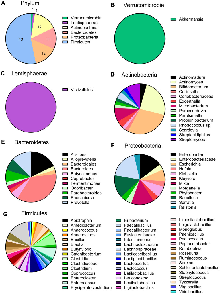
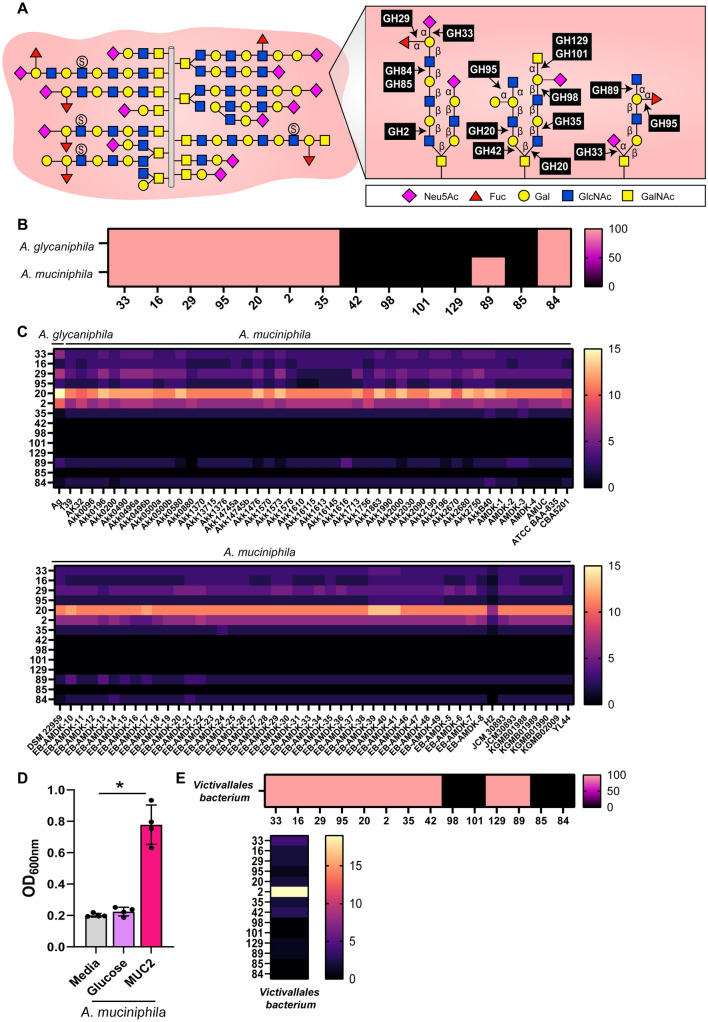
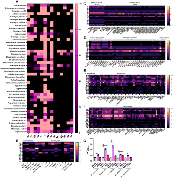
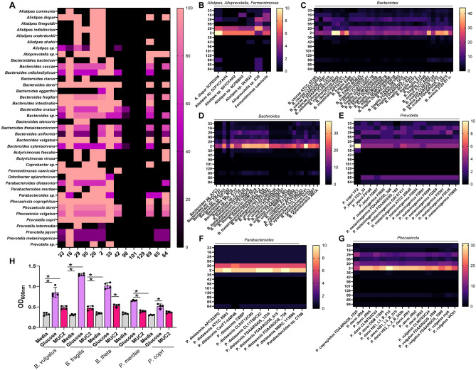
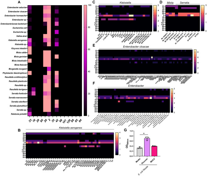
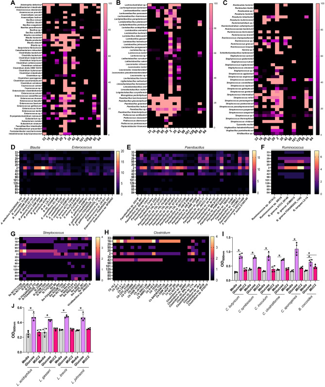
Mucin-degrading microbes are known to harbor glycosyl hydrolases (GHs) which cleave specific glycan linkages. Although several microbial species have been identified as mucin degraders, there are likely many other members of the healthy gut community with the capacity to degrade mucins. The aim of the present study was to systematically examine the CAZyme mucin-degrading profiles of the human gut microbiota. Within the Verrucomicrobia phylum, all Akkermansia glycaniphila and muciniphila genomes harbored multiple gene copies of mucin-degrading GHs. The only representative of the Lentisphaerae phylum, Victivallales, harbored a GH profile that closely mirrored Akkermansia. In the Actinobacteria phylum, we found several Actinomadura, Actinomyces, Bifidobacterium, Streptacidiphilus and Streptomyces species with mucin-degrading GHs. Within the Bacteroidetes phylum, Alistipes, Alloprevotella, Bacteroides, Fermenitomonas Parabacteroides, Prevotella and Phocaeicola species had mucin degrading GHs. Firmicutes contained Abiotrophia, Blautia, Enterococcus, Paenibacillus, Ruminococcus, Streptococcus, and Viridibacillus species with mucin-degrading GHs. Interestingly, far fewer mucin-degrading GHs were observed in the Proteobacteria phylum and were found in Klebsiella, Mixta, Serratia and Enterobacter species. We confirmed the mucin-degrading capability of 23 representative gut microbes using a chemically defined media lacking glucose supplemented with porcine intestinal mucus. These data greatly expand our knowledge of microbial-mediated mucin degradation within the human gut microbiota.
Overview
The claims and statistics of this paper haven’t been checked yet.
Run the check — free, no account needed. It reads the open-access text and checks the sources it cites.
Integrity signals
Registry
Cites
Statistics
Figures
Verification
Claims the paper makes
The claims and statistics of this paper haven’t been checked yet.
Run the check — free, no account needed. It reads the open-access text and checks the sources it cites.
Claims other papers make about this one
Each is checked against this paper’s own text: does it say what the citing paper says it does?
In the citing paper
“Host substrates, such as mucin and bile salts, can also be metabolized by the gut microbiota [18, 19].”
From Role of the gut microbiota in nutrient competition and protection against intestinal pathogen colonization (2023) · cites this paper as [19]
Self-check
Percentages, sums and reported differences re-computed from the paper’s own numbers.
The self-check needs the paper’s full text.
Citations
Cites
The carbohydrate-active enzymes database (CAZy) in 2013
Lombard, V., Ramulu, H. G., Drula, É., Coutinho, P. M., & Henrissat, B. (2013) · Nucleic Acids Research · doi:10.1093/nar/gkt1178 · Cited by 6,452
The Carbohydrate-Active EnZymes database (CAZy): an expert resource for Glycogenomics
Cantarel, B. L., Coutinho, P. M., Rancurel, C., Bernard, T., Lombard, V., & Henrissat, B. (2008) · Nucleic Acids Research · doi:10.1093/nar/gkn663 · Cited by 6,108
Dynamics and Stabilization of the Human Gut Microbiome during the First Year of Life
Bäckhed, F., Roswall, J., Peng, Y., Feng, Q., Jia, H., Kovatcheva-Datchary, P., Li, Y., Xia, Y., Xie, H., Zhong, H., et al. (2015) · Cell Host & Microbe · doi:10.1016/j.chom.2015.04.004 · Cited by 3,315
A classification of glycosyl hydrolases based on amino acid sequence similarities.
Henrissat, B. (1991) · Biochemical Journal · doi:10.1042/bj2800309 · Cited by 3,188
Desai, M. S., Seekatz, A. M., Koropatkin, N. M., Kamada, N., Hickey, C. A., Wolter, M., Pudlo, N. A., Kitamoto, S., Terrapon, N., Muller, A., et al. (2016) · Cell · doi:10.1016/j.cell.2016.10.043 · Cited by 2,805
Akkermansia muciniphila gen. nov., sp. nov., a human intestinal mucin-degrading bacterium
Derrien, M., Vaughan, E. E., Plugge, C. M., & de Vos, W. M. (2004) · International Journal of Systematic and Evolutionary Microbiology · doi:10.1099/ijs.0.02873-0 · Cited by 2,094
New families in the classification of glycosyl hydrolases based on amino acid sequence similarities
Henrissat, B., & Bairoch, A. (1993) · Biochemical Journal · doi:10.1042/bj2930781 · Cited by 1,974
The abundance and variety of carbohydrate-active enzymes in the human gut microbiota
Kaoutari, A. E., Armougom, F., Gordon, J. I., Raoult, D., & Henrissat, B. (2013) · Nature Reviews Microbiology · doi:10.1038/nrmicro3050 · Cited by 1,683
Mucin dynamics and enteric pathogens
McGuckin, M. A., Lindén, S. K., Sutton, P., & Florin, T. H. (2011) · Nature Reviews Microbiology · doi:10.1038/nrmicro2538 · Cited by 1,428
Png, C. W., Lindén, S. K., Gilshenan, K. S., Zoetendal, E. G., McSweeney, C. S., Sly, L. I., McGuckin, M. A., & Florin, T. H. J. (2010) · American Journal of Gastroenterology · doi:10.1038/ajg.2010.281 · Cited by 1,364
Glycan foraging in vivo by an intestine-adapted bacterial symbiont
Sonnenburg, J. L., Xu, J., Leip, D. D., Chen, C.-H., Westover, B. P., Weatherford, J., Buhler, J. D., & Gordon, J. I. (2005) · Science · doi:10.1126/science.1109051 · Cited by 1,184
Goblet cells deliver luminal antigen to CD103+ dendritic cells in the small intestine
Mcdole, J. R., Wheeler, L. W., McDonald, K. G., Wang, B., Konjufca, V. H., Knoop, K. A., Newberry, R. D., & Miller, M. J. (2012) · Nature · doi:10.1038/nature10863 · Cited by 976
Martens, E. C., Chiang, H. C., & Gordon, J. I. (2008) · Cell Host & Microbe · doi:10.1016/j.chom.2008.09.007 · Cited by 954
Mucin glycan foraging in the human gut microbiome
Tailford, L. E., Crost, E. H., Kavanaugh, D. W., & Juge, N. (2015) · Frontiers in Genetics · doi:10.3389/fgene.2015.00081 · Cited by 881
Early life dynamics of the human gut virome and bacterial microbiome in infants
Lim, E. S., Zhou, Y., Zhao, G., Bauer, I. K., Droit, L., Ndao, I. M., Warner, B. B., Tarr, P. I., Wang, D., & Holtz, L. R. (2015) · Nature Medicine · doi:10.1038/nm.3950 · Cited by 670
Fermentation of mucins and plant polysaccharides by anaerobic bacteria from the human colon
Salyers, A. A., West, S. E. H., Vercellotti, J. R., & Wilkins, T. D. (1977) · Applied and Environmental Microbiology · doi:10.1128/aem.34.5.529-533.1977 · Cited by 471
The IMG/M data management and analysis system v.6.0: new tools and advanced capabilities
Chen, I.-M. A., Chu, K., Palaniappan, K., Ratner, A., Huang, J., Huntemann, M., Hajek, P. P., Ritter, S., Varghese, N. J., Seshadri, R., et al. (2020) · Nucleic Acids Research · doi:10.1093/nar/gkaa939 · Cited by 433
Complex Carbohydrate Utilization by the Healthy Human Microbiome
Cantarel, B. L., Lombard, V., & Henrissat, B. (2012) · PLoS ONE · doi:10.1371/journal.pone.0028742 · Cited by 378
Turroni, F., Bottacini, F., Foroni, E., Mulder, I., Kim, J.-H., Zomer, A., Sánchez, B., Bidossi, A., Ferrarini, A., Giubellini, V., et al. (2010) · Proceedings of the National Academy of Sciences · doi:10.1073/pnas.1011100107 · Cited by 364
Bjursell, M. K., Martens, E. C., & Gordon, J. I. (2006) · Journal of Biological Chemistry · doi:10.1074/jbc.m606509200 · Cited by 338
Utilisation of Mucin Glycans by the Human Gut Symbiont Ruminococcus gnavus Is Strain-Dependent
Crost, E. H., Tailford, L. E., Gall, G. L., Fons, M., Henrissat, B., & Juge, N. (2013) · PLoS ONE · doi:10.1371/journal.pone.0076341 · Cited by 325
Park, B. H., Karpinets, T. V., Syed, M. H., Leuze, M. R., & Uberbacher, E. C. (2010) · Glycobiology · doi:10.1093/glycob/cwq106 · Cited by 304
Makino, H., Kushiro, A., Ishikawa, E., Kubota, H., Gawad, A., Sakai, T., Oishi, K., Martı́n, R., Amor, K. B., Knol, J., et al. (2013) · PLoS ONE · doi:10.1371/journal.pone.0078331 · Cited by 304
Mucin Degradation in Human Colon Ecosystems
Hoskins, L. C., & Boulding, E. T. (1981) · Journal of Clinical Investigation · doi:10.1172/jci110009 · Cited by 276
Corfield, A. P., Wagner, S. A., Clamp, J. R., Kriaris, M. S., & Hoskins, L. C. (1992) · Infection and Immunity · doi:10.1128/iai.60.10.3971-3978.1992 · Cited by 267
Slimy partners: the mucus barrier and gut microbiome in ulcerative colitis
Fang, J. C., Wang, H., Zhou, Y., Zhang, H., Zhou, H., & Zhang, X. (2021) · Experimental & Molecular Medicine · doi:10.1038/s12276-021-00617-8 · Cited by 251
Mucin Degradation by Bifidobacterium Strains Isolated from the Human Intestinal Microbiota
Ruas‐Madiedo, P., Gueimonde, M., Fernández-García, M., Reyes‐Gavilán, C. G. D. L., & Margollés, A. (2008) · Applied and Environmental Microbiology · doi:10.1128/aem.02509-07 · Cited by 222
Trefoil Factor Peptides and Gastrointestinal Function
Aihara, E., Engevik, K. A., & Montrose, M. H. (2016) · Annual Review of Physiology · doi:10.1146/annurev-physiol-021115-105447 · Cited by 201
Symbiotic Human Gut Bacteria with Variable Metabolic Priorities for Host Mucosal Glycans
Pudlo, N. A., Urs, K., Kumar, S. S., German, J. B., Mills, D. A., & Martens, E. C. (2015) · mBio · doi:10.1128/mbio.01282-15 · Cited by 187
A refined palate: Bacterial consumption of host glycans in the gut
Marcobal, A. M., Southwick, A. M., Earle, K. A., & Sonnenburg, J. L. (2013) · Glycobiology · doi:10.1093/glycob/cwt040 · Cited by 178
Identification of mucin degraders of the human gut microbiota
Raimondi, S., Musmeci, E., Candeliere, F., Amaretti, A., & Rossi, M. (2021) · Scientific Reports · doi:10.1038/s41598-021-90553-4 · Cited by 173
Mucin degradation in human colon ecosystems
Miller, R. S., & Hoskins, L. C. (1981) · Gastroenterology · doi:10.1016/0016-5085(81)90503-5 · Cited by 165
Mucin Cross-Feeding of Infant Bifidobacteria and Eubacterium hallii
Bunešová, V., Lacroix, C., & Schwab, C. (2017) · Microbial Ecology · doi:10.1007/s00248-017-1037-4 · Cited by 151
Crouch, L. I., Liberato, M. V., Urbanowicz, P. A., Baslé, A., Lamb, C. A., Stewart, C. J., Cooke, K., Doona, M., Needham, S. J., Brady, R. R. W., et al. (2020) · Nature Communications · doi:10.1038/s41467-020-17847-5 · Cited by 143
Ravcheev, D. A., & Thiele, I. (2017) · Frontiers in Genetics · doi:10.3389/fgene.2017.00111 · Cited by 139
Bell, A., Brunt, J., Crost, E. H., Vaux, L., Nepravishta, R., Owen, C. D., Latousakis, D., Xiao, A., Li, W., Chen, X., et al. (2019) · Nature Microbiology · doi:10.1038/s41564-019-0590-7 · Cited by 137
Zhang, G., Mills, D. A., & Block, D. E. (2008) · Applied and Environmental Microbiology · doi:10.1128/aem.01416-08 · Cited by 133
Importance of Glycans to the Host-Bacteroides Mutualism in the Mammalian Intestine
Comstock, L. E. (2009) · Cell Host & Microbe · doi:10.1016/j.chom.2009.05.010 · Cited by 118
Mucosal glycan degradation of the host by the gut microbiota
Bell, A., & Juge, N. (2020) · Glycobiology · doi:10.1093/glycob/cwaa097 · Cited by 116
Trastoy, B., Naegeli, A., Anso, I., Sjögren, J., & Guerin, M. E. (2020) · Nature Communications · doi:10.1038/s41467-020-18696-y · Cited by 104
Kiyohara, M., Nakatomi, T., Kurihara, S., Fushinobu, S., Suzuki, H., Tanaka, T., Shoda, S., Kitaoka, M., Katayama, T., Yamamoto, K., et al. (2011) · Journal of Biological Chemistry · doi:10.1074/jbc.m111.277384 · Cited by 97
Establishment of a Mucin-Degrading Intestinal Microflora During the First Two Years of Human Life
Midtvedt, A., Carlstedt‐Duke, B., & Midtvedt, T. (1994) · Journal of Pediatric Gastroenterology and Nutrition · doi:10.1097/00005176-199404000-00012 · Cited by 61
Low, K. E., Smith, S. P., Abbott, D. W., & Boraston, A. B. (2020) · Glycobiology · doi:10.1093/glycob/cwaa050 · Cited by 41
Draft genome sequence of Fermentimonas caenicola strain SIT8, isolated from the human gut
Beye, M., Bakour, S., Traore, S. I., Rathored, J., Labas, N., Raoult, D., & Fournier, P. (2018) · Standards in Genomic Sciences · doi:10.1186/s40793-018-0310-6 · Cited by 12
Plugge, C. M., & Zoetendal, E. G. (2014) · Springer eBooks · doi:10.1007/978-3-642-38954-2_150 · Cited by 7
Cited by
Sorted by citations
Gut microbiota-derived short-chain fatty acids and their role in human health and disease
Mukhopadhya, I., & Louis, P. (2025) · Nature Reviews Microbiology · doi:10.1038/s41579-025-01183-w · Cited by 628
The gut microbiota and its biogeography
McCallum, G., & Tropini, C. (2023) · Nature Reviews Microbiology · doi:10.1038/s41579-023-00969-0 · Cited by 361
Holmberg, S. M., Feeney, R. H., K., V., Puértolas-Balint, F., Singh, D. K., Wongkuna, S., Zandbergen, L. M., Hauner, H., Brandl, B., Nieminen, A. I., et al. (2024) · Nature Communications · doi:10.1038/s41467-024-47594-w · Cited by 262
Shuoker, B., Pichler, M. J., Jin, C., Sakanaka, H., Wu, H., Gascueña, A. M., Liu, J., Nielsen, T. S., Holgersson, J., Karlsson, E. N., et al. (2023) · Nature Communications · doi:10.1038/s41467-023-37533-6 · Cited by 196
Host control of the microbiome: Mechanisms, evolution, and disease
Wilde, J., Slack, E. M. C., & Foster, K. R. (2024) · Science · doi:10.1126/science.adi3338 · Cited by 172
Metadata
- Authors
- Glover, J. S., Ticer, T. D., & Engevik, M. A.
- Affiliations
- Medical University of South Carolina (US)
- Corresponding author
- Engevik, Melinda A.
- Venue
- Scientific Reports
- Published
- 2022-05-19
- Volume
- 12
- Issue
- 1
- Pages
- 8456-8456
- ISSN
- 2045-2322 (ISSN-L)
- PMC
- PMC9120202
- OpenAlex
- W4280576091
- Cited by
- 307
- References
- 45
- Citations by year
- 2026: 68 · 2025: 90 · 2024: 90 · 2023: 48 · 2022: 11
- FWCI
- 19.84
- Type
- journal article
- Language
- en
- Open access
- gold
- doi.org
- Locations
- Scientific Reports (publishedVersion, cc-by)
- PubMed Central (publishedVersion, cc-by)
- PubMed (publishedVersion)
- DOAJ (DOAJ: Directory of Open Access Journals) (submittedVersion)
- Funders
- U.S. Department of Health & Human Services | NIH | National Institute of General Medical Sciences (1T32GM132055-01)
- U.S. Department of Health & Human Services | NIH | National Institute of Diabetes and Digestive and Kidney Diseases (1T32DK124191-01A1, DK123195-01)
- Topics
- Life Sciences › Biochemistry, Genetics and Molecular Biology › Molecular Biology › Gut microbiota and health
- Health Sciences › Dentistry › Periodontics › Oral microbiology and periodontitis research
- Health Sciences › Nursing › Nutrition and Dietetics › Infant Nutrition and Health
- Keywords
- mucin degradation, human gut microbiome, glycoside hydrolases, carbohydrate-active enzymes, Akkermansia, Verrucomicrobia, Bacteroidetes, Firmicutes, actinobacteria, mucus
- MeSH terms
- Clostridiales, Eubacteriales, Gastrointestinal Microbiome, Animals, Humans, Mucins, Polysaccharides, Swine, Verrucomicrobia
About these checks
Registry, retraction, Cites and statistics results are computed, not judged, with one exception: a printed reference that no index lists is read once by a model before it counts as a citation error. Claims are extracted by a model and kept only when their quote appears verbatim in the text; each cited source is then checked, with a model’s help, for whether it says what the claim relies on. Those verdicts can be wrong — read the claim’s quote and the cited source before relying on one.
Statistics and arithmetic are recomputed, not judged.
Confidence bands are shown only for fields calibrated on our annotated test set. None is calibrated yet, so no band is shown.
What counts as a claim ›How we measure confidence ›How we check this ›
Are you an author, or do you see a problem with this page? Request a review
