

The DOI matches its Crossref record; 2 of the 2 references it cites were found in a registry or an index.
Verteporfin-induced proteotoxicity impairs cell homeostasis and survival in neuroblastoma subtypes independent of YAP/TAZ expression
Open accessPublished version of a preprint
Abstract
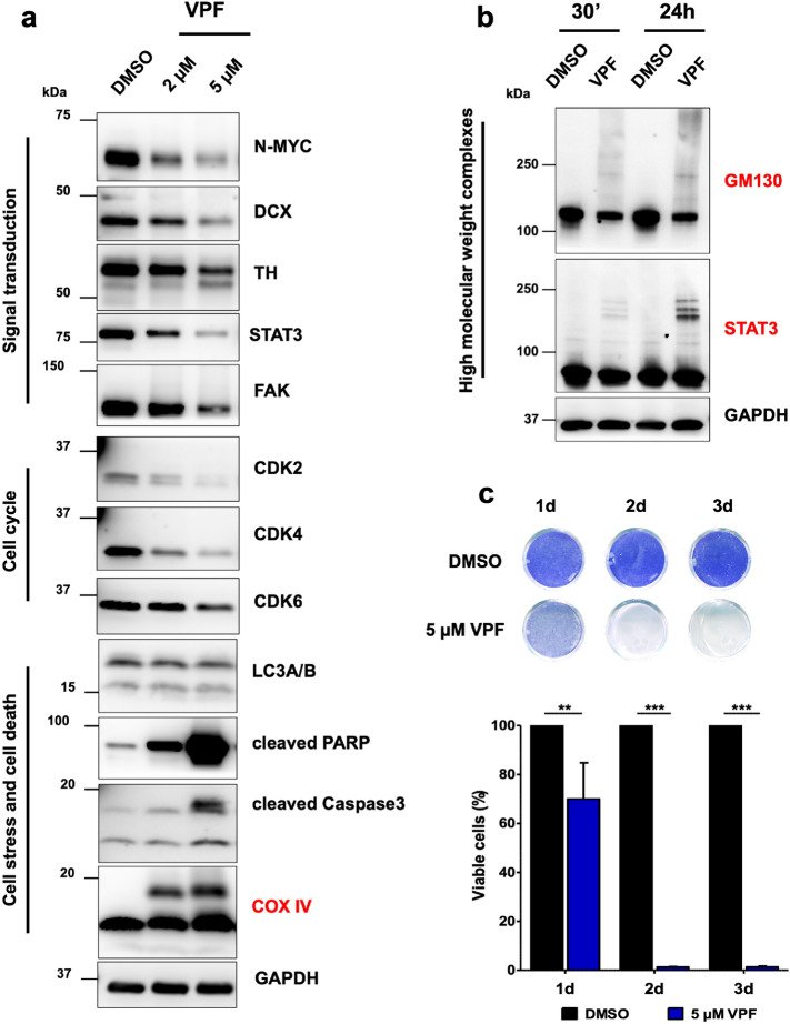
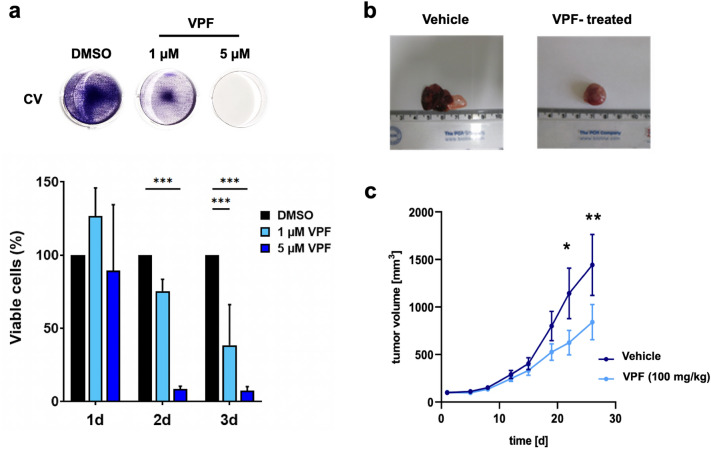
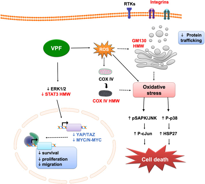
Neuroblastoma (NB) is a highly aggressive extracranial solid tumor in children. Due to its heterogeneity, NB remains a therapeutic challenge. Several oncogenic factors, including the Hippo effectors YAP/TAZ, are associated with NB tumorigenesis. Verteporfin (VPF) is an FDA-approved drug shown to directly inhibit YAP/TAZ activity. Our study aimed to investigate VPF’s potential as a therapeutic agent in NB. We show that VPF selectively and efficiently impairs the viability of YAP/TAZ-expressing NB GI-ME-N and SK-N-AS cells, but not of non-malignant fibroblasts. To investigate whether VPF-mediated NB cell killing is YAP-dependent, we tested VPF potency in CRISPR-mediated YAP/TAZ knock-out GI-ME-N cells, and BE(2)-M17 NB cells (a MYCN- amplified, predominantly YAP-negative NB subtype). Our data shows that VPF-mediated NB cell killing is not dependent on YAP expression. Moreover, we determined that the formation of higher molecular weight (HMW) complexes is an early and shared VPF-induced cytotoxic mechanism in both YAP-positive and YAP-negative NB models. The accumulation of HMW complexes, involving STAT3, GM130 and COX IV proteins, impaired cell homeostasis and triggered cell stress and cell death mechanisms. Altogether, our study shows significant in vitro and in vivo VPF-induced suppression of NB growth, making VPF a potential therapeutic candidate against NB.
Overview
The claims and statistics of this paper haven’t been checked yet.
Run the check — free, no account needed. It reads the open-access text and checks the sources it cites.
Integrity signals
Registry
Cites
Statistics
Figures
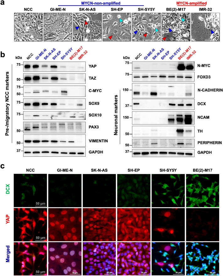
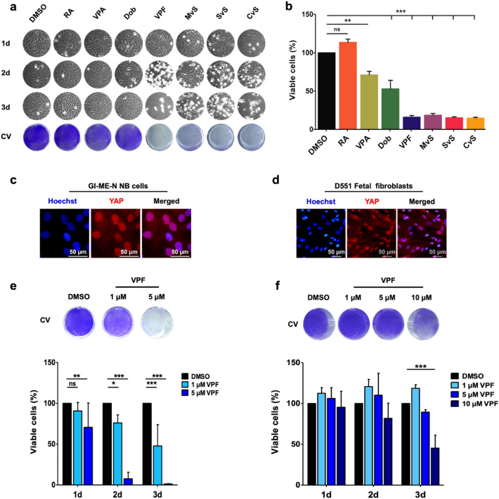
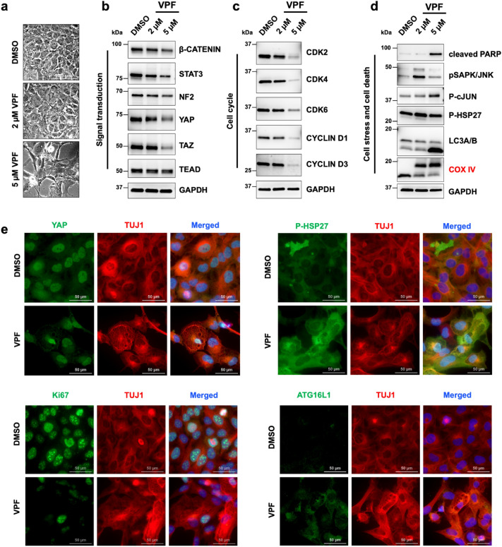
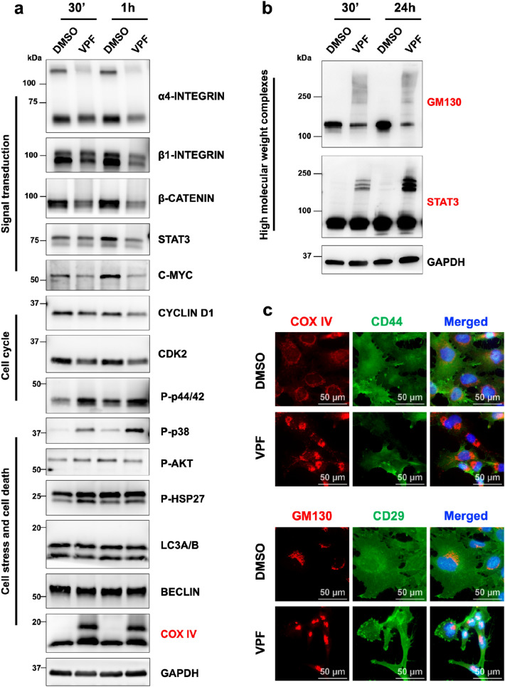
Verification
Claims the paper makes
The claims and statistics of this paper haven’t been checked yet.
Run the check — free, no account needed. It reads the open-access text and checks the sources it cites.
Claims other papers make about this one
Each is checked against this paper’s own text: does it say what the citing paper says it does?
The cited source says what the claim attributes to it.
In the citing paper
“TUBB protein levels were also downregulated at 2.5 μm of VP, likely as a consequence of its proteotoxic effects by promoting higher molecular weight complexes [45, 46] (Fig. 8C).”
From Mechanical stiffness orchestrates distinct regulation of MRTFs and YAP / TAZ transcriptional cofactors in hepatocytes (2026) · cites this paper as [45]
Self-check
Percentages, sums and reported differences re-computed from the paper’s own numbers.
The self-check needs the paper’s full text.
Citations
Cites
YAP/TAZ upstream signals and downstream responses
Totaro, A., Panciera, T., & Piccolo, S. (2018) · Nature Cell Biology · doi:10.1038/s41556-018-0142-z · Cited by 1,086
Drugging the ‘undruggable’ cancer targets
Dang, C. V., Reddy, E. P., Shokat, K. M., & Soucek, L. (2017) · Nature Reviews Cancer · doi:10.1038/nrc.2017.36 · Cited by 927
Research works printed but not linked: 58
- Maguire, L. H., Thomas, A. R. & Goldstein, A. M. Tumors of the neural crest: Common themes in development and cancer. Dev. Dyn. 244, 311–322 (2015). · DOI
- Delloye-Bourgeois, C. & Castellani, V. Hijacking of embryonic programs by neural crest-derived neuroblastoma: From physiological migration to metastatic dissemination. Front. Mol. Neurosci. 12, 52 (2019). · DOI
- Qiu, B. & Matthay, K. K. Advancing therapy for neuroblastoma. Nat. Rev. Clin. Oncol. 19, 515–533 (2022). · DOI
- Pugh, T. J. et al. The genetic landscape of high-risk neuroblastoma. Nat. Genet. 45, 279–284 (2013). · DOI
- Reynolds, C. P., Matthay, K. K., Villablanca, J. G. & Maurer, B. J. Retinoid therapy of high-risk neuroblastoma. Cancer Lett. 197, 185–192 (2003). · DOI
- Fletcher, J. I. et al. Too many targets, not enough patients: Rethinking neuroblastoma clinical trials. Nat. Rev. Cancer 18, 389–400 (2018). · DOI
- Westermann, F. et al. Distinct transcriptional MYCN/c-MYC activities are associated with spontaneous regression or malignant progression in neuroblastomas. Genome Biol. 9, 1–14 (2008). · DOI
- Breit, S. & Schwab, M. Suppression of MYC by high expression of NMYC in human neuroblastoma cells. J. Neurosci. Res. 24, 21–28 (1989). · DOI
- Zimmerman, M. W. et al. MYC drives a subset of high-risk pediatric neuroblastomas and is activated through mechanisms including enhancer hijacking and focal enhancer amplification. Cancer Discov. 8, 320–335 (2018). · DOI
- Boeva, V. et al. Heterogeneity of neuroblastoma cell identity defined by transcriptional circuitries. Nat. Genet. 49, 1408–1413 (2017). · DOI
- van Groningen, T. et al. Neuroblastoma is composed of two super-enhancer-associated differentiation states. Nat. Genet. 49, 1261–1266 (2017). · DOI
- Shim, J. et al. YAP-mediated repression of HRK regulates tumor growth, therapy response, and survival under tumor environmental stress in neuroblastoma. Cancer Res. 80, 4741–4753 (2020). · DOI
- Coggins, G. E. et al. YAP1 mediates resistance to MEK1/2 inhibition in neuroblastomas with hyperactivated RAS signaling. Cancer Res. 79, 6204–6214 (2019). · DOI · Paper page
- Fusco, P. et al. Verteporfin induces apoptosis and reduces the stem cell-like properties in Neuroblastoma tumour-initiating cells through inhibition of the YAP/TAZ pathway. Eur. J. Pharmacol. 893, 173829 (2021). · DOI
- Yang, C., Tan, J., Zhu, J., Wang, S. & Wei, G. YAP promotes tumorigenesis and cisplatin resistance in neuroblastoma. Oncotarget 8, 37154–37163 (2017). · DOI
- Shim, J. & Goldsmith, K. C. A new player in neuroblastoma: YAP and its role in the neuroblastoma microenvironment. Cancers (Basel) 13, 4650 (2021). · DOI
- Shen, X. et al. YAP promotes the proliferation of neuroblastoma cells through decreasing the nuclear location of p27 Kip1 mediated by Akt. Cell Prolif. 53, e12734 (2020). · DOI
- Takemoto, M. et al. The synergistic antitumor effect of combination therapy with a MEK inhibitor and YAP inhibitor on pERK-positive neuroblastoma. Biochem. Biophys. Res. Commun. 570, 41–46 (2021). · DOI
- Wang, Q. et al. TAZ promotes epithelial to mesenchymal transition via the upregulation of connective tissue growth factor expression in neuroblastoma cells. Mol. Med. Rep. 11, 982–988 (2015). · DOI
- Wang, M. et al. Transcriptional co-activator TAZ sustains proliferation and tumorigenicity of neuroblastoma by targeting CTGF and PDGF-β. Oncotarget 6, 9517–9530 (2015). · DOI
- Schramm, A. et al. Mutational dynamics between primary and relapse neuroblastomas. Nat. Genet. 47, 872–877 (2015). · DOI
- Hindley, C. J. et al. The Hippo pathway member YAP enhances human neural crest cell fate and migration. Sci. Rep. 6, 1–9 (2016). · DOI
- Sorrentino, G. et al. Metabolic control of YAP and TAZ by the mevalonate pathway. Nat. Cell Biol. 16, 357–366 (2014). · DOI
- Scott, L. J. & Goa, K. L. Verteporfin. Drugs Aging 16, 139–146 (2000). · DOI
- Giraud, J. et al. Verteporfin targeting YAP1/TAZ-TEAD transcriptional activity inhibits the tumorigenic properties of gastric cancer stem cells. Int. J. Cancer 146, 2255–2267 (2020). · DOI
- Liu-Chittenden, Y. et al. Genetic and pharmacological disruption of the TEAD-YAP complex suppresses the oncogenic activity of YAP. Genes Dev. 26, 1300–1305 (2012). · DOI · Paper page
- Wei, C. & Li, X. The role of photoactivated and non-photoactivated verteporfin on tumor. Front. Pharmacol. 11, 1626 (2020). · DOI
- Lukoseviciute, M. et al. From pioneer to repressor: Bimodal foxd3 activity dynamically remodels neural crest regulatory landscape in vivo. Dev. Cell 47, 608-628.e6 (2018). · DOI · Paper page
- Ferlemann, F. C., Menon, V., Condurat, A. L., Rößler, J. & Pruszak, J. Surface marker profiling of SH-SY5Y cells enables small molecule screens identifying BMP4 as a modulator of neuroblastoma differentiation. Sci. Rep. 7, 13612 (2017). · DOI
- Huang, Y., Tsubota, S., Nishio, N., Takahashi, Y. & Kadomatsu, K. Combination of tumor necrosis factor-α and epidermal growth factor induces the adrenergic-to-mesenchymal transdifferentiation in SH-SY5Y neuroblastoma cells. Cancer Sci. 112, 715–724 (2021). · DOI
- Cerami, E. et al. The cBio cancer genomics portal: An open platform for exploring multidimensional cancer genomics data. Cancer Discov. 2, 401–404 (2012). · DOI · Paper page
- Gao, J. et al. Integrative analysis of complex cancer genomics and clinical profiles using the cBioPortal. Sci. Signal 6, 269 (2013). · DOI · Paper page
- AlAmri, M. A., Kadri, H., Alderwick, L. J., Jeeves, M. & Mehellou, Y. The photosensitising clinical agent verteporfin is an inhibitor of SPAK and OSR1 kinases. ChemBioChem 19, 2072–2080 (2018). · DOI
- Gibault, F. et al. Non-photoinduced biological properties of verteporfin. Curr. Med. Chem. 23, 1171–1184 (2016). · DOI
- Zhang, H. et al. Tumor-selective proteotoxicity of verteporfin inhibits colon cancer progression independently of YAP1. Sci. Signal 8, 98 (2015). · DOI · Paper page
- Gavini, J. et al. Verteporfin-induced lysosomal compartment dysregulation potentiates the effect of sorafenib in hepatocellular carcinoma. Cell Death Dis. 10, 1–17 (2019). · DOI
- Lu, J. et al. Verteporfin- and sodium porfimer-mediated photodynamic therapy enhances pancreatic cancer cell death without activating stromal cells in the microenvironment. J. Biomed. Opt. 24, 1–11 (2019). · DOI
- Konstantinou, E. K. et al. Verteporfin-induced formation of protein cross-linked oligomers and high molecular weight complexes is mediated by light and leads to cell toxicity. Sci. Rep. 7, 46581 (2017). · DOI
- Read, R. D. Repurposing the drug verteporfin as anti-neoplastic therapy for glioblastoma. Neuro Oncol. 24, 708–710 (2022). · DOI
- Huggett, M. T. et al. Phase I/II study of verteporfin photodynamic therapy in locally advanced pancreatic cancer. Br. J. Cancer 110, 1698–1704 (2014). · DOI
- Kuramoto, K. et al. Verteporfin inhibits oxidative phosphorylation and induces cell death specifically in glioma stem cells. FEBS J. 287, 2023–2036 (2020). · DOI
- Eales, K. L., Wilkinson, E. A., Cruickshank, G., Tucker, J. H. R. & Tennant, D. A. Verteporfin selectively kills hypoxic glioma cells through iron-binding and increased production of reactive oxygen species. Sci. Rep. 8, 14358 (2018). · DOI
- Yin, L. & Chen, G. Verteporfin promotes the apoptosis and inhibits the proliferation, migration, and invasion of cervical cancer cells by downregulating SULT2B1 expression. Med. Sci. Monit. 26, e926780-1 (2020). · DOI
- Wei, H. et al. Verteporfin suppresses cell survival, angiogenesis and vasculogenic mimicry of pancreatic ductal adenocarcinoma via disrupting the YAP-TEAD complex. Cancer Sci. 108, 478–487 (2017). · DOI
- Morscher, R. J. et al. Inhibition of neuroblastoma tumor growth by ketogenic diet and/or calorie restriction in a CD1-Nu mouse model. PLoS ONE 10, e0129802 (2015). · DOI
- Hanahan, D. & Weinberg, R. A. Hallmarks of cancer: The next generation. Cell 144, 646–674 (2011). · DOI · Paper page
- Kraus, D., Palasuberniam, P. & Chen, B. Therapeutic Enhancement of verteporfin-mediated photodynamic therapy by mTOR inhibitors. Photochem. Photobiol. 96, 358–364 (2020). · DOI
- Zanconato, F., Cordenonsi, M. & Piccolo, S. YAP/TAZ at the Roots of cancer. Cancer Cell 29, 783–803 (2016). · DOI · Paper page
- Barrette, A. M. et al. Anti-invasive efficacy and survival benefit of the YAP-TEAD inhibitor verteporfin in preclinical glioblastoma models. Neuro Oncol. 24, 694–707 (2022). · DOI
- Zhu, L. et al. Prp19 is an independent prognostic marker and promotes neuroblastoma metastasis by regulating the hippo-YAP signaling pathway. Front. Oncol. 1, 575366 (2020).
- Liang, J. et al. Verteporfin Inhibits PD-L1 through autophagy and the STAT1-IRF1-TRIM28 signaling axis, exerting antitumor efficacy A C. Cancer Immunol. Res. 8, 952–965 (2020). · DOI
- Li, Y., Park, J. S., Deng, J. H. & Bai, Y. Cytochrome c oxidase subunit IV is essential for assembly and respiratory function of the enzyme complex. J. Bioenerg. Biomembr. 38, 283–291 (2006). · DOI
- Chang, S. H. et al. GOLGA2/GM130, cis-Golgi matrix protein, is a novel target of anticancer gene therapy. Mol. Ther. 20, 2052–2063 (2012). · DOI
- Bui, S., Mejia, I., Díaz, B. & Wang, Y. Adaptation of the Golgi apparatus in cancer cell invasion and metastasis. Front. Cell Dev. Biol. 9, 806482 (2021). · DOI
- Shah, S. R. et al. Verteporfin-loaded polymeric microparticles for intratumoral treatment of brain cancer. Mol. Pharm. 16, 1433–1443 (2019). · DOI
- Pellosi, D. S., Paula, L. B., de Melo, M. T. & Tedesco, A. C. Targeted and synergic glioblastoma treatment: Multifunctional nanoparticles delivering verteporfin as adjuvant therapy for temozolomide chemotherapy. Mol. Pharm. 16, 1009–1024 (2019). · DOI
- Thomas, R., Menon, V., Mani, R. & Pruszak, J. Glycan epitope and integrin expression dynamics characterize neural crest epithelial-to-mesenchymal transition (EMT) in human pluripotent stem cell differentiation. Stem Cell Rev. Rep. 18, 2952–2965 (2022). · DOI
- Menon, V. et al. Comprehensive cell surface antigen analysis identifies transferrin receptor protein-1 (CD71) as a negative selection marker for human neuronal cells. Stem Cells 37, 1293–1306 (2019). · DOI
Cited by
Sorted by citations
Torres‐Ortiz, B. S., Pérez‐Calixto, D., Carretero‐Ortega, J., Guzman‐López, A. G., Pinto‐Dueñas, D. C., Hernández‐Juárez, M., Jiménez‐Escobar, A., Rodríguez‐Hernández, A., Díaz‐Bello, B., Serna‐Márquez, N., et al. (2026) · The FEBS Journal · doi:10.1111/febs.70570
Metadata
- Authors
- Condurat, A.-L., Aminzadeh-Gohari, S., Malnar, M., Schider, N., Opitz, L., Thomas, R., Menon, V., Kofler, B., & Pruszak, J.
- Affiliations
- University of Freiburg (DE)
- Paracelsus Medical University (AT)
- Corresponding author
- Pruszak, Jan
- ORCID
- Venue
- Scientific Reports
- Published
- 2023-03-07
- Volume
- 13
- Issue
- 1
- Pages
- 3760-3760
- ISSN
- 2045-2322 (ISSN-L)
- PMC
- PMC9992669
- OpenAlex
- W4323359919
- Cited by
- 14
- References
- 2
- Citations by year
- 2026: 5 · 2025: 4 · 2024: 4 · 2023: 1
- FWCI
- 2.02
- Type
- journal article
- Language
- en
- Open access
- gold
- doi.org
- Locations
- Scientific Reports (publishedVersion, cc-by)
- Research Square (acceptedVersion, cc-by)
- PubMed Central (publishedVersion, cc-by)
- PubMed (publishedVersion)
- Amsterdam UMC (VU Amsterdam) - Institutional Repository (publishedVersion, other-oa)
- Pure Amsterdam UMC (submittedVersion, cc-by)
- DOAJ (DOAJ: Directory of Open Access Journals) (submittedVersion, cc-by-sa)
- Funders
- Excellence Initiative of the German Research Foundation
- Faculty of Medicine – University of Freiburg
- Austrian Science Fund (P31228-B33)
- German Research Foundation Emmy Noether (PR1132/3-1)
- Topics
- Life Sciences › Biochemistry, Genetics and Molecular Biology › Cell Biology › Hippo pathway signaling and YAP/TAZ
- Health Sciences › Medicine › Oncology › Neuroblastoma Research and Treatments
- Life Sciences › Biochemistry, Genetics and Molecular Biology › Molecular Biology › Cell death mechanisms and regulation
- Keywords
- verteporfin, neuroblastoma, YAP/TAZ, proteotoxicity, high molecular weight protein complexes, STAT3, GM130, cytochrome c oxidase subunit IV, cellular stress, cell death, MYCN-amplified neuroblastoma, cellular homeostasis, drug repurposing
- MeSH terms
- Verteporfin, Child, Homeostasis, Humans, Neuroblastoma, Carcinogenesis
About these checks
Registry, retraction, Cites and statistics results are computed, not judged, with one exception: a printed reference that no index lists is read once by a model before it counts as a citation error. Claims are extracted by a model and kept only when their quote appears verbatim in the text; each cited source is then checked, with a model’s help, for whether it says what the claim relies on. Those verdicts can be wrong — read the claim’s quote and the cited source before relying on one.
Statistics and arithmetic are recomputed, not judged.
Confidence bands are shown only for fields calibrated on our annotated test set. None is calibrated yet, so no band is shown.
What counts as a claim ›How we measure confidence ›How we check this ›
Are you an author, or do you see a problem with this page? Request a review
