

The DOI matches its Crossref record; 2 of the 2 references it cites were found in a registry or an index.
VirtuousPocketome: a computational tool for screening protein–ligand complexes to identify similar binding sites
Open accessPublished version of a preprint
Abstract
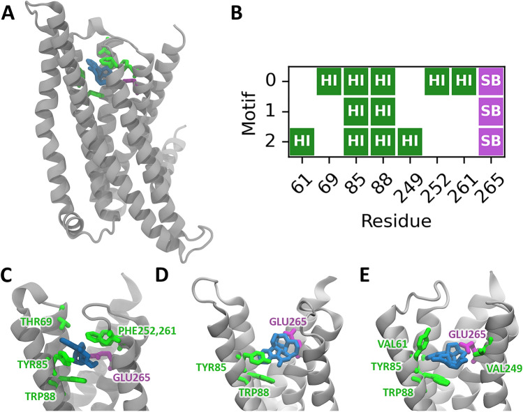
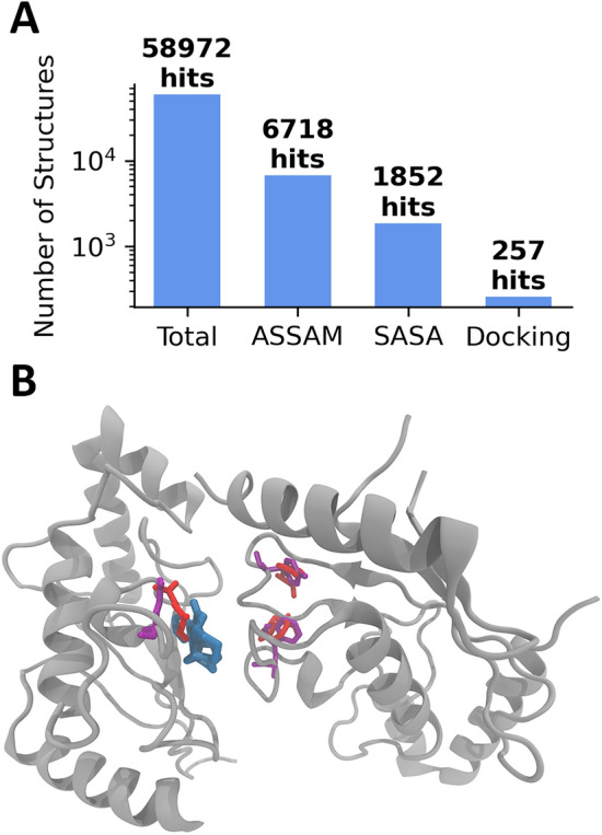
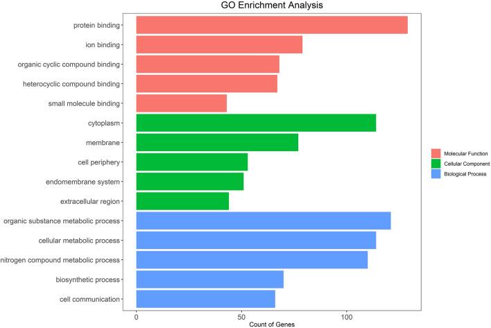
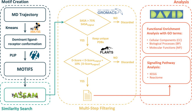
Protein residues within binding pockets play a critical role in determining the range of ligands that can interact with a protein, influencing its structure and function. Identifying structural similarities in proteins offers valuable insights into their function and activation mechanisms, aiding in predicting protein–ligand interactions, anticipating off-target effects, and facilitating the development of therapeutic agents. Numerous computational methods assessing global or local similarity in protein cavities have emerged, but their utilization is impeded by complexity, impractical automation for amino acid pattern searches, and an inability to evaluate the dynamics of scrutinized protein–ligand systems. Here, we present a general, automatic and unbiased computational pipeline, named VirtuousPocketome , aimed at screening huge databases of proteins for similar binding pockets starting from an interested protein–ligand complex. We demonstrate the pipeline's potential by exploring a recently-solved human bitter taste receptor, i.e. the TAS2R46, complexed with strychnine. We pinpointed 145 proteins sharing similar binding sites compared to the analysed bitter taste receptor and the enrichment analysis highlighted the related biological processes, molecular functions and cellular components. This work represents the foundation for future studies aimed at understanding the effective role of tastants outside the gustatory system: this could pave the way towards the rationalization of the diet as a supplement to standard pharmacological treatments and the design of novel tastants-inspired compounds to target other proteins involved in specific diseases or disorders. The proposed pipeline is publicly accessible, can be applied to any protein–ligand complex, and could be expanded to screen any database of protein structures.
Overview
The claims and statistics of this paper haven’t been checked yet.
Run the check — free, no account needed. It reads the open-access text and checks the sources it cites.
Integrity signals
Registry
Cites
Statistics
Figures
Verification
Claims the paper makes
The claims and statistics of this paper haven’t been checked yet.
Run the check — free, no account needed. It reads the open-access text and checks the sources it cites.
Self-check
Percentages, sums and reported differences re-computed from the paper’s own numbers.
The self-check needs the paper’s full text.
Citations
Cites
Highly accurate protein structure prediction with AlphaFold
Jumper, J., Evans, R., Pritzel, A., Green, T., Figurnov, M., Ronneberger, O., Tunyasuvunakool, K., Bates, R., Žídek, A., Potapenko, A., et al. (2021) · Nature · doi:10.1038/s41586-021-03819-2 · Cited by 43,764
KEGG for taxonomy-based analysis of pathways and genomes
Kanehisa, M., Furumichi, M., Sato, Y., Kawashima, M., & Ishiguro-Watanabe, M. (2023) · Nucleic Acids Research · doi:10.1093/nar/gkac963 · Cited by 5,833
Research works printed but not linked: 119
- Artymiuk, P. J., Poirrette, A. R., Rice, D. W. & Willett, P. A polymerase I palm in adenylyl cyclase?. Nature 388, 33–34 (1997). · DOI
- Ehrt, C., Brinkjost, T. & Koch, O. A benchmark driven guide to binding site comparison: An exhaustive evaluation using tailor-made data sets (ProSPECCTs). PLoS Comput. Biol. 14, e1006483 (2018). · DOI
- Hodos, R. A., Kidd, B. A., Shameer, K., Readhead, B. P. & Dudley, J. T. In silico methods for drug repurposing and pharmacology. WIREs Mech. Dis. 8, 186–210 (2016). · DOI
- Independent Interdisciplinary Consultant & Flower, D. Drug discovery: Today and tomorrow. Bioinformation 16, 1–3 (2020). · DOI
- Kabir, A. & Muth, A. Polypharmacology: The science of multi-targeting molecules. Pharmacol. Res. 176, 106055 (2022). · DOI
- Chaudhari, R., Fong, L. W., Tan, Z., Huang, B. & Zhang, S. An up-to-date overview of computational polypharmacology in modern drug discovery. Expert Opin. Drug Discov. 15, 1025–1044 (2020). · DOI
- Cannariato, M., Miceli, M. & Deriu, M. A. In silico investigation of Alsin RLD conformational dynamics and phosphoinositides binding mechanism. PLoS ONE 17, e0270955 (2022). · DOI
- Eguida, M. & Rognan, D. Estimating the similarity between protein pockets. IJMS 23, 12462 (2022). · DOI
- Jambon, M., Imberty, A., Deléage, G. & Geourjon, C. A new bioinformatic approach to detect common 3D sites in protein structures: Detection of 3D sites in protein structures. Proteins 52, 137–145 (2003). · DOI
- Stark, A., Sunyaev, S. & Russell, R. B. A model for statistical significance of local similarities in structure. J. Mol. Biol. 326, 1307–1316 (2003). · DOI
- Kinoshita, K. Identification of protein biochemical function by searching the similar shape and electrostatic potential on the molecular surface of proteins. Seibutsu Butsuri 44, 150–154 (2004). · DOI
- Zhang, Y. TM-align: A protein structure alignment algorithm based on the TM-score. Nucleic Acids Res. 33, 2302–2309 (2005). · DOI · Paper page
- Shulman-Peleg, A., Nussinov, R. & Wolfson, H. J. SiteEngines: Recognition and comparison of binding sites and protein-protein interfaces. Nucleic Acids Res. 33, W337–W341 (2005). · DOI
- Lisewski, A. M. & Lichtarge, O. Rapid detection of similarity in protein structure and function through contact metric distances. Nucleic Acids Res. 34, e152–e152 (2006). · DOI
- Nagarajan, D. & Chandra, N. PocketMatch (version 2.0): A parallel algorithm for the detection of structural similarities between protein ligand binding-sites. in 2013 National Conference on Parallel Computing Technologies (PARCOMPTECH) 1–6 (IEEE, 2013). https://doi.org/10.1109/ParCompTech.2013.6621397. · DOI
- Yeturu, K. & Chandra, N. PocketMatch: A new algorithm to compare binding sites in protein structures. BMC Bioinform. 9, 543 (2008). · DOI
- Schalon, C., Surgand, J., Kellenberger, E. & Rognan, D. A simple and fuzzy method to align and compare druggable ligand-binding sites. Proteins 71, 1755–1778 (2008). · DOI
- Shulman-Peleg, A., Shatsky, M., Nussinov, R. & Wolfson, H. J. MultiBind and MAPPIS: Webservers for multiple alignment of protein 3D-binding sites and their interactions. Nucleic Acids Res. 36, W260–W264 (2008). · DOI
- Tseng, Y. Y., Dundas, J. & Liang, J. Predicting protein function and binding profile via matching of local evolutionary and geometric surface patterns. J. Mol. Biol. 387, 451–464 (2009). · DOI
- Tseng, Y. Y., Chen, Z. J. & Li, W.-H. f POP: Footprinting functional pockets of proteins by comparative spatial patterns. Nucleic Acids Res. 38, D288–D295 (2010). · DOI
- Xie, L., Xie, L. & Bourne, P. E. A unified statistical model to support local sequence order independent similarity searching for ligand-binding sites and its application to genome-based drug discovery. Bioinformatics 25, i305–i312 (2009). · DOI
- Das, S., Krein, M. P. & Breneman, C. M. PESDserv: A server for high-throughput comparison of protein binding site surfaces. Bioinformatics 26, 1913–1914 (2010). · DOI
- Standley, D. M., Yamashita, R., Kinjo, A. R., Toh, H. & Nakamura, H. SeSAW: Balancing sequence and structural information in protein functional mapping. Bioinformatics 26, 1258–1259 (2010). · DOI
- Moll, M., Bryant, D. H. & Kavraki, L. E. The LabelHash algorithm for substructure matching. BMC Bioinform. 11, 555 (2010). · DOI
- Weill, N. & Rognan, D. Alignment-free ultra-high-throughput comparison of druggable protein−ligand binding sites. J. Chem. Inf. Model. 50, 123–135 (2010). · DOI
- Desaphy, J., Azdimousa, K., Kellenberger, E. & Rognan, D. Comparison and druggability prediction of protein-ligand binding sites from pharmacophore-annotated cavity shapes. J. Chem. Inf. Model. 52, 2287–2299 (2012). · DOI
- Wood, D. J., Vlieg, J. D., Wagener, M. & Ritschel, T. Pharmacophore fingerprint-based approach to binding site subpocket similarity and its application to bioisostere replacement. J. Chem. Inf. Model. 52, 2031–2043 (2012). · DOI
- Konc, J. & Janezic, D. ProBiS-2012: Web server and web services for detection of structurally similar binding sites in proteins. Nucleic Acids Res. 40, W214–W221 (2012). · DOI
- Ito, J.-I., Tabei, Y., Shimizu, K., Tsuda, K. & Tomii, K. PoSSuM: A database of similar protein-ligand binding and putative pockets. Nucleic Acids Res. 40, D541–D548 (2012). · DOI
- Roy, A., Yang, J. & Zhang, Y. COFACTOR: An accurate comparative algorithm for structure-based protein function annotation. Nucleic Acids Res. 40, W471–W477 (2012). · DOI
- Nadzirin, N., Gardiner, E. J., Willett, P., Artymiuk, P. J. & Firdaus-Raih, M. SPRITE and ASSAM: Web servers for side chain 3D-motif searching in protein structures. Nucleic Acids Res. 40, W380–W386 (2012). · DOI
- Lin, Y., Yoo, S. & Sanchez, R. SiteComp: A server for ligand binding site analysis in protein structures. Bioinformatics 28, 1172–1173 (2012). · DOI
- Kurbatova, N., Chartier, M., Zylber, M. I. & Najmanovich, R. IsoCleft Finder: A web-based tool for the detection and analysis of protein binding-site geometric and chemical similarities. F1000Res 2, 117 (2013). · DOI
- Kirshner, D. A., Nilmeier, J. P. & Lightstone, F. C. Catalytic site identification: A web server to identify catalytic site structural matches throughout PDB. Nucleic Acids Res. 41, W256–W265 (2013). · DOI
- Nadzirin, N., Willett, P., Artymiuk, P. J. & Firdaus-Raih, M. IMAAAGINE: A webserver for searching hypothetical 3D amino acid side chain arrangements in the Protein Data Bank. Nucleic Acids Res. 41, W432–W440 (2013). · DOI
- Gao, M. & Skolnick, J. APoc: Large-scale identification of similar protein pockets. Bioinformatics 29, 597–604 (2013). · DOI
- Desaphy, J., Raimbaud, E., Ducrot, P. & Rognan, D. Encoding protein-ligand interaction patterns in fingerprints and graphs. J. Chem. Inf. Model. 53, 623–637 (2013). · DOI
- Caprari, S., Toti, D., Viet Hung, L., Di Stefano, M. & Polticelli, F. ASSIST: A fast versatile local structural comparison tool. Bioinformatics 30, 1022–1024 (2014). · DOI
- Batista, J., Hawkins, P. C., Tolbert, R. & Geballe, M. T. SiteHopper: A unique tool for binding site comparison. J. Cheminform. 6, 57 (2014). · DOI
- Chartier, M., Adriansen, E. & Najmanovich, R. IsoMIF Finder: Online detection of binding site molecular interaction field similarities. Bioinformatics 32, 621–623 (2016). · DOI
- Chartier, M. & Najmanovich, R. Detection of binding site molecular interaction field similarities. J. Chem. Inf. Model. 55, 1600–1615 (2015). · DOI
- Krotzky, T., Grunwald, C., Egerland, U. & Klebe, G. Large-scale mining for similar protein binding pockets: With RAPMAD retrieval on the fly becomes real. J. Chem. Inf. Model. 55, 165–179 (2015). · DOI
- Lee, H. S. & Im, W. G-LoSA: An efficient computational tool for local structure-centric biological studies and drug design: G-LoSA. Protein Sci. 25, 865–876 (2016). · DOI
- Núñez-Vivanco, G., Valdés-Jiménez, A., Besoaín, F. & Reyes-Parada, M. Geomfinder: a multi-feature identifier of similar three-dimensional protein patterns: A ligand-independent approach. J. Cheminform. 8, 19 (2016). · DOI
- Rey, J., Rasolohery, I., Tufféry, P., Guyon, F. & Moroy, G. PatchSearch: A web server for off-target protein identification. Nucleic Acids Res. 47, W365–W372 (2019). · DOI
- Ab Ghani, N. S., Ramlan, E. I. & Firdaus-Raih, M. Drug ReposER: A web server for predicting similar amino acid arrangements to known drug binding interfaces for potential drug repositioning. Nucleic Acids Res. 47, W350–W356 (2019). · DOI
- Simonovsky, M. & Meyers, J. DeeplyTough: Learning structural comparison of protein binding sites. J. Chem. Inf. Model. 60, 2356–2366 (2020). · DOI
- Breslin, P. A. S. An evolutionary perspective on food and human taste. Curr. Biol. 23, R409–R418 (2013). · DOI
- Ho, H. K. et al. Functionally expressed bitter taste receptor TAS2R14 in human epidermal keratinocytes serves as a chemosensory receptor. Exp. Dermatol. 30, 216–225 (2021). · DOI
- Shaw, L. et al. Personalized expression of bitter ‘taste’ receptors in human skin. PLoS ONE 13, e0205322 (2018). · DOI
- Singh, N., Vrontakis, M., Parkinson, F. & Chelikani, P. Functional bitter taste receptors are expressed in brain cells. Biochem. Biophys. Res. Commun. 406, 146–151 (2011). · DOI
- Kyriazis, G. A., Soundarapandian, M. M. & Tyrberg, B. Sweet taste receptor signaling in beta cells mediates fructose-induced potentiation of glucose-stimulated insulin secretion. Proc. Natl. Acad. Sci. USA. 109, 1115 (2012). · DOI
- Kyriazis, G. A., Smith, K. R., Tyrberg, B., Hussain, T. & Pratley, R. E. Sweet taste receptors regulate basal insulin secretion and contribute to compensatory insulin hypersecretion during the development of diabetes in male mice. Endocrinology 155, 2112–2121 (2014). · DOI
- Foster, S. R. et al. Expression, regulation and putative nutrient-sensing function of taste GPCRs in the heart. PLoS ONE 8, e64579 (2013). · DOI
- Foster, S. R. et al. Bitter taste receptor agonists elicit G-protein-dependent negative inotropy in the murine heart. FASEB J. 28, 4497–4508 (2014). · DOI
- Deckmann, K. et al. Bitter triggers acetylcholine release from polymodal urethral chemosensory cells and bladder reflexes. Proc. Natl. Acad. Sci. USA. 111, 8287–8292 (2014). · DOI
- Deshpande, D. A. et al. Bitter taste receptors on airway smooth muscle bronchodilate by localized calcium signaling and reverse obstruction. Nat. Med. 16, 1299–1304 (2010). · DOI
- Avau, B. et al. Targeting extra-oral bitter taste receptors modulates gastrointestinal motility with effects on satiation. Sci. Rep. 5, 1–12 (2015). · DOI
- Iwatsuki, K. & Uneyama, H. Sense of taste in the gastrointestinal tract. J. Pharmacol. Sci. 118, 123–128 (2012). · DOI
- Raka, F., Farr, S., Kelly, J., Stoianov, A. & Adeli, K. Metabolic control via nutrient-sensing mechanisms: Role of taste receptors and the gut-brain neuroendocrine axis. Am. J. Physiol. Endocrinol. Metab. 317, E559–E572 (2019). · DOI
- Carey, R. M. & Lee, R. J. Taste receptors in upper airway innate immunity. Nutrients 11, 2017 (2019). · DOI
- Behrens, M. & Lang, T. Extra-oral taste receptors: Function, disease, and perspectives. Front. Nutr. 9, 881177 (2022). · DOI
- Zhai, K. et al. Activation of bitter taste receptors (tas2rs) relaxes detrusor smooth muscle and suppresses overactive bladder symptoms. Oncotarget 7, 21156–21167 (2016). · DOI
- Chandrashekar, J. et al. T2Rs function as bitter taste receptors. Cell 100, 703–711 (2000). · DOI
- Sutkeviciute, I. & Vilardaga, J.-P. Structural insights into emergent signaling modes of G protein–coupled receptors. J. Biol. Chem. 295, 11626–11642 (2020). · DOI
- Di Pizio, A. et al. Comparing class A GPCRs to bitter taste receptors. Biophys. Methods Cell Biol. 132, 401–427 (2016). · DOI
- Brockhoff, A., Behrens, M., Niv, M. Y. & Meyerhof, W. Structural requirements of bitter taste receptor activation. Proc. Natl. Acad. Sci. USA 107, 11110–11115 (2010). · DOI
- Slack, J. P. et al. Modulation of bitter taste perception by a small molecule hTAS2R antagonist. Curr. Biol. 20, 1104–1109 (2010). · DOI
- Meyerhof, W. et al. The molecular receptive ranges of human TAS2R bitter taste receptors. Chem. Sens. 35, 157–170 (2009). · DOI
- Gowers, R. et al. MDAnalysis: A Python Package for the Rapid Analysis of Molecular Dynamics Simulations, 98–105 (2016). https://doi.org/10.25080/Majora-629e541a-00e. · DOI
- Xu, W. et al. Structural basis for strychnine activation of human bitter taste receptor TAS2R46. Science 377, 1298–1304 (2022). · DOI
- Born, S., Levit, A., Niv, M. Y., Meyerhof, W. & Behrens, M. The human bitter taste receptor TAS2R10 is tailored to accommodate numerous diverse ligands. J. Neurosci. 33, 201–213 (2013). · DOI
- Jensen, A. A., Gharagozloo, P., Birdsall, N. J. M. & Zlotos, D. P. Pharmacological characterisation of strychnine and brucine analogues at glycine and α7 nicotinic acetylcholine receptors. Eur. J. Pharmacol. 539, 27–33 (2006). · DOI
- DiPizio, A. et al. Rational design of agonists for bitter taste receptor TAS2R14: From modeling to bench and back. Cell. Mol. Life Sci. 77, 531 (2020). · DOI
- Fierro, F., Giorgetti, A., Carloni, P., Meyerhof, W. & Alfonso-Prieto, M. Dual binding mode of “bitter sugars” to their human bitter taste receptor target. Sci. Rep. 9, 1–16 (2019). · DOI
- Nicoli, A., Dunkel, A., Giorgino, T., de Graaf, C. & Di Pizio, A. Classification model for the second extracellular loop of class A GPCRs. J. Chem. Inf. Model. 62, 511–522 (2022). · DOI
- Topin, J. et al. Functional molecular switches of mammalian G protein-coupled bitter-taste receptors. Cell. Mol. Life Sci. 6, 348706 (2021).
- Kuipers, W. et al. Study of the interaction between aryloxypropanolamines and Asn386 in helix VII of the human 5-hydroxytryptamine1A receptor. Mol. Pharmacol. 51, 889–896 (1997). · DOI
- Oksenberg, D. et al. A single amino-acid difference confers major pharmacological variation between human and rodent 5-HT1B receptors. Nature 360, 161–163 (1992). · DOI
- Suryanarayana, S., Daunt, D. A., Von Zastrow, M. & Kobilka, B. K. A point mutation in the seventh hydrophobic domain of the alpha 2 adrenergic receptor increases its affinity for a family of beta receptor antagonists. J. Biol. Chem. 266, 15488–15492 (1991). · DOI
- Suryanarayana, S. & Kobilka, B. K. Amino acid substitutions at position 312 in the seventh hydrophobic segment of the beta 2-adrenergic receptor modify ligand-binding specificity. Mol. Pharmacol. 44, 111–114 (1993).
- Erb, L. et al. Site-directed mutagenesis of P2U purinoceptors: Positively charged amino acids in transmembrane helices 6 and 7 affect agonist potency and specificity. J. Biol. Chem. 270, 4185–4188 (1995). · DOI
- Jiang, Q. et al. A mutational analysis of residues essential for ligand recognition at the human P2Y1 receptor. Mol. Pharmacol. 52, 499–507 (1997). · DOI
- Kopin, A. S., McBride, E. W., Quinn, S. M., Kolakowski, L. F. & Beinborn, M. The role of the cholecystokinin-B/gastrin receptor transmembrane domains in determining affinity for subtype-selective ligands. J. Biol. Chem. 270, 5019–5023 (1995). · DOI
- Pronin, A. N. Identification of ligands for two human bitter T2R receptors. Chem. Sens. 29, 583–593 (2004). · DOI
- Kuijpers, G. A. J., Vergara, L. A., Calvo, S. & Yadid, G. Inhibitory effect of strychnine on acetylcholine receptor activation in bovine adrenal medullary chromaffin cells. Br. J. Pharmacol. 113, 471–478 (1994). · DOI
- García-Colunga, J. & Miledi, R. Modulation of nicotinic acetylcholine receptors by strychnine. Proc. Natl. Acad. Sci. USA 96, 4113–4118 (1999). · DOI
- Zlotos, D. P., Mandour, Y. M. & Jensen, A. A. Strychnine and its mono- and dimeric analogues: A pharmaco-chemical perspective. Nat. Prod. Rep. 39, 1910–1937 (2022). · DOI
- Abraham, M. J. et al. GROMACS: High performance molecular simulations through multi-level parallelism from laptops to supercomputers. SoftwareX 1–2, 19–25 (2015). · DOI · Paper page
- Rodrigues, J. P. G. L. M., Teixeira, J. M. C., Trellet, M. & Bonvin, A. M. J. J. pdb-tools: A swiss army knife for molecular structures. F1000 Res. 7, 1961 (2018). · DOI
- Adasme, M. F. et al. PLIP 2021: Expanding the scope of the protein–ligand interaction profiler to DNA and RNA. Nucleic Acids Res. 49, W530–W534 (2021). · DOI
- Spriggs, R. V., Artymiuk, P. J. & Willett, P. Searching for patterns of amino acids in 3D protein structures. J. Chem. Inf. Comput. Sci. 43, 412–421 (2003). · DOI
- Bron, C. & Kerbosch, J. Algorithm 457: Finding all cliques of an undirected graph. Commun. ACM 16, 575–577 (1973). · DOI
- Korb, O., Stützle, T. & Exner, T. E. PLANTS: Application of ant colony optimization to structure-based drug design. In Ant Colony Optimization and Swarm Intelligence Vol. 4150 (eds Dorigo, M. et al.) 247–258 (Springer, 2006). · DOI
- Korb, O., Stützle, T. & Exner, T. E. An ant colony optimization approach to flexible protein–ligand docking. Swarm. Intell. 1, 115–134 (2007). · DOI
- Çınaroğlu, S. S. & Timuçin, E. Comparative assessment of seven docking programs on a nonredundant metalloprotein subset of the PDBbind refined. J. Chem. Inf. Model. 59, 3846–3859 (2019). · DOI
- Eldridge, M. D., Murray, C. W., Auton, T. R., Paolini, G. V. & Mee, R. P. Empirical scoring functions: I. The development of a fast empirical scoring function to estimate the binding affinity of ligands in receptor complexes. J. Comput. Aid. Mol. Des. 11, 425–445 (1997). · DOI
- Sirci, F. et al. Virtual fragment screening: Discovery of histamine H3 receptor ligands using ligand-based and protein-based molecular fingerprints. J. Chem. Inf. Model. 52, 3308–3324 (2012). · DOI
- Kooistra, A. J., Leurs, R., De Esch, I. J. P. & De Graaf, C. Structure-based prediction of G-protein-coupled receptor ligand function: A β-adrenoceptor case study. J. Chem. Inf. Model. 55, 1045–1061 (2015). · DOI
- Bassani, D., Pavan, M., Sturlese, M. & Moro, S. Sodium or not sodium: Should its presence affect the accuracy of pose prediction in docking GPCR antagonists?. Pharmaceuticals 15, 346 (2022). · DOI
- Kooistra, A. J. et al. Function-specific virtual screening for GPCR ligands using a combined scoring method. Sci. Rep. 6, 28288 (2016). · DOI
- De Graaf, C. et al. Crystal structure-based virtual screening for fragment-like ligands of the human histamine H1 receptor. J. Med. Chem. 54, 8195–8206 (2011). · DOI
- Huang, D. W., Sherman, B. T. & Lempicki, R. A. Systematic and integrative analysis of large gene lists using DAVID bioinformatics resources. Nat. Protoc. 4, 44–57 (2009). · DOI · Paper page
- Sherman, B. T. et al. DAVID: A web server for functional enrichment analysis and functional annotation of gene lists (2021 update). Nucleic Acids Res. 50, W216–W221 (2022). · DOI
- The Gene Ontology Consortium. The gene ontology project in 2008. Nucleic Acids Res. 36, D440–D444 (2008). · DOI
- Gillespie, M. et al. The reactome pathway knowledgebase 2022. Nucleic Acids Res. 50, D687–D692 (2022). · DOI
- Kanehisa, M. KEGG: Kyoto encyclopedia of genes and genomes. Nucleic Acids Res. 28, 27–30 (2000). · DOI · Paper page
- Ferreira, J. A. & Zwinderman, A. H. On the Benjamini–Hochberg method. Ann. Stat. 34, 1827–1849 (2006). · DOI
- NCBI Resource Coordinators et al. Database resources of the National center for biotechnology information. Nucleic Acids Res. 46, D8–D13 (2018). · DOI
- Berman, H. M. The protein data bank. Nucleic Acids Res. 28, 235–242 (2000). · DOI · Paper page
- Jo, S., Kim, T., Iyer, V. G. & Im, W. CHARMM-GUI: A web-based graphical user interface for CHARMM. J. Comput. Chem. 29, 1859–1865 (2008). · DOI · Paper page
- Dickson, C. J., Walker, R. C. & Gould, I. R. Lipid21: Complex lipid membrane simulations with AMBER. J. Chem. Theory Comput. 18, 1726–1736 (2022). · DOI
- Tian, C. et al. ff19SB: Amino-acid-specific protein backbone parameters trained against quantum mechanics energy surfaces in solution. J. Chem. Theory Comput. 16, 528–552 (2020). · DOI
- Wang, J., Wolf, R. M., Caldwell, J. W., Kollman, P. A. & Case, D. A. Development and testing of a general amber force field. J. Comput. Chem. 25, 1157–1174 (2004). · DOI
- Berendsen, H. J. C., Postma, J. P. M., van Gunsteren, W. F., DiNola, A. & Haak, J. R. Molecular dynamics with coupling to an external bath. J. Chem. Phys. 81, 3684–3690 (1984). · DOI
- Nosé, S. A unified formulation of the constant temperature molecular dynamics methods. J. Chem. Phys. 81, 511–519 (1984). · DOI
- Parrinello, M. & Rahman, A. Polymorphic transitions in single crystals: A new molecular dynamics method. J. Appl. Phys. 52, 7182–7190 (1981). · DOI
- Hess, B., Bekker, H., Berendsen, H. J. C. & Fraaije, J. G. E. M. LINCS: A linear constraint solver for molecular simulations. J. Comput. Chem. 18, 1463–1472 (1997). · DOI
- Humphrey, W., Dalke, A. & Schulten, K. VMD: Visual molecular dynamics. J. Mol. Graph. 14, 33–38 (1996). · DOI · Paper page
Metadata
- Authors
- Pallante, L., Cannariato, M., Androutsos, L., Zizzi, E. A., Bompotas, A., Hada, X., Grasso, G., Kalogeras, A., Mavroudi, S., Di Benedetto, G., Theofilatos, K., & Deriu, M. A.
- Affiliations
- Politecnico di Torino (IT)
- Industrial Systems Institute (GR)
- University of Applied Sciences and Arts of Southern Switzerland (CH)
- University of Patras (GR)
- Corresponding author
- Deriu, Marco A.
- ORCID
- Venue
- Scientific Reports
- Published
- 2024-03-15
- Volume
- 14
- Issue
- 1
- Pages
- 6296-6296
- ISSN
- 2045-2322 (ISSN-L)
- PMC
- PMC10943019
- OpenAlex
- W4392847330
- Cited by
- 12
- References
- 2
- Citations by year
- 2026: 1 · 2025: 6 · 2024: 5
- FWCI
- 2.54
- Type
- journal article
- Language
- en
- Open access
- gold
- doi.org
- Locations
- Scientific Reports (publishedVersion, cc-by)
- bioRxiv (Cold Spring Harbor Laboratory) (acceptedVersion, cc-by-nc-nd)
- PubMed Central (publishedVersion, cc-by)
- PubMed (publishedVersion)
- PORTO@Iris (submittedVersion, cc-by)
- DOAJ (DOAJ: Directory of Open Access Journals) (submittedVersion)
- Funders
- H2020 Marie Skłodowska-Curie Actions (872181)
- Topics
- Life Sciences › Pharmacology, Toxicology and Pharmaceutics › Drug Discovery › Computational Drug Discovery Methods
- Life Sciences › Biochemistry, Genetics and Molecular Biology › Molecular Biology › Protein Structure and Dynamics
- Life Sciences › Biochemistry, Genetics and Molecular Biology › Structural Biology › Enzyme Structure and Function
- Keywords
- protein pockets, protein-ligand complex, binding site similarity, protein structure comparison, strychnine, off-target effects
- MeSH terms
- Binding Sites, Humans, Ligands, Protein Binding, Proteins, Taste, Taste Buds
About these checks
Registry, retraction, Cites and statistics results are computed, not judged, with one exception: a printed reference that no index lists is read once by a model before it counts as a citation error. Claims are extracted by a model and kept only when their quote appears verbatim in the text; each cited source is then checked, with a model’s help, for whether it says what the claim relies on. Those verdicts can be wrong — read the claim’s quote and the cited source before relying on one.
Statistics and arithmetic are recomputed, not judged.
Confidence bands are shown only for fields calibrated on our annotated test set. None is calibrated yet, so no band is shown.
What counts as a claim ›How we measure confidence ›How we check this ›
Are you an author, or do you see a problem with this page? Request a review
How to cite
Cite CoChat for the checks on this page, and the paper for its own findings.
- This page
- CoChat. (2026). Citation check: VirtuousPocketome: a computational tool for screening protein–ligand complexes to identify similar binding sites. https://app.cochat.ai/papers/10.1038/s41598-024-56893-7
- The paper
- Pallante, L., Cannariato, M., Androutsos, L., Zizzi, E. A., Bompotas, A., Hada, X., Grasso, G., Kalogeras, A., Mavroudi, S., Di Benedetto, G., Theofilatos, K., & Deriu, M. A. (2024). VirtuousPocketome: a computational tool for screening protein–ligand complexes to identify similar binding sites. Scientific Reports. https://doi.org/10.1038/s41598-024-56893-7
