

The DOI matches its Crossref record.
Dual pH/redox-responsive hyperbranched polymeric nanocarriers with TME-trigger size shrinkage and charge reversible ability for amplified chemotherapy of breast cancer
Abstract
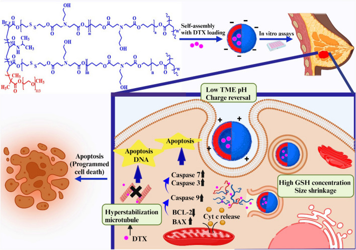
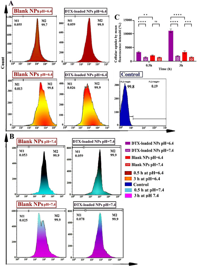
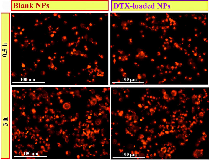
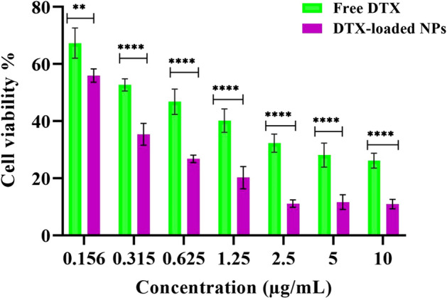
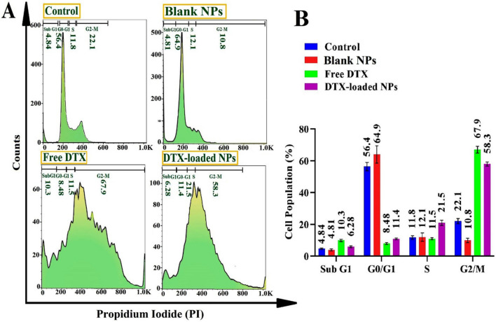
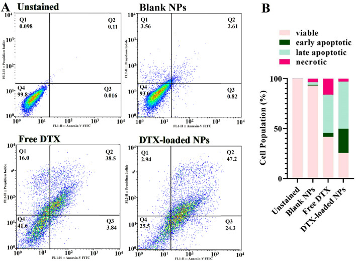
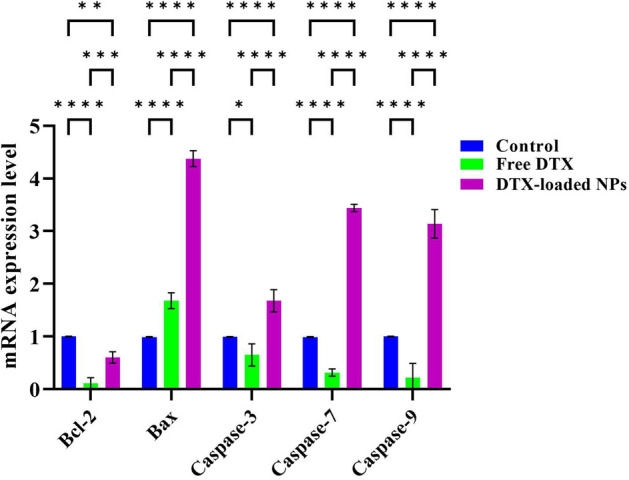
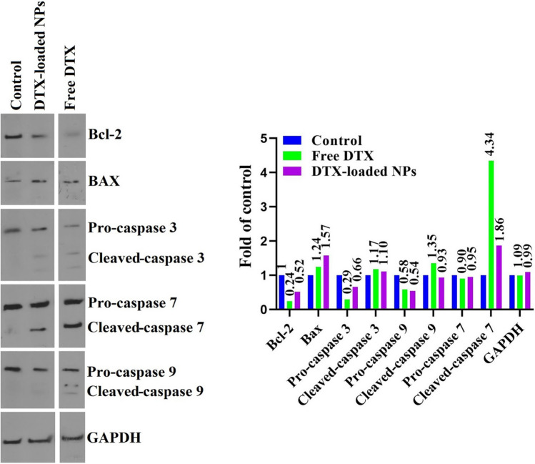
A novel pH/redox-responsive hyperbranched MeO-PEG-b-(NIPAAm-co-PBAE) nanoparticles (NPs) were designed with size shrinkage and charge-reversible potential for targeted delivery of docetaxel (DTX) to MDA-MB-231 cell lines. In the tumor microenvironment (TME), amine protonation induces charge reversal and disulfide bond cleavage under high TME GSH concentration causing size shrinkage, improved deep tumor penetration, and active targeting of the therapeutic agents. These nano drug delivery systems (NDDSs) significantly promoted cancer cell uptake (~ 100% at 0.5 h), facilitating site-specific delivery and deep tumor penetration. The MTT assay revealed significantly higher cytotoxicity ( P value < 0.0001) for DTX-loaded NPs compared to free DTX. Cell cycle analysis revealed G2/M (58.3 ± 2.1%) and S (21.5 ± 1.3%) arrest for DTX-loaded NPs, while free DTX caused G2/M (67.9 ± 1.1%) and sub-G1 (10.3 ± 0.8%) arrest. DTX-loaded NPs induced higher apoptosis ( P value < 0.001) in MDA-MB-231 cells (71.5 ± 2.8%) compared to free DTX (42.3 ± 3.1%). Western blotting and RT-PCR assays confirmed significant up-regulation of protein levels and apoptotic genes by DTX-loaded NPs compared to free DTX. In conclusion, TME-responsive charge reversal and size-shrinkable smart NDDSs designed based on low pH, and high glutathione (GSH), offer more effective site-specific delivery of therapeutic agents to tumors.
Overview
The claims and statistics of this paper haven’t been checked yet.
Run the check — free, no account needed. It reads the open-access text and checks the sources it cites.
Integrity signals
Registry
Cites
Statistics
Figures
Verification
Claims the paper makes
The claims and statistics of this paper haven’t been checked yet.
Run the check — free, no account needed. It reads the open-access text and checks the sources it cites.
Claims other papers make about this one
Each is checked against this paper’s own text: does it say what the citing paper says it does?
In the citing paper
“To address this issue, researchers have developed transformable nanocarriers whose size or surface charge can dynamically respond to specific TME stimuli, such as pH, enzymes, or GSH [20].”
From Overcoming tumor microenvironment barriers: transformable and bioinspired nanomedicine strategies for deep tumor penetration (2026) · cites this paper as [20]
In the citing paper
“These systems typically adopt a “long-circulation, local-shrinkage” strategy, where large particles contract or disassemble at the tumor site, improving extravasation and deep tissue diffusion.”
From Overcoming tumor microenvironment barriers: transformable and bioinspired nanomedicine strategies for deep tumor penetration (2026) · cites this paper as [20]
Self-check
Percentages, sums and reported differences re-computed from the paper’s own numbers.
The self-check needs the paper’s full text.
Citations
Cites
No references could be resolved yet.
Research works printed but not linked: 60
- Xu, M. & Li, S. Nano-drug delivery system targeting tumor microenvironment: A prospective strategy for melanoma treatment. Cancer Lett. 216397 (2023). · DOI
- Cheng, R. & Santos, H. A. Smart nanoparticle-based platforms for regulating tumor microenvironment and cancer immunotherapy. Adv. Healthc. Mater. 12, 2202063 (2023). · DOI
- Wong, K. H., Yang, D., Chen, S., He, C. & Chen, M. Development of nanoscale drug delivery systems of dihydroartemisinin for cancer therapy: A review. Asian J. Pharm. Sci. 17, 475–490 (2022). · DOI
- Liu, J. et al. Application of nano drug delivery system (NDDS) in cancer therapy: A perspective. Recent Pat. Anticancer Drug Discov. 18, 125–132 (2023). · DOI
- Chidambaram, M., Manavalan, R. & Kathiresan, K. Nanotherapeutics to overcome conventional cancer chemotherapy limitations. J. Pharm. Pharm. Sci. 14, 67–77 (2011). · DOI
- Zhang, Y., Li, M., Gao, X., Chen, Y. & Liu, T. Nanotechnology in cancer diagnosis: Progress, challenges and opportunities. J. Hematol. Oncol. 12, 1–13 (2019). · DOI · Paper page
- Guragain, S., Bastakoti, B. P., Malgras, V., Nakashima, K. & Yamauchi, Y. Multi-stimuli-responsive polymeric materials. Chem. Eur. J. 21, 13164–13174 (2015). · DOI
- Cheng, R., Meng, F., Deng, C., Klok, H.-A. & Zhong, Z. Dual and multi-stimuli responsive polymeric nanoparticles for programmed site-specific drug delivery. Biomaterials 34, 3647–3657 (2013). · DOI
- Fadeel, B. & Garcia-Bennett, A. E. Better safe than sorry: Understanding the toxicological properties of inorganic nanoparticles manufactured for biomedical applications. Adv. Drug Deliv. Rev. 62, 362–374 (2010). · DOI
- Wang, R. et al. Impacts of particle size on the cytotoxicity, cellular internalization, pharmacokinetics and biodistribution of betulinic acid nanosuspensions in combined chemotherapy. Int. J. Pharms. 588, 119799 (2020). · DOI
- Ding, L. et al. Surface charge of supramolecular nanosystems for in vivo biodistribution: A MicroSPECT/CT imaging study. Small 16, 2003290 (2020). · DOI
- Zhou, Q., Zhang, L., Yang, T. & Wu, H. Stimuli-responsive polymeric micelles for drug delivery and cancer therapy. Int. J. Nanomedicine. 13, 2921 (2018). · DOI
- Guo, X., Wang, L., Wei, X. & Zhou, S. Polymer-based drug delivery systems for cancer treatment. J. Polym. Sci. A: Polym. Chem. 54, 3525–3550 (2016). · DOI
- Jin, M.-Z. & Jin, W.-L. The updated landscape of tumor microenvironment and drug repurposing. Signal Transduct. Target. Ther. 5, 166 (2020). · DOI · Paper page
- Aghaz, F. et al. Codelivery of resveratrol melatonin utilizing pH responsive sericin based nanocarriers inhibits the proliferation of breast cancer cell line at the different pH. Sci. Rep. 13, 11090 (2023). · DOI
- Zhang, P., Chen, D., Li, L. & Sun, K. Charge reversal nano-systems for tumor therapy. J. Nanobiotechnology 20, 1–27 (2022). · DOI
- Li, Z. et al. Charge-reversal nanomedicines as a smart bullet for deep tumor penetration. Smart Mater. Med. 3, 243–253 (2022). · DOI
- Swietach, P., Hulikova, A., Patiar, S., Vaughan-Jones, R. D. & Harris, A. L. Importance of intracellular pH in determining the uptake and efficacy of the weakly basic chemotherapeutic drug, doxorubicin. PLoS One 7, e35949 (2012). · DOI · Paper page
- Berrino, E. & Supuran, C. T. Novel approaches for designing drugs that interfere with pH regulation. Expert Opin. Drug Discov. 14, 231–248 (2019). · DOI
- Shu, M. et al. Tumor microenvironment triple-responsive nanoparticles enable enhanced tumor penetration and synergetic chemo-photodynamic therapy. Biomaterials 268, 120574 (2021). · DOI
- Estrela, J. M., Ortega, A. & Obrador, E. Glutathione in cancer biology and therapy. Crit. Rev. Clin. Lab Sci. 43, 143–181 (2006). · DOI · Paper page
- Cheng, R. et al. Glutathione-responsive nano-vehicles as a promising platform for targeted intracellular drug and gene delivery. J. Control Release 152, 2–12 (2011). · DOI
- Fu, S., Rempson, C. M., Puche, V., Zhao, B. & Zhang, F. Construction of disulfide containing redox-responsive polymeric nanomedicine. Methods 199, 67–79 (2022). · DOI
- Hashizume, H. et al. Openings between defective endothelial cells explain tumor vessel leakiness. Am. J. Pathol. 156, 1363–1380 (2000). · DOI · Paper page
- Sousa-Pimenta, M. et al. Chemotherapeutic properties and side-effects associated with the clinical practice of terpene alkaloids: Paclitaxel, docetaxel, and cabazitaxel. Front. Pharmacol. 14, 1157306 (2023). · DOI
- Chaurawal, N. & Raza, K. Nano-interventions for the drug delivery of docetaxel to cancer cells. Health Sci. Rev. Oxf. 18, 100101 (2023). · DOI
- Moloney, C. et al. Chain-extension in hyperbranched polymers alters tissue distribution and cytotoxicity profiles in orthotopic models of triple negative breast cancers. Biomater. Sci. 11, 6545–6560 (2023). · DOI
- Bal-Öztürk, A. et al. Hyperbranched polymer-based nanoparticle drug delivery platform for the nucleus-targeting in cancer therapy. J. Drug Deliv. Sci. Technol. 81, 104195 (2023). · DOI
- Indumathy, B. et al. A comprehensive review on processing, development and applications of organofunctional silanes and silane-based hyperbranched polymers. Polymers 15, 2517 (2023). · DOI
- Chen, J. & Zhang, Y. Hyperbranched polymers: Recent advances in photodynamic therapy against cancer. Pharmaceutics 15, 2222 (2023). · DOI
- Matyjaszewski, K. Atom transfer radical polymerization (ATRP): Current status and future perspectives. Macromolecules 45, 4015–4039 (2012). · DOI
- Taghipour, Y. D. et al. Dual targeting Salinomycin-loaded smart nanomicelles for enhanced accumulation and therapeutic outcome in breast cancer. Int. J. Pharm. 642, 123095 (2023). · DOI
- Chu, B. et al. Synthesis, characterization and drug loading property of Monomethoxy-Poly (ethylene glycol)-Poly (ε-caprolactone)-Poly (D, L-lactide)(MPEG-PCLA) copolymers. Sci. Rep. 6, 34069 (2016). · DOI
- Wagner, J. G. Interpretation of percent dissolved-time plots derived from in vitro testing of conventional tablets and capsules. J. Pharm. Sci. 58, 1253–1257 (1969). · DOI
- Sabzi, A. et al. Targeted co-delivery of curcumin and doxorubicin by citric acid functionalized Poly (ε-caprolactone) based micelle in MDA-MB-231 cell. Colloids Surf. B: Biointerfaces 194, 111225 (2020). · DOI
- Sadr, S. H., Davaran, S., Alizadeh, E., Salehi, R. & Ramazani, A. PLA-based magnetic nanoparticles armed with thermo/pH responsive polymers for combination cancer chemotherapy. J. Drug Deliv. Sci. Technol. 45, 240–254 (2018). · DOI
- Honary, S. & Zahir, F. Effect of zeta potential on the properties of nano-drug delivery systems-a review (Part 2). Trop. J. Pharm. Res. 12, 265–273 (2013).
- Subhan, M. A., Yalamarty, S. S. K., Filipczak, N., Parveen, F. & Torchilin, V. P. Recent advances in tumor targeting via EPR effect for cancer treatment. J. Pers. Med. 11, 571 (2021). · DOI · Paper page
- Kim, J. et al. Verteporfin-loaded poly (ethylene glycol)-poly (beta-amino ester)-poly (ethylene glycol) triblock micelles for cancer therapy. Biomacromolecules 19, 3361–3370 (2018). · DOI
- Ni, N., Wang, W., Sun, Y., Sun, X. & Leong, D. T. Inducible endothelial leakiness in nanotherapeutic applications. Biomaterials 287, 121640 (2022). · DOI
- Kim, J., Wilson, D. R., Zamboni, C. G. & Green, J. J. Targeted polymeric nanoparticles for cancer gene therapy. J. Drug Target. 23, 627–641 (2015). · DOI
- Li, J. et al. A reduction and pH dual-sensitive polymeric vector for long-circulating and tumor-targeted siRNA delivery. Adv. Mater. 26, 8217–8224 (2014). · DOI
- Sun, M. et al. Arginine-modified nanostructured lipid carriers with charge-reversal and pH-sensitive membranolytic properties for anticancer drug delivery. Adv. Healthc. Mater. 6, 1600693 (2017). · DOI
- He, C., Hu, Y., Yin, L., Tang, C. & Yin, C. Effects of particle size and surface charge on cellular uptake and biodistribution of polymeric nanoparticles. Biomaterials 31, 3657–3666 (2010). · DOI · Paper page
- Zhang, S. et al. pH-and redox-responsive nanoparticles composed of charge-reversible pullulan-based shells and disulfide-containing poly (ss-amino ester) cores for co-delivery of a gene and chemotherapeutic agent. Nanotechnology 29, 325101 (2018). · DOI
- Li, F. et al. Co-delivery of VEGF siRNA and etoposide for enhanced anti-angiogenesis and anti-proliferation effect via multi-functional nanoparticles for orthotopic non-small cell lung cancer treatment. Theranostics 9, 5886 (2019). · DOI
- Zhao, X. et al. Polymerization-induced self-assembly to produce prodrug nanoparticles with reduction-responsive camptothecin release and pH-responsive charge-reversible property. Macromol. Rapid Commun. 41, 2000260 (2020). · DOI
- Lv, Y. et al. Visual validation of the measurement of entrapment efficiency of drug nanocarriers. Int. J. Pharm. 547, 395–403 (2018). · DOI
- Zhuang, W. et al. Redox and pH dual-responsive polymeric micelles with aggregation-induced emission feature for cellular imaging and chemotherapy. ACS Appl. Mater. Interfaces 10, 18489–18498 (2018). · DOI
- Francia, V. et al. Corona composition can affect the mechanisms cells use to internalize nanoparticles. ACS Nano 13, 11107–11121 (2019). · DOI
- Chen, L. et al. Tumor-acidity activated surface charge conversion of two-photon fluorescent nanoprobe for enhanced cellular uptake and targeted imaging of intracellular hydrogen peroxide. Chem. Sci. 10, 9351–9357 (2019). · DOI
- Arora, S. et al. Translocation and toxicity of docetaxel multi-walled carbon nanotube conjugates in mammalian breast cancer cells. J. Biomed. Nanotechnol. 10, 3601–3609 (2014). · DOI
- Xiao, Y. et al. Multifunctional unimolecular micelles for cancer-targeted drug delivery and positron emission tomography imaging. Biomaterials 33, 3071–3082 (2012). · DOI
- Ghassami, E., Varshosaz, J., Mirian, M. & Jahanian-Najafabadi, A. HER-2 aptamer-targeted Ecoflex® nanoparticles loaded with docetaxel promote breast cancer cells apoptosis and anti-metastatic effect. IET Nanobiotechnol. 13, 428–434 (2019). · DOI
- Chen, Z. et al. Aptamer-mediated delivery of docetaxel to prostate cancer through polymeric nanoparticles for enhancement of antitumor efficacy. Eur. J. Pharm. Biopharm. 107, 130–141 (2016). · DOI
- Ramasamy, T. et al. pH sensitive polyelectrolyte complex micelles for highly effective combination chemotherapy. J. Mater. Chem. B. 2, 6324–6333 (2014). · DOI
- Zhao, S. et al. pH-sensitive docetaxel-loaded d-α-tocopheryl polyethylene glycol succinate–poly (β-amino ester) copolymer nanoparticles for overcoming multidrug resistance. Biomacromolecules 14, 2636–2646 (2013). · DOI
- Zhu, S. et al. Synergistic antitumor activities of docetaxel and octreotide associated with apoptotic-upregulation in castration-resistant prostate cancer. PLoS One 9, e91817 (2014). · DOI
- Safi, A., Heidarian, E. & Ahmadi, R. Quercetin synergistically enhances the anticancer efficacy of docetaxel through induction of apoptosis and modulation of PI3K/AKT, MAPK/ERK, and JAK/STAT3 signaling pathways in MDA-MB-231 breast cancer cell line. Int. J. Mol. Cell. Med. 10, 11 (2021).
- Dey, G., Bharti, R., Das, A. K., Sen, R. & Mandal, M. Resensitization of Akt induced docetaxel resistance in breast cancer by ‘Iturin A’a lipopeptide molecule from marine bacteria Bacillus megaterium. Sci. rep. 7, 1–11 (2017). · DOI
Cited by
Sorted by citations
Sheng, J., Yuan, W., Zhang, M., Chen, Z., Qi, Y., Zhao, Y., & Zhang, S. (2026) · Journal of Nanobiotechnology · doi:10.1186/s12951-026-04028-7 · Cited by 35
Metadata
- Authors
- Badparvar, F., Marjani, A. P., Salehi, R., & Ramezani, F.
- Venue
- Scientific Reports
- Published
- 2024-04-12
- Volume
- 14
- Issue
- 1
- ISSN
- 2045-2322
- PMC
- PMC11349913
- Cited by
- 48
- References
- 0
- Language
- en
About these checks
Registry, retraction, Cites and statistics results are computed, not judged, with one exception: a printed reference that no index lists is read once by a model before it counts as a citation error. Claims are extracted by a model and kept only when their quote appears verbatim in the text; each cited source is then checked, with a model’s help, for whether it says what the claim relies on. Those verdicts can be wrong — read the claim’s quote and the cited source before relying on one.
Statistics and arithmetic are recomputed, not judged.
Confidence bands are shown only for fields calibrated on our annotated test set. None is calibrated yet, so no band is shown.
What counts as a claim ›How we measure confidence ›How we check this ›
Are you an author, or do you see a problem with this page? Request a review
