

The DOI matches its Crossref record; 4 of the 4 references it cites were found in a registry or an index.
Interpretable machine learning for predicting sepsis risk in emergency triage patients
Open accessAbstract
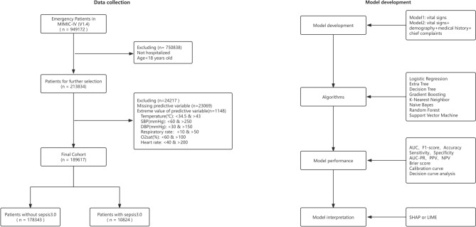
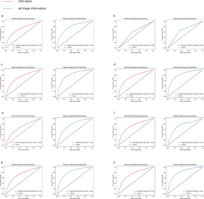
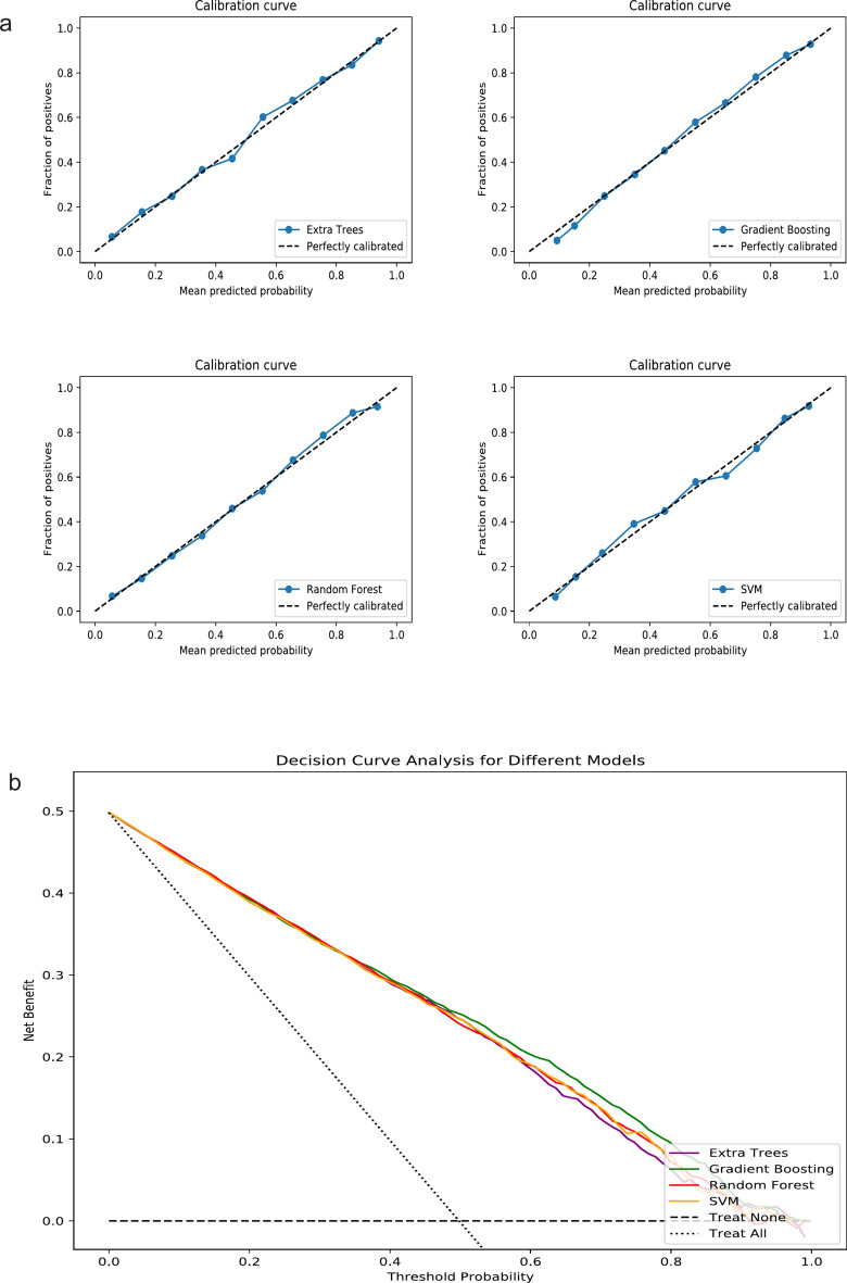
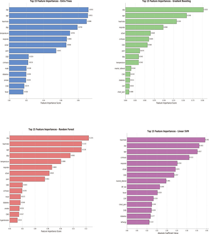
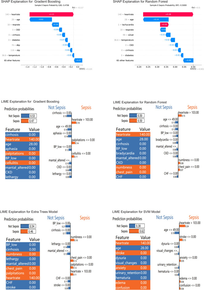
The study aimed to develop and validate a sepsis prediction model using structured electronic medical records (sEMR) and machine learning (ML) methods in emergency triage. The goal was to enhance early sepsis screening by integrating comprehensive triage information beyond vital signs. This retrospective cohort study utilized data from the MIMIC-IV database. Two models were developed: Model 1 based on vital signs alone, and Model 2 incorporating vital signs, demographic characteristics, medical history, and chief complaints. Eight ML algorithms were employed, and model performance was evaluated using metrics such as AUC, F1 Score, and calibration curves. SHapley Additive exPlanations (SHAP) and Local Interpretable Model-agnostic Explanations (LIME) methods were used to enhance model interpretability. The study included 189,617 patients, with 5.95% diagnosed with sepsis. Model 2 consistently outperformed Model 1 across most algorithms. In Model 2, Gradient Boosting achieved the highest AUC of 0.83, followed by Extra Tree, Random Forest, and Support Vector Machine (all 0.82). The SHAP method provided more comprehensible explanations for the Gradient Boosting algorithm. Modeling with comprehensive triage information using sEMR and ML methods was more effective in predicting sepsis at triage compared to using vital signs alone. Interpretable ML enhanced model transparency and provided sepsis prediction probabilities, offering a feasible approach for early sepsis screening and aiding healthcare professionals in making informed decisions during the triage process.
Overview
The claims and statistics of this paper haven’t been checked yet.
Run the check — free, no account needed. It reads the open-access text and checks the sources it cites.
Integrity signals
Registry
Cites
Statistics
Figures
Verification
Claims the paper makes
The claims and statistics of this paper haven’t been checked yet.
Run the check — free, no account needed. It reads the open-access text and checks the sources it cites.
Claims other papers make about this one
Each is checked against this paper’s own text: does it say what the citing paper says it does?
The source backs part of the claim, not all of it — or backs it for a different population, condition or comparison.
In the citing paper
“For example, Zhang et al. applied multiple machine learning models to the MIMIC‑IV dataset, with the Random Forest classifier achieving an AUROC of up to 0.94 in internal evaluations, emphasizing that both the choice of algorithm and the selection of predictive features play a key role in determining model effectiveness [36].”
From Explainable AI for critical care: a systematic review of interpretable models for sepsis and ICU mortality prediction (2026) · cites this paper as [36]
Experimental — accuracy not yet measured
Self-check
Percentages, sums and reported differences re-computed from the paper’s own numbers.
The self-check needs the paper’s full text.
Citations
Cites
Surviving Sepsis Campaign: International guidelines for management of sepsis and septic shock: 2016.
Rhodes, A., Evans, L. E., Alhazzani, W., Levy, M. M., Antonelli, M., Ferrer, R., Kumar, A., Sevransky, J. E., Sprung, C. L., Nunnally, M. E., et al. (2017) · Intensive Care Medicine · doi:10.1007/s00134-017-4683-6 · Cited by 6,621
Collins, G. S., Reitsma, J. B., Altman, D. G., & Moons, K. G. M. (2015) · BMJ · doi:10.1136/bmj.g7594 · Cited by 4,388
Surviving Sepsis Campaign: International Guidelines for Management of Sepsis and Septic Shock 2021.
Evans, L., Rhodes, A., Alhazzani, W., Antonelli, M., Coopersmith, C. M., French, C., Machado, F. R., Mcintyre, L., Ostermann, M., Prescott, H. C., et al. (2021) · Critical Care Medicine · doi:10.1097/ccm.0000000000005337 · Cited by 2,935
Christodoulou, E., Ma, J., Collins, G. S., Steyerberg, E. W., Verbakel, J. Y., & Van Calster, B. (2019) · Journal of Clinical Epidemiology · doi:10.1016/j.jclinepi.2019.02.004 · Cited by 2,119
Research works printed but not linked: 40
- Yealy, D. M. et al. Early care of adults with suspected sepsis in the emergency department and out-of-hospital environment: A consensus-based task force report. Ann. Emerg. Med. 78, 1–19. https://doi.org/10.1016/j.annemergmed.2021.02.006 (2021). · DOI
- Kalich, B. A. et al. Impact of an antibiotic-specific sepsis bundle on appropriate and timely antibiotic administration for severe sepsis in the emergency department. J. Emerg. Med. 50, 79-88.e71. https://doi.org/10.1016/j.jemermed.2015.09.007 (2016). · DOI
- Levy, M. M., Evans, L. E. & Rhodes, A. The Surviving Sepsis Campaign Bundle: 2018 update. Intensive Care Med. 44, 925–928. https://doi.org/10.1007/s00134-018-5085-0 (2018). · DOI · Paper page
- Wang, H. E., Jones, A. R. & Donnelly, J. P. Revised national estimates of emergency department visits for sepsis in the United States. Crit. Care Med. 45, 1443–1449. https://doi.org/10.1097/ccm.0000000000002538 (2017). · DOI
- Rhee, C. et al. Incidence and trends of sepsis in US hospitals using clinical vs claims data, 2009–2014. JAMA 318, 1241–1249. https://doi.org/10.1001/jama.2017.13836 (2017). · DOI · Paper page
- Wang, H. E., Weaver, M. D., Shapiro, N. I. & Yealy, D. M. Opportunities for Emergency Medical Services care of sepsis. Resuscitation 81, 193–197. https://doi.org/10.1016/j.resuscitation.2009.11.008 (2010). · DOI
- Femling, J., Weiss, S., Hauswald, E. & Tarby, D. EMS patients and walk-in patients presenting with severe sepsis: Differences in management and outcome. South Med. J. 107, 751–756. https://doi.org/10.14423/smj.0000000000000206 (2014). · DOI
- Rahmatinejad, Z. et al. Comparing in-hospital mortality prediction by senior emergency resident’s judgment and prognostic models in the emergency department. Biomed. Res. Int. 2023, 6042762. https://doi.org/10.1155/2023/6042762 (2023). · DOI
- Wattanasit, P. & Khwannimit, B. Comparison the accuracy of early warning scores with qSOFA and SIRS for predicting sepsis in the emergency department. Am. J. Emerg. Med. 46, 284–288. https://doi.org/10.1016/j.ajem.2020.07.077 (2021). · DOI
- Oduncu, A. F., Kıyan, G. S. & Yalçınlı, S. Comparison of qSOFA, SIRS, and NEWS scoring systems for diagnosis, mortality, and morbidity of sepsis in emergency department. Am. J. Emerg. Med. 48, 54–59. https://doi.org/10.1016/j.ajem.2021.04.006 (2021). · DOI
- Sabir, L., Ramlakhan, S. & Goodacre, S. Comparison of qSOFA and Hospital Early Warning Scores for prognosis in suspected sepsis in emergency department patients: A systematic review. Emerg. Med. J. EMJ 39, 284–294. https://doi.org/10.1136/emermed-2020-210416 (2022). · DOI
- Usman, O. A., Usman, A. A. & Ward, M. A. Comparison of SIRS, qSOFA, and NEWS for the early identification of sepsis in the Emergency Department. Am. J. Emerg. Med. 37, 1490–1497. https://doi.org/10.1016/j.ajem.2018.10.058 (2019). · DOI
- van der Woude, S. W., van Doormaal, F. F., Hutten, B. A., Nellen, F. J. & Holleman, F. Classifying sepsis patients in the emergency department using SIRS, qSOFA or MEWS. Netherlands J. Med. 76, 158–166 (2018).
- Brink, A. et al. Predicting mortality in patients with suspected sepsis at the Emergency Department; A retrospective cohort study comparing qSOFA, SIRS and National Early Warning Score. PLoS One 14, e0211133. https://doi.org/10.1371/journal.pone.0211133 (2019). · DOI
- Loritz, M., Busch, H. J., Helbing, T. & Fink, K. Prospective evaluation of the quickSOFA score as a screening for sepsis in the emergency department. Intern. Emerg. Med. 15, 685–693. https://doi.org/10.1007/s11739-019-02258-2 (2020). · DOI
- Rodriguez, R. M. et al. Comparison of qSOFA with current emergency department tools for screening of patients with sepsis for critical illness. Emerg. Med. J. EMJ 35, 350–356. https://doi.org/10.1136/emermed-2017-207383 (2018). · DOI
- Monclús Cols, E. et al. Comparison of the Quick Sepsis-related Organ Dysfunction score and severity levels assigned with the Andorran Triage Model in an urban tertiary care hospital emergency department. Emergencias revista de la Sociedad Espanola de Medicina de Emergencias 30, 400–404 (2018).
- Baig, M. A. et al. Comparison of qSOFA and SOFA score for predicting mortality in severe sepsis and septic shock patients in the emergency department of a low middle income country. Turk. J. Emerg. Med. 18, 148–151. https://doi.org/10.1016/j.tjem.2018.08.002 (2018). · DOI
- Evans, L. et al. Surviving sepsis campaign: international guidelines for management of sepsis and septic shock 2021. Intens. Care Med. 47, 1181–1247. https://doi.org/10.1007/s00134-021-06506-y (2021). · DOI · Paper page
- Fernandes, M. et al. Clinical decision support systems for triage in the emergency department using intelligent systems: A review. Artif. Intell. Med. 102, 101762. https://doi.org/10.1016/j.artmed.2019.101762 (2020). · DOI
- Mueller, B., Kinoshita, T., Peebles, A., Graber, M. A. & Lee, S. Artificial intelligence and machine learning in emergency medicine: A narrative review. Acute Med. Surg. 9, e740. https://doi.org/10.1002/ams2.740 (2022). · DOI
- Yun, H., Choi, J. & Park, J. H. Prediction of critical care outcome for adult patients presenting to emergency department using initial triage information: An XGBoost algorithm analysis. JMIR Med. Inform. 9, e30770. https://doi.org/10.2196/30770 (2021). · DOI
- De Hond, A. et al. Machine learning for developing a prediction model of hospital admission of emergency department patients: Hype or hope?. Int. J. Med. Inform. 152, 104496. https://doi.org/10.1016/j.ijmedinf.2021.104496 (2021). · DOI · Paper page
- Jin, Y. & Kattan, M. W. Methodologic issues specific to prediction model development and evaluation. Chest 164, 1281–1289. https://doi.org/10.1016/j.chest.2023.06.038 (2023). · DOI
- Freitag, A., Constanti, M., O’Flynn, N. & Faust, S. N. Suspected sepsis: Summary of NICE guidance. BMJ (Clin. Res. Ed.) 354, i4030. https://doi.org/10.1136/bmj.i4030 (2016). · DOI
- Filbin, M. R. et al. Challenges and opportunities for emergency department sepsis screening at triage. Sci. Rep. 8, 11059. https://doi.org/10.1038/s41598-018-29427-1 (2018). · DOI
- Rahmatinejad, Z. et al. A comparative study of explainable ensemble learning and logistic regression for predicting in-hospital mortality in the emergency department. Sci. Rep. 14, 3406. https://doi.org/10.1038/s41598-024-54038-4 (2024). · DOI
- Zaboli, A. et al. Triage of patients with fever: The Manchester triage system’s predictive validity for sepsis or septic shock and seven-day mortality. J. Crit. Care 59, 63–69. https://doi.org/10.1016/j.jcrc.2020.05.019 (2020). · DOI
- Heffner, A. C., Horton, J. M., Marchick, M. R. & Jones, A. E. Etiology of illness in patients with severe sepsis admitted to the hospital from the emergency department. Clin. Infect. Dis. 50, 814–820. https://doi.org/10.1086/650580 (2010). · DOI · Paper page
- Klein Klouwenberg, P. M. et al. Likelihood of infection in patients with presumed sepsis at the time of intensive care unit admission: A cohort study. Crit. Care 19, 319. https://doi.org/10.1186/s13054-015-1035-1 (2015). · DOI · Paper page
- Molnár, G. et al. Differentiating sepsis from similar groups of symptoms at triage level in emergency care. Physiol. Int. https://doi.org/10.1556/2060.2021.00005 (2021). · DOI
- Smyth, M. A. et al. Derivation and internal validation of the screening to enhance prehospital identification of sepsis (SEPSIS) score in adults on arrival at the emergency department. Scand. J. Trauma Resuscitat. Emerg. Med. 27, 67. https://doi.org/10.1186/s13049-019-0642-2 (2019). · DOI
- Liu, B. et al. Development and internal validation of a simple prognostic score for early sepsis risk stratification in the emergency department. BMJ Open 11, e046009. https://doi.org/10.1136/bmjopen-2020-046009 (2021). · DOI
- Petruniak, L., El-Masri, M. & Fox-Wasylyshyn, S. Exploring the Predictors of Emergency Department Triage Acuity Assignment in Patients With Sepsis. Can. J. Nurs. Res. Revue canadienne de recherche en sciences infirmieres 50, 81–88. https://doi.org/10.1177/0844562118766178 (2018). · DOI
- Shibata, J. et al. Risk factors of sepsis among patients with qSOFA<2 in the emergency department. Am. J. Emerg. Med. 50, 699–706. https://doi.org/10.1016/j.ajem.2021.09.035 (2021). · DOI
- Luo, Y., Wang, Z. & Wang, C. Improvement of APACHE II score system for disease severity based on XGBoost algorithm. BMC Med. Inform. Decis. Mak. 21, 237. https://doi.org/10.1186/s12911-021-01591-x (2021). · DOI
- Olza, A., Millán, E. & Rodríguez-Álvarez, M. X. Development and validation of predictive models for unplanned hospitalization in the Basque Country: Analyzing the variability of non-deterministic algorithms. BMC Med. Inform. Decis. Mak. 23, 152. https://doi.org/10.1186/s12911-023-02226-z (2023). · DOI
- Klug, M. et al. A gradient boosting machine learning model for predicting early mortality in the emergency department triage: Devising a nine-point triage score. J. General Intern. Med. 35, 220–227. https://doi.org/10.1007/s11606-019-05512-7 (2020). · DOI
- Ali, S. et al. The enlightening role of explainable artificial intelligence in medical & healthcare domains: A systematic literature review. Comput. Biol. Med. 166, 107555. https://doi.org/10.1016/j.compbiomed.2023.107555 (2023). · DOI
- Rahmatinejad, Z. et al. Comparison of six scoring systems for predicting in-hospital mortality among patients with SARS-COV2 presenting to the emergency department. Indian J. Crit. Care Med. 27, 416–425. https://doi.org/10.5005/jp-journals-10071-24463 (2023). · DOI
Cited by
Sorted by citations
Kabir, S., Hossain, M. S., & Andersson, K. (2025) · Algorithms · doi:10.3390/a18090556 · Cited by 44
Boussi Rahmouni, H., Hassine, N. B. E. H., Chouchen, M., Ceylan, H. İ., Muntean, R. I., Bragazzi, N. L., & Dergaa, I. (2025) · Healthcare · doi:10.3390/healthcare13202553 · Cited by 25
Athukorala, V. S., & Ilmini, W. M. K. S. (2026) · BMC Medical Informatics and Decision Making · doi:10.1186/s12911-026-03344-0 · Cited by 10
“[36].”
Metadata
- Authors
- Liu, Z., Shu, W., Li, T., Zhang, X., & Chong, W.
- Affiliations
- First Hospital of China Medical University (CN)
- Corresponding author
- Chong, Wei
- Venue
- Scientific Reports
- Published
- 2025-01-06
- Volume
- 15
- Issue
- 1
- Pages
- 887-887
- ISSN
- 2045-2322 (ISSN-L)
- PMC
- PMC11704257
- OpenAlex
- W4406081467
- Cited by
- 43
- References
- 4
- Citations by year
- 2026: 31 · 2025: 12
- FWCI
- 29.80
- Type
- journal article
- Language
- en
- Open access
- gold
- doi.org
- Licence
- Locations
- Scientific Reports (publishedVersion, cc-by-nc-nd)
- PubMed Central (publishedVersion, cc-by-nc-nd)
- PubMed (publishedVersion)
- DOAJ (DOAJ: Directory of Open Access Journals) (submittedVersion, cc-by-sa)
- Funders
- Education Department of Liaoning Province, China (LJ232410159024)
- Topics
- Health Sciences › Medicine › Critical Care and Intensive Care Medicine › Sepsis Diagnosis and Treatment
- Physical Sciences › Computer Science › Artificial Intelligence › Machine Learning in Healthcare
- Health Sciences › Health Professions › Health Information Management › Artificial Intelligence in Healthcare
- Keywords
- sepsis prediction, emergency department triage, model interpretability, SHAP, lime, vital signs, electronic health records, gradient boosting, MIMIC-IV, clinical decision support
- MeSH terms
- Machine Learning, Adult, Aged, Algorithms, Emergency Service, Hospital, Female, Humans, Male, Middle Aged, Retrospective Studies, Triage, Risk Assessment, Sepsis, Vital Signs, Electronic Health Records
About these checks
Registry, retraction, Cites and statistics results are computed, not judged, with one exception: a printed reference that no index lists is read once by a model before it counts as a citation error. Claims are extracted by a model and kept only when their quote appears verbatim in the text; each cited source is then checked, with a model’s help, for whether it says what the claim relies on. Those verdicts can be wrong — read the claim’s quote and the cited source before relying on one.
Statistics and arithmetic are recomputed, not judged.
Confidence bands are shown only for fields calibrated on our annotated test set. None is calibrated yet, so no band is shown.
What counts as a claim ›How we measure confidence ›How we check this ›
Are you an author, or do you see a problem with this page? Request a review
