Cited by 14Mosquito-borne diseases and controlViral Infections and VectorsSARS-CoV-2 and COVID-19 Research

The DOI matches its Crossref record; 1 of the 1 references it cites were found in a registry or an index.

Antibodies from dengue patients with prior exposure to Japanese encephalitis virus are broadly neutralizing against Zika virus

Open access

Published version of a preprint

Abstract

Exposure to multiple mosquito-borne flaviviruses within a lifetime is not uncommon; however, how sequential exposures to different flaviviruses shape the cross-reactive humoral response against an antigen from a different serocomplex has yet to be explored. Here, we report that dengue-infected individuals initially primed with the Japanese encephalitis virus (JEV) showed broad, highly neutralizing potencies against Zika virus (ZIKV). We also identified a rare class of ZIKV-cross-reactive human monoclonal antibodies with increased somatic hypermutation and broad neutralization against multiple flaviviruses. One huMAb, K8b, binds quaternary epitopes with heavy and light chains separately interacting with overlapping envelope protein dimer units spanning domains I, II, and III through cryo-electron microscopy and structure-based mutagenesis. JEV virus-like particle immunization in mice further confirmed that such cross-reactive antibodies, mainly IgG3 isotype, can be induced and proliferate through heterologous dengue virus (DENV) serotype 2 virus-like particle stimulation. Our findings highlight the role of prior immunity in JEV and DENV in shaping the breadth of humoral response and provide insights for future vaccination strategies in flavivirus-endemic countries.

Overview

The claims and statistics of this paper haven’t been checked yet.

Run the check — free, no account needed. It reads the open-access text and checks the sources it cites.

A few free checks a day · papers already verified open instantly · results are public

Integrity signals

Registry

Crossref · 10.1038/s42003-023-05661-w

Cites

1 of 1 references verified0 citation errors · 0 not checkable

Statistics

Figures

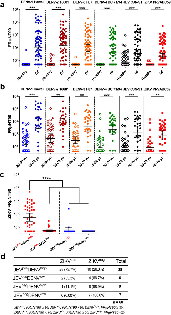
Fig. 1 Dengue-immune donors with prior JEV exposure have elevated ZIKV-neutralizing titers. a Neutralization antibody profile against the four serotypes of DENV, JEV, and ZIKV of healthy (n = 80) and dengue-infected febrile (DF, n = 60) individuals. Two-fold serially diluted plasma samples from healthy and DF individuals were evaluated for neutralization against the prototype strains of DENV-1 to -4 serotypes, JEV, and ZIKV using FRμNT90. Empty and shaded circles represent the FRμNT90 values of the healthy and DF individuals, respectively. b Comparison of the neutralization antibody profile of DF individuals against DENV-1 to -4 serotypes, JEV, and ZIKV prototype virions based on age classifications: 20–30 years old (n = 30) and 50–70 years old (n = 30). Empty and shaded circles represent the FRμNT90 values of the 20–30 and 50–70 age groups, respectively. c Assessment of the breadth of ZIKV neutralization among the four groups of donors dichotomized according to the presence (pos) or absence (neg) of protective or high neutralizing FRμNT90 titers against JEV and DENV, respectively. JEV-immune individuals with high (JEVposDENVhigh; n = 39) or low DENV immunity (JEVposDENVlow; n = 24) are represented by red filled or empty squares, respectively. JEV-negative donors with high (JEVnegDENVhigh ; n = 12) or low DENV (JEVnegDENVlow; n = 65) immunity are shown in blue filled or empty squares, respectively. d Effect of JEV and DENV immunity in DF individuals to ZIKV-neutralizing antibody titers. Fisher’s exact test was performed in n = 60 with a p-value of 0.000013. All data are representative of three independent experiments. In (a–c), the black solid horizontal bars represent the geometric mean titers (GMT) of samples with SD analyzed within each group. FRμNT90 titer <10 was represented with 5 for graphic display and statistical analysis. Significant differences were tested using one-way ANOVA followed by Tukey’s multiple comparisons post-test, with levels of significance defined by ***P < 0.0001; **P < 0.001. Each point shows the mean of data from two independent experiments. DENV dengue virus, JEV Japanese encephalitis virus, ZIKV Zika virus, FRμNT focus-reduction microneutralization test, VLP virus-like particle, ANOVA analysis of variance, SD standard deviation.
Fig. 1 Dengue-immune donors with prior JEV exposure have elevated ZIKV-neutralizing titers. a Neutralization antibody profile against the four serotypes of DENV, JEV, and ZIKV of healthy (n = 80) and dengue-infected febrile (DF, n = 60) individuals. Two-fold serially diluted plasma samples from healthy and DF individuals were evaluated for neutralization against the prototype strains of DENV-1 to -4 serotypes, JEV, and ZIKV using FRμNT90. Empty and shaded circles represent the FRμNT90 values of the healthy and DF individuals, respectively. b Comparison of the neutralization antibody profile of DF individuals against DENV-1 to -4 serotypes, JEV, and ZIKV prototype virions based on age classifications: 20–30 years old (n = 30) and 50–70 years old (n = 30). Empty and shaded circles represent the FRμNT90 values of the 20–30 and 50–70 age groups, respectively. c Assessment of the breadth of ZIKV neutralization among the four groups of donors dichotomized according to the presence (pos) or absence (neg) of protective or high neutralizing FRμNT90 titers against JEV and DENV, respectively. JEV-immune individuals with high (JEVposDENVhigh; n = 39) or low DENV immunity (JEVposDENVlow; n = 24) are represented by red filled or empty squares, respectively. JEV-negative donors with high (JEVnegDENVhigh ; n = 12) or low DENV (JEVnegDENVlow; n = 65) immunity are shown in blue filled or empty squares, respectively. d Effect of JEV and DENV immunity in DF individuals to ZIKV-neutralizing antibody titers. Fisher’s exact test was performed in n = 60 with a p-value of 0.000013. All data are representative of three independent experiments. In (a–c), the black solid horizontal bars represent the geometric mean titers (GMT) of samples with SD analyzed within each group. FRμNT90 titer <10 was represented with 5 for graphic display and statistical analysis. Significant differences were tested using one-way ANOVA followed by Tukey’s multiple comparisons post-test, with levels of significance defined by ***P < 0.0001; **P < 0.001. Each point shows the mean of data from two independent experiments. DENV dengue virus, JEV Japanese encephalitis virus, ZIKV Zika virus, FRμNT focus-reduction microneutralization test, VLP virus-like particle, ANOVA analysis of variance, SD standard deviation.CC BY · PubMed Central
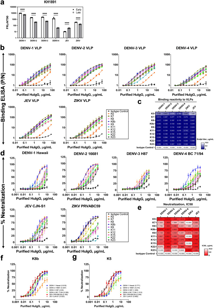
Fig. 2 Functional characterization of anti-ZIKV human monoclonal antibodies (huMAbs) from donor KH1891. a Neutralizing antibody titers of donor KH1891 against the prototype strains of the DENV serotypes 1–4, JEV, and ZIKV at less than 1 month and 18 months post-infection plasma collection, representing the early and late convalescent stages, respectively. Significant differences were tested using two-way ANOVA followed by Bonferroni post-tests from three independent experiments. ****P < 0.001. b–e Characterization of purified huMAbs, expressed as IgG1, isolated from donor KH1891. b The binding reactivity against virus-like particles (VLPs) and d in vitro neutralization activities of 10 purified huMAbs against DENV-1 to -4, JEV, and ZIKV were measured by ELISA and FRμNT, respectively. Each VLP was properly titrated to obtain equimolar concentrations per well before use in antigen-capture ELISAs. Each point shows the mean ± SD of data from two and three independent experiments for binding ELISA and microneutralization assays, respectively. A nonlinear curve was generated for all antibody dilution series for all of the assays. The corresponding antibody identities are shown in the box. c, e Heatmap of VLP-binding profile (shades of blue) (c) and of neutralization profile (shades of red) (e) of the 11 huMAbs against recombinantly produced DENV-1 to -4, JEV, and ZIKV VLPs and prototype strains of DENVs, JEV, and ZIKV virus particles, respectively. The heatmaps were generated based on the minimal binding concentration (or endpoint titer values) against each VLP and the IC50 values for each virion; exact numerical values expressed in μg/mL are shown in each box. Each huMAb was tested in duplicates for each of the binding ELISA assays, while FRμNT data were representative of three independent experiments. A purified human IgG1 was used as an isotype control in all assays. Data are representative of three independent experiments. f, g FRμNT50 (μg/mL) values of K8b (f) and K5 (g) for neutralization of the prototype strains of the four DENV serotypes, JEV, and ZIKV. Each point shows the mean ± SD of data from three independent experiments. The geometric mean inhibitory concentration (μg/mL) at 50% (IC50) is indicated in parenthesis. DENV dengue virus, JEV Japanese encephalitis virus, ZIKV Zika virus, ELISA enzyme-linked immunosorbent assay, FRμNT focus-reduction microneutralization test, VLP virus-like particle, ANOVA analysis of variance, SD standard deviation.
Fig. 2 Functional characterization of anti-ZIKV human monoclonal antibodies (huMAbs) from donor KH1891. a Neutralizing antibody titers of donor KH1891 against the prototype strains of the DENV serotypes 1–4, JEV, and ZIKV at less than 1 month and 18 months post-infection plasma collection, representing the early and late convalescent stages, respectively. Significant differences were tested using two-way ANOVA followed by Bonferroni post-tests from three independent experiments. ****P < 0.001. b–e Characterization of purified huMAbs, expressed as IgG1, isolated from donor KH1891. b The binding reactivity against virus-like particles (VLPs) and d in vitro neutralization activities of 10 purified huMAbs against DENV-1 to -4, JEV, and ZIKV were measured by ELISA and FRμNT, respectively. Each VLP was properly titrated to obtain equimolar concentrations per well before use in antigen-capture ELISAs. Each point shows the mean ± SD of data from two and three independent experiments for binding ELISA and microneutralization assays, respectively. A nonlinear curve was generated for all antibody dilution series for all of the assays. The corresponding antibody identities are shown in the box. c, e Heatmap of VLP-binding profile (shades of blue) (c) and of neutralization profile (shades of red) (e) of the 11 huMAbs against recombinantly produced DENV-1 to -4, JEV, and ZIKV VLPs and prototype strains of DENVs, JEV, and ZIKV virus particles, respectively. The heatmaps were generated based on the minimal binding concentration (or endpoint titer values) against each VLP and the IC50 values for each virion; exact numerical values expressed in μg/mL are shown in each box. Each huMAb was tested in duplicates for each of the binding ELISA assays, while FRμNT data were representative of three independent experiments. A purified human IgG1 was used as an isotype control in all assays. Data are representative of three independent experiments. f, g FRμNT50 (μg/mL) values of K8b (f) and K5 (g) for neutralization of the prototype strains of the four DENV serotypes, JEV, and ZIKV. Each point shows the mean ± SD of data from three independent experiments. The geometric mean inhibitory concentration (μg/mL) at 50% (IC50) is indicated in parenthesis. DENV dengue virus, JEV Japanese encephalitis virus, ZIKV Zika virus, ELISA enzyme-linked immunosorbent assay, FRμNT focus-reduction microneutralization test, VLP virus-like particle, ANOVA analysis of variance, SD standard deviation.CC BY · PubMed Central
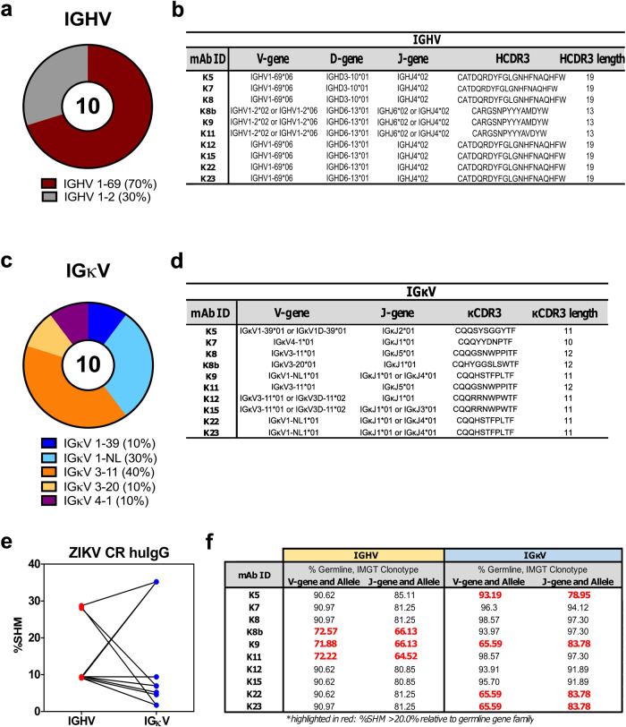
Fig. 3 Defining the human Ig genes and properties of broadly neutralizing ZIKV-CR huMAbs. a, c Pie plots showing immunoglobulin heavy (a) and kappa light chain (c) gene family utilization and distribution of ZIKV-CR neutralizing huMAbs isolated from KH1891. The proportion of IGHV and IGκV gene usage is reported beside the different color schemes. b, d Gene family assignments for the 10 purified IgG1 B cell clones from KH1891. IMGT was used to assign the germline reference sequence for IGHV (b) and IGκV (d) and the relative similarity with the germline clonotype expressed in %. e Relative percentage of somatic hypermutation (%SHM) in the paired IGHV and IGκV genes of ZIKV-CR huMAbs. f Comparison of the V-J genes and alleles of heavy and light chains of ZIKV-CR huMAbs. Values highlighted in red show increased SHM (>20%) relative to the germline gene family. ZIKV Zika virus.
Fig. 3 Defining the human Ig genes and properties of broadly neutralizing ZIKV-CR huMAbs. a, c Pie plots showing immunoglobulin heavy (a) and kappa light chain (c) gene family utilization and distribution of ZIKV-CR neutralizing huMAbs isolated from KH1891. The proportion of IGHV and IGκV gene usage is reported beside the different color schemes. b, d Gene family assignments for the 10 purified IgG1 B cell clones from KH1891. IMGT was used to assign the germline reference sequence for IGHV (b) and IGκV (d) and the relative similarity with the germline clonotype expressed in %. e Relative percentage of somatic hypermutation (%SHM) in the paired IGHV and IGκV genes of ZIKV-CR huMAbs. f Comparison of the V-J genes and alleles of heavy and light chains of ZIKV-CR huMAbs. Values highlighted in red show increased SHM (>20%) relative to the germline gene family. ZIKV Zika virus.CC BY · PubMed Central
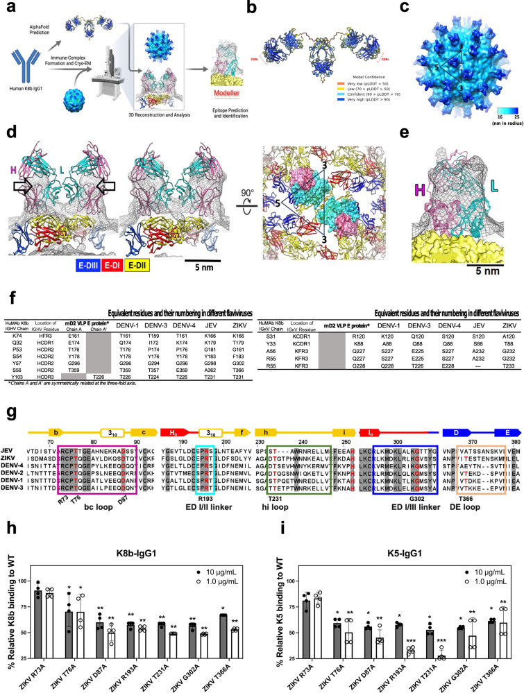
Fig. 4 Mapping of target flavivirus epitopes of huMAb K8b. a Structure-based strategy and workflow for the epitope mapping of K8b using mature DENV-2 virus-like particles (mD2VLP). AlphaFold was used to predict the three-dimensional (3D) structure of huMAb K8b expressed as IgG1. K8b-IgG1 was utilized to form an immune complex with mD2VLPs and 3D reconstructed by cryo-EM. The derived structure was further improved by MODELLER to predict the key epitopes and build epitope models. The antibody and apparatus icons were sourced icons and created with biorender.com. b AlphaFold model structure for K8b-IgG1 with coloration based on model confidence. The complementarity determining regions (CDRs) were indicated. c The cryo-EM structure of K8b-IgG1 and mD2VLP complex revealed the arrangement of E glycoprotein at T = 1 icosahedral symmetry. In the configuration, 60 F(ab) molecules (depicted in blue) were bound to 60 envelope proteins on the VLP surface (shown in cyan), while the Fc region was not resolved in the structure. d Fitting the predicted structure of K8b-IgG1 into cryo-EM density maps is presented, with contour levels of 3.7 σ (left) and 1.2 σ (middle). Two perspectives are shown: side view (d, left and middle) and top view (d, right). The separation of variable domains from the constant domain was clearly defined in the density map at higher contour levels, as indicated by the arrows. The heavy and light chains of K8b-IgG1 were shown in purple and cyan, respectively. Heavy and light chain pairs of K8b-IgG1 bound to E proteins at the inter-dimer surfaces. Densities of F(ab) pairs of K8b-IgG1 were shaded in gray. The residues on the E protein interacting with the heavy and light chains of F(ab) were shown by purple and cyan spheres, respectively. Envelope domains, E-DI, E-DII, and E-DIII, are colored in red, yellow, and blue, respectively. The triangle represents an asymmetric unit. e A flexible refinement of immune complex structure into cryo-EM map showing ten conformers of K8b-IgG1 predicted by MODELLER. The heavy (H) and light (L) chain variable domains are represented by pink and cyan ribbons, respectively. The mD2VLP is shown as a yellow surface, and the EM map (0.1 σ) is represented as black meshes. The cross-correlation function (CCF) is 0.72. For (d) and (e), a scale bar of 5 nm is shown. f Interacting or contact residues between the E proteins on an mD2VLP and the heavy or light chain of K8b-IgG1, including the equivalent residues (and their AA positions) in other flaviviruses, predicted by MODELLER. g A representative amino acid sequence alignment of the E proteins among prototype flaviviruses including selected DENV serotypes, JEV, and ZIKV using Clustal Omega and visualized by JalView 2.11.1.4. Potential binding sites of K8b are highlighted in boxes: bc loop (magenta), ED I/II linker (cyan), hi loop (green), ED I/III linker (blue), and DE loop (flesh). Residues in bold and in red are surface-exposed (solvent-accessible surface area, QSASA > 0.30). Sequence identities were normalized by aligned length, while the positions of the amino acids in E were labeled according to ZIKV PRVABC59. The shaded residues show 100% conservancy among the selected flavivirus genera. Above the sequences, the arrows indicate β-strands, rectangles represent the helices, and lines show the spanning loops and strands in the E structure. The E domains I, II, and III are represented by arrows and lines in red, yellow, and blue, respectively. h, i Binding ELISA reactivity profile of purified K8b-IgG1 (h) and K5-IgG1 (i) at 10 and 1.0 μg/mL antibody concentrations against seven single-site mutants relative to the wild-type ZIKV VLP in ELISA. The consensus amino acid residues in the bc loop (ZIKV R73, T76, and D87), ED I/II linker (ZIKV R193), hi loop (ZIKV T231), ED I/III linker (ZIKV G302), and ED III DE loop (ZIKV T366) were mutated to alanine. For (h, i), the statistical differences were determined by two-way ANOVA with Tukey’s multiple comparisons test relative to the binding reactivity to the ZIKV WT VLP, and are defined as significant at: *P < 0.05, **P < 0.001, and ***P < 0.0001. The binding reactivity ELISA data shown in (h, i) are representative of four independent experiments, expressed as geometric means ± SD. DENV dengue virus, JEV Japanese encephalitis virus, ZIKV Zika virus, ELISA enzyme-linked immunosorbent assay, FRμNT focus-reduction microneutralization test, ANOVA analysis of variance, SD standard deviation.
Fig. 4 Mapping of target flavivirus epitopes of huMAb K8b. a Structure-based strategy and workflow for the epitope mapping of K8b using mature DENV-2 virus-like particles (mD2VLP). AlphaFold was used to predict the three-dimensional (3D) structure of huMAb K8b expressed as IgG1. K8b-IgG1 was utilized to form an immune complex with mD2VLPs and 3D reconstructed by cryo-EM. The derived structure was further improved by MODELLER to predict the key epitopes and build epitope models. The antibody and apparatus icons were sourced icons and created with biorender.com. b AlphaFold model structure for K8b-IgG1 with coloration based on model confidence. The complementarity determining regions (CDRs) were indicated. c The cryo-EM structure of K8b-IgG1 and mD2VLP complex revealed the arrangement of E glycoprotein at T = 1 icosahedral symmetry. In the configuration, 60 F(ab) molecules (depicted in blue) were bound to 60 envelope proteins on the VLP surface (shown in cyan), while the Fc region was not resolved in the structure. d Fitting the predicted structure of K8b-IgG1 into cryo-EM density maps is presented, with contour levels of 3.7 σ (left) and 1.2 σ (middle). Two perspectives are shown: side view (d, left and middle) and top view (d, right). The separation of variable domains from the constant domain was clearly defined in the density map at higher contour levels, as indicated by the arrows. The heavy and light chains of K8b-IgG1 were shown in purple and cyan, respectively. Heavy and light chain pairs of K8b-IgG1 bound to E proteins at the inter-dimer surfaces. Densities of F(ab) pairs of K8b-IgG1 were shaded in gray. The residues on the E protein interacting with the heavy and light chains of F(ab) were shown by purple and cyan spheres, respectively. Envelope domains, E-DI, E-DII, and E-DIII, are colored in red, yellow, and blue, respectively. The triangle represents an asymmetric unit. e A flexible refinement of immune complex structure into cryo-EM map showing ten conformers of K8b-IgG1 predicted by MODELLER. The heavy (H) and light (L) chain variable domains are represented by pink and cyan ribbons, respectively. The mD2VLP is shown as a yellow surface, and the EM map (0.1 σ) is represented as black meshes. The cross-correlation function (CCF) is 0.72. For (d) and (e), a scale bar of 5 nm is shown. f Interacting or contact residues between the E proteins on an mD2VLP and the heavy or light chain of K8b-IgG1, including the equivalent residues (and their AA positions) in other flaviviruses, predicted by MODELLER. g A representative amino acid sequence alignment of the E proteins among prototype flaviviruses including selected DENV serotypes, JEV, and ZIKV using Clustal Omega and visualized by JalView 2.11.1.4. Potential binding sites of K8b are highlighted in boxes: bc loop (magenta), ED I/II linker (cyan), hi loop (green), ED I/III linker (blue), and DE loop (flesh). Residues in bold and in red are surface-exposed (solvent-accessible surface area, QSASA > 0.30). Sequence identities were normalized by aligned length, while the positions of the amino acids in E were labeled according to ZIKV PRVABC59. The shaded residues show 100% conservancy among the selected flavivirus genera. Above the sequences, the arrows indicate β-strands, rectangles represent the helices, and lines show the spanning loops and strands in the E structure. The E domains I, II, and III are represented by arrows and lines in red, yellow, and blue, respectively. h, i Binding ELISA reactivity profile of purified K8b-IgG1 (h) and K5-IgG1 (i) at 10 and 1.0 μg/mL antibody concentrations against seven single-site mutants relative to the wild-type ZIKV VLP in ELISA. The consensus amino acid residues in the bc loop (ZIKV R73, T76, and D87), ED I/II linker (ZIKV R193), hi loop (ZIKV T231), ED I/III linker (ZIKV G302), and ED III DE loop (ZIKV T366) were mutated to alanine. For (h, i), the statistical differences were determined by two-way ANOVA with Tukey’s multiple comparisons test relative to the binding reactivity to the ZIKV WT VLP, and are defined as significant at: *P < 0.05, **P < 0.001, and ***P < 0.0001. The binding reactivity ELISA data shown in (h, i) are representative of four independent experiments, expressed as geometric means ± SD. DENV dengue virus, JEV Japanese encephalitis virus, ZIKV Zika virus, ELISA enzyme-linked immunosorbent assay, FRμNT focus-reduction microneutralization test, ANOVA analysis of variance, SD standard deviation.CC BY · PubMed Central
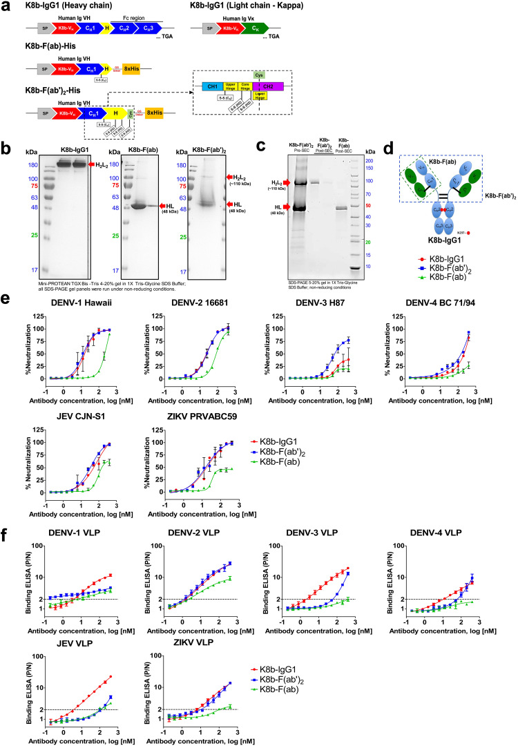
Fig. 5 Generation and functional characterization of purified K8b-IgG1, K8b-F(ab’)2, and K8b-F(ab). a Schematic representation of the designed gene constructs coding for full-length human IgG1, F(ab’)2, and F(ab) encoding the Ig variable heavy chain genes of K8b (K8b-VH), and hence termed as pVCHIg-hG1-K8b, pVCHIg-K8b-F(ab’)2-His and pVCHIg-K8b-F(ab)-His, respectively. The schematic diagram of the expression vector, pVCLIg-hκ, encoding the variable Ig kappa light chain domain of K8b (K8b-Vκ) is also shown in the upper right panel. Each of the molecules was expressed via co-transfection using the kappa light chain expressing plasmid, pVCLIg-hκ-K8b. Inset shows the detailed schematic map of the human IgƔ constant domains 1 (CH1) and 2 (CH2), including the IgG1 hinge regions and the actual location where cysteine (Cys) was introduced for increased thermal stability. CH1–3, human IgƔ constant domains 1–3; Cκ, human Igκ constant domain; 8x-His, poly-Histidine tag; SP, signal peptide; H, hinge; TGA, stop codon are highlighted. b SDS-PAGE of purified human monoclonal antibody, K8b, expressed as full-length IgG1, F(ab), and F(ab’)2 molecules visualized under nonreducing conditions. Red arrow marks show the dimeric species of K8b-IgG1 at 150 kDa, monovalent K8b-F(ab) at 48 kDa, and the bivalent K8b-F(ab’)2 at 110 kDa. The molecular mass of the protein marker is indicated on the left side, lane 1, for each gel panel. c Electrophoretic mobility of purified K8b-F(ab’)2 after size-exclusion chromatography (SEC) showing the separation of K8b-F(ab’)2 and K8b-F(ab) molecules from the pooled pre-SEC K8b-F(ab’)2 fractions in lane 1. Lanes 2 and 3 show the non-reduced form of K8b-F(ab’)2 post-SEC and lane 4 shows the K8b-F(ab) molecules post-SEC purity separation. The molecular mass of the protein marker is indicated on the right side, lane 5. d Cartoon representation of K8b IgG1, F(ab), and F(ab’)2 molecules. e Neutralization profile of K8b expressed as full-length IgG1, F(ab), and F(ab’)2. Prototype virus strains of DENV-1 to -4, JEV, and ZIKV were used for the FRμNT. f Binding profile of K8b expressed as full-length IgG1, F(ab), and F(ab’)2. Each VLP was properly titrated to obtain equimolar concentrations per well before use in antigen-capture ELISAs. In (e) and (f), each point shows the mean ± SD of data from two independent experiments. A nonlinear curve was generated for all antibody dilution series for all of the assays. DENV dengue virus, JEV Japanese encephalitis virus, ZIKV Zika virus, ELISA enzyme-linked immunosorbent assay, FRμNT focus-reduction microneutralization test, SD standard deviation, SASA solvent-accessible surface area.
Fig. 5 Generation and functional characterization of purified K8b-IgG1, K8b-F(ab’)2, and K8b-F(ab). a Schematic representation of the designed gene constructs coding for full-length human IgG1, F(ab’)2, and F(ab) encoding the Ig variable heavy chain genes of K8b (K8b-VH), and hence termed as pVCHIg-hG1-K8b, pVCHIg-K8b-F(ab’)2-His and pVCHIg-K8b-F(ab)-His, respectively. The schematic diagram of the expression vector, pVCLIg-hκ, encoding the variable Ig kappa light chain domain of K8b (K8b-Vκ) is also shown in the upper right panel. Each of the molecules was expressed via co-transfection using the kappa light chain expressing plasmid, pVCLIg-hκ-K8b. Inset shows the detailed schematic map of the human IgƔ constant domains 1 (CH1) and 2 (CH2), including the IgG1 hinge regions and the actual location where cysteine (Cys) was introduced for increased thermal stability. CH1–3, human IgƔ constant domains 1–3; Cκ, human Igκ constant domain; 8x-His, poly-Histidine tag; SP, signal peptide; H, hinge; TGA, stop codon are highlighted. b SDS-PAGE of purified human monoclonal antibody, K8b, expressed as full-length IgG1, F(ab), and F(ab’)2 molecules visualized under nonreducing conditions. Red arrow marks show the dimeric species of K8b-IgG1 at 150 kDa, monovalent K8b-F(ab) at 48 kDa, and the bivalent K8b-F(ab’)2 at 110 kDa. The molecular mass of the protein marker is indicated on the left side, lane 1, for each gel panel. c Electrophoretic mobility of purified K8b-F(ab’)2 after size-exclusion chromatography (SEC) showing the separation of K8b-F(ab’)2 and K8b-F(ab) molecules from the pooled pre-SEC K8b-F(ab’)2 fractions in lane 1. Lanes 2 and 3 show the non-reduced form of K8b-F(ab’)2 post-SEC and lane 4 shows the K8b-F(ab) molecules post-SEC purity separation. The molecular mass of the protein marker is indicated on the right side, lane 5. d Cartoon representation of K8b IgG1, F(ab), and F(ab’)2 molecules. e Neutralization profile of K8b expressed as full-length IgG1, F(ab), and F(ab’)2. Prototype virus strains of DENV-1 to -4, JEV, and ZIKV were used for the FRμNT. f Binding profile of K8b expressed as full-length IgG1, F(ab), and F(ab’)2. Each VLP was properly titrated to obtain equimolar concentrations per well before use in antigen-capture ELISAs. In (e) and (f), each point shows the mean ± SD of data from two independent experiments. A nonlinear curve was generated for all antibody dilution series for all of the assays. DENV dengue virus, JEV Japanese encephalitis virus, ZIKV Zika virus, ELISA enzyme-linked immunosorbent assay, FRμNT focus-reduction microneutralization test, SD standard deviation, SASA solvent-accessible surface area.CC BY · PubMed Central
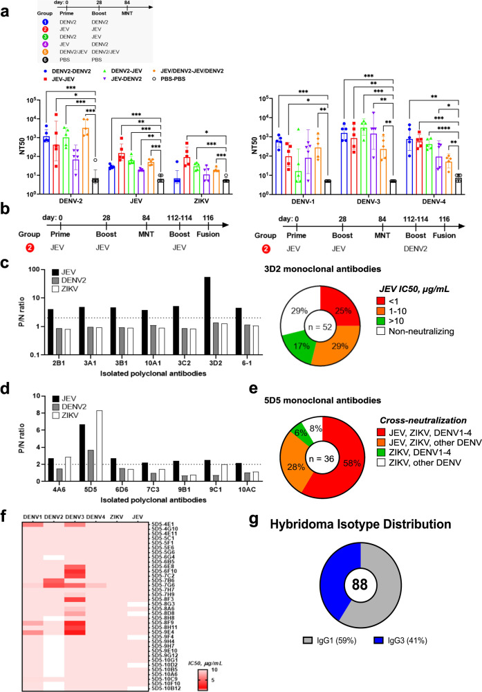
Fig. 6 Neutralization antibody profile of murine sera and murine monoclonal antibodies (mAbs) generated from various DENV-2 and JEV VLP prime-boost immunizations. a BALB/c mice (n = 5) were primed and boosted at 0 and 28 days, respectively, with various combinations of mature DENV-2 VLP (mD2VLP) and JEV VLP antigens. Mice were bled at 84 days post-vaccination, and individual serum specimens were evaluated for 50% focus-reduction microneutralization (FRµNT50) activity against DENV, JEV, and ZIKV. The bar plots show GMT ± SD for each group. The p values (*P < 0.05, **P < 0.01, ***P < 0.001, ****P < 0.0001) were determined on log-transformed data using one-way ANOVA with Tukey’s multiple comparisons post-test. b Schedule of boost and myeloma fusion experiments performed in representative mice from the homologous JEV VLP prime-boost immunization. c, d Representative mice (JEV FRµNT50 > 120) from JEV prime-boost immunization were further boosted for three consecutive times (112–114 days post-vaccination) with JEV VLP (c) or mD2VLP (d) and the splenocytes were harvested two days after the last boost and then individually fused with myeloma cells for hybridoma generation. Culture supernatants from selected polyclonal hybridomas were tested for positive VLP ELISA reactivity (P/N ratio ≥ 2) and further subcloned by limiting dilution to isolate monoclonal cells. c 71% of the 52 JEV-specific monoclones isolated from 3D2 polyclone secreted JEV-neutralizing mAbs with varying potencies at 50% inhibitory concentration (IC50, µg/mL) of unpurified supernatant. c Right: Pie plot showing murine monoclones selected from polyclonal hybridomas and the distribution of 52 type-specific 3D2 mAbs with varying breadth of neutralization. d A total of 36 monoclones were isolated from 5D5 polyclone. Percentages represent the proportions of secreted mAbs according to their breadth of cross-neutralization within the different serocomplexes. e Pie plot showing murine monoclones selected from polyclonal hybridomas and the distribution of 36 cross-neutralizing 5D5 monoclonal antibodies. The relative proportion of monoclones with varying breadth of neutralization is reported beside the different color schemes. f Heatmap of DENV, JEV, and ZIKV IC50 (µg/mL, unpurified supernatant) neutralization titers of the 36 cross-reactive mAbs. Data are representative of two independent experiments. g Distribution of immunoglobulin (IgG) isotypes identified from hybridoma screening. DENV dengue virus, JEV Japanese encephalitis virus, ZIKV Zika virus, VLP virus-like particle, ELISA enzyme-linked immunosorbent assay, GMT geometric mean titer, ANOVA analysis of variance, SD standard deviation.
Fig. 6 Neutralization antibody profile of murine sera and murine monoclonal antibodies (mAbs) generated from various DENV-2 and JEV VLP prime-boost immunizations. a BALB/c mice (n = 5) were primed and boosted at 0 and 28 days, respectively, with various combinations of mature DENV-2 VLP (mD2VLP) and JEV VLP antigens. Mice were bled at 84 days post-vaccination, and individual serum specimens were evaluated for 50% focus-reduction microneutralization (FRµNT50) activity against DENV, JEV, and ZIKV. The bar plots show GMT ± SD for each group. The p values (*P < 0.05, **P < 0.01, ***P < 0.001, ****P < 0.0001) were determined on log-transformed data using one-way ANOVA with Tukey’s multiple comparisons post-test. b Schedule of boost and myeloma fusion experiments performed in representative mice from the homologous JEV VLP prime-boost immunization. c, d Representative mice (JEV FRµNT50 > 120) from JEV prime-boost immunization were further boosted for three consecutive times (112–114 days post-vaccination) with JEV VLP (c) or mD2VLP (d) and the splenocytes were harvested two days after the last boost and then individually fused with myeloma cells for hybridoma generation. Culture supernatants from selected polyclonal hybridomas were tested for positive VLP ELISA reactivity (P/N ratio ≥ 2) and further subcloned by limiting dilution to isolate monoclonal cells. c 71% of the 52 JEV-specific monoclones isolated from 3D2 polyclone secreted JEV-neutralizing mAbs with varying potencies at 50% inhibitory concentration (IC50, µg/mL) of unpurified supernatant. c Right: Pie plot showing murine monoclones selected from polyclonal hybridomas and the distribution of 52 type-specific 3D2 mAbs with varying breadth of neutralization. d A total of 36 monoclones were isolated from 5D5 polyclone. Percentages represent the proportions of secreted mAbs according to their breadth of cross-neutralization within the different serocomplexes. e Pie plot showing murine monoclones selected from polyclonal hybridomas and the distribution of 36 cross-neutralizing 5D5 monoclonal antibodies. The relative proportion of monoclones with varying breadth of neutralization is reported beside the different color schemes. f Heatmap of DENV, JEV, and ZIKV IC50 (µg/mL, unpurified supernatant) neutralization titers of the 36 cross-reactive mAbs. Data are representative of two independent experiments. g Distribution of immunoglobulin (IgG) isotypes identified from hybridoma screening. DENV dengue virus, JEV Japanese encephalitis virus, ZIKV Zika virus, VLP virus-like particle, ELISA enzyme-linked immunosorbent assay, GMT geometric mean titer, ANOVA analysis of variance, SD standard deviation.CC BY · PubMed Central

Verification

Claims the paper makes

The claims and statistics of this paper haven’t been checked yet.

Run the check — free, no account needed. It reads the open-access text and checks the sources it cites.

A few free checks a day · papers already verified open instantly · results are public

Self-check

Percentages, sums and reported differences re-computed from the paper’s own numbers.

The self-check needs the paper’s full text.

Citations

Cites

Research works printed but not linked: 111

  1. Pierson, T. C. & Diamond, M. S. The continued threat of emerging flaviviruses. Nat. Microbiol. 5, 796–812 (2020). · DOI
  2. Kuno, G., Chang, G.-J. J., Tsuchiya, R., Karabatsos, N. & Cropp, C. B. Phylogeny of the genus flavivirus. J. Virol. 72, 73–83 (1998). · DOI
  3. Heinz, F. X. & Stiasny, K. Flaviviruses and flavivirus vaccines. Vaccine 30, 4301–4306 (2012). · DOI
  4. Mackenzie, J. S., Gubler, D. J. & Petersen, L. R. Emerging flaviviruses: the spread and resurgence of Japanese encephalitis, West Nile and dengue viruses. Nat. Med. 10, S98–S109 (2004). · DOI
  5. deAlwis, R. et al. Identification of human neutralizing antibodies that bind to complex epitopes on dengue virions. Proc. Natl Acad. Sci. USA. 109, 7439–7444 (2012). · DOI
  6. Collins, M. H. et al. Lack of durable cross-neutralizing antibodies against Zika virus from dengue virus infection. Emerg. Infect. Dis. 23, 773–781 (2017). · DOI
  7. Priyamvada, L. et al. Human antibody responses after dengue virus infection are highly cross-reactive to Zika virus. Proc. Natl Acad. Sci. USA. 113, 7852–7857 (2016). · DOI
  8. Dejnirattisai, W. et al. Dengue virus sero-cross-reactivity drives antibody-dependent enhancement of infection with Zika virus. Nat. Immunol. 17, 1102–1108 (2016). · DOI
  9. Robbiani, D. F. et al. Recurrent potent human neutralizing antibodies to Zika virus in Brazil and Mexico. Cell 169, 597–609.e11 (2017). · DOI
  10. Xu, M. et al. Plasmablasts generated during repeated dengue infection are virus glycoprotein–specific and bind to multiple virus serotypes. J. Immunol. 189, 5877–5885 (2012). · DOI
  11. Perng, G. C. et al. Seroprevalence of Zika and dengue virus antibodies among migrant workers, Taiwan, 2017. Emerg. Infect. Dis. 25, 2017–2019 (2019). · DOI
  12. Chien, Y. W. et al. Low seroprevalence of Zika virus infection among adults in Southern Taiwan. BMC Infect. Dis. 19, 884 (2019). · DOI
  13. Huang, A. S. E., Shu, P. Y. & Yang, C. H. A new reportable disease is born: Taiwan Centers for Disease Control’s response to emerging Zika virus infection. J. Formos. Med. Assoc. 115, 223–225 (2016). · DOI
  14. Hsu, L. C. et al. The incidence of Japanese encephalitis in Taiwan—a population-based study. PLoS Negl. Trop. Dis. 8, e3030 (2014). · DOI
  15. Wu, Y. C. et al. The epidemiology of Japanese encephalitis on Taiwan during 1966-1997. Am. J. Trop. Med. Hyg. 61, 78–84 (1999). · DOI
  16. Chang, C. J., Chen, C. S., Tien, C. J. & Lu, M. R. Epidemiological, clinical and climatic characteristics of dengue fever in Kaohsiung City, Taiwan with implication for prevention and control. PLoS One 13, 1–15 (2018).
  17. King, C. et al. Major epidemics of dengue in Taiwan in 1981-2000: related to intensive virus activities in Asia. Dengue Bull. 24, 1–10 (2000).
  18. Andrade, P. et al. Primary and secondary dengue virus infections elicit similar memory b-cell responses, but breadth to other serotypes and cross-reactivity to zika virus is higher in secondary dengue. J. Infect. Dis. 222, 590–600 (2020). · DOI
  19. Beltramello, M. et al. The human immune response to dengue virus is dominated by highly cross-reactive antibodies endowed with neutralizing and enhancing activity. Cell Host Microbe 8, 271–283 (2010). · DOI
  20. Montoya, M. et al. Longitudinal analysis of antibody cross-neutralization following Zika virus and dengue virus infection in Asia and the Americas. J. Infect. Dis. 218, 536–545 (2018). · DOI
  21. Huang, J. et al. Isolation of human monoclonal antibodies from peripheral blood B cells. Nat. Protoc. 8, 1907–1915 (2013). · DOI
  22. Durham, N. D. et al. Broadly neutralizing human antibodies against dengue virus identified by single B cell transcriptomics. Elife 8, 1–29 (2019). · DOI
  23. Wrammert, J. et al. Rapid cloning of high-affinity human monoclonal antibodies against influenza virus. Nature 453, 667–671 (2008). · DOI
  24. Horns, F., Dekker, C. L. & Quake, S. R. Memory B cell activation, broad anti-influenza antibodies, and bystander activation revealed by single-cell transcriptomics. Cell Rep. 30, 905–913.e6 (2020). · DOI
  25. Methot, S. P. & DiNoia, J. M. Molecular mechanisms of somatic hypermutation and class switch recombination. Adv. Immunol. 133, 37–87 (2017). · DOI
  26. Shen, W. F. et al. Epitope resurfacing on dengue virus-like particle vaccine preparation to induce broad neutralizing antibody. Elife 7, 1–24 (2018). · DOI
  27. Saron, W. A. A. et al. Flavivirus serocomplex cross-reactive immunity is protective by activating heterologous memory CD4 T cells. Sci. Adv. 4, 1–15 (2018). · DOI
  28. Lima, N. S. et al. Pre-existing immunity to Japanese encephalitis virus alters CD4 T cell responses to Zika virus inactivated vaccine. Front. Immunol. 12, 1–14 (2021). · DOI
  29. Kuadkitkan, A., Wikan, N., Sornjai, W. & Smith, D. R. Zika virus and microcephaly in Southeast Asia: a cause for concern?. J. Infect. Public Health 13, 11–15 (2020). · DOI
  30. Duong, V., Dussart, P. & Buchy, P. Zika virus in Asia. Int. J. Infect. Dis. 54, 121–128 (2017). · DOI
  31. Wang, W. H. et al. An epidemiological survey of the current status of Zika and the immune interaction between dengue and Zika infection in Southern Taiwan. Int. J. Infect. Dis. 93, 151–159 (2020). · DOI
  32. Gordon, A. et al. Prior dengue virus infection and risk of Zika: a pediatric cohort in Nicaragua. PLoS Med. 16, 1–16 (2019). · DOI
  33. Katzelnick, L. C. et al. Zika virus infection enhances future risk of severe dengue disease. Science 369, 1123–1128 (2020). · DOI
  34. Rodriguez-Barraquer, I. et al. Impact of preexisting dengue immunity on Zika virus emergence in a dengue endemic region. Science 363, 607–610 (2019). · DOI
  35. Katzelnick, L. C. et al. Dengue and Zika virus infections in children elicit cross-reactive protective and enhancing antibodies that persist long term. Sci. Transl. Med. 13, eabg9478 (2021). · DOI
  36. Bardina, S. V. et al. Enhancement of Zika virus pathogenesis by preexisting antiflavivirus immunity. Science 356, 175–180 (2017). · DOI
  37. McCracken, M. K. et al. Impact of prior flavivirus immunity on Zika virus infection in rhesus macaques. PLoS Pathog. 13, 1–22 (2017). · DOI
  38. Hou, B., Chen, H., Gao, N. & An, J. Cross-reactive immunity among five medically important mosquito-borne flaviviruses related to human diseases. Viruses 14, 1–13 (2022). · DOI
  39. Castanha, P. M. S. et al. Force of infection of dengue serotypes in a population-based study in the northeast of Brazil. Epidemiol. Infect. 141, 1080–1088 (2013). · DOI
  40. He, X. et al. The effects of Japanese encephalitis virus antibodies on Zika virus infection. Med. Microbiol. Immunol. 209, 177–188 (2020). · DOI
  41. Rodriguez-Barraquer, I. et al. From re-emergence to hyperendemicity: the natural history of the dengue epidemic in Brazil. PLoS Negl. Trop. Dis. 5, 1–7 (2011). · DOI
  42. Smith, S. A. et al. Persistence of circulating memory B cell clones with potential for dengue virus disease enhancement for decades following infection. J. Virol. 86, 2665–2675 (2012). · DOI
  43. Sierra, B. et al. Secondary heterologous dengue infection risk: disequilibrium between immune regulation and inflammation? Cell. Immunol. 262, 134–140 (2010). · DOI
  44. Halstead, S. B. Neutralization and antibody-dependent enhancement of dengue viruses. Adv. Virus Res. 60, 421–467 (2003). · DOI
  45. Zhang, W. et al. The pre-existing cellular immunity to Japanese encephalitis virus heterotypically protects mice from Zika virus infection. Sci. Bull. 65, 402–409 (2020). · DOI
  46. Konishi, E., Yamaoka, M., Khin-Sane-Win, Kurane, I. & Mason, P. W. Induction of protective immunity against Japanese encephalitis in mice by immunization with a plasmid encoding Japanese encephalitis virus premembrane and envelope genes. J. Virol. 72, 4925–4930 (1998). · DOI
  47. Sevvana, M. & Kuhn, R. J. Mapping the diverse structural landscape of the flavivirus antibody repertoire. Curr. Opin. Virol. 45, 51–64 (2020). · DOI
  48. Lorenz, C. & Chiaravalloti-Neto, F. Why are there no human West Nile virus outbreaks in South America? Lancet Reg. Health Am. 12, 100276 (2022).
  49. Victora, G. D. & Nussenzweig, M. C. Germinal centers. Annu. Rev. Immunol. 30, 429–457 (2012). · DOI · Paper page
  50. Feroldi, E. et al. Memory immune response and safety of a booster dose of Japanese encephalitis chimeric virus vaccine (JE-CV) in JE-CV-primed children. Hum. Vaccin. Immunother. 9, 889–897 (2013). · DOI
  51. Sohn, Y. M., Tandan, J. B., Yoksan, S., Ji, M. & Ohrr, H. A 5-year follow-up of antibody response in children vaccinated with single dose of live attenuated SA14-14-2 Japanese encephalitis vaccine: Immunogenicity and anamnestic responses. Vaccine 26, 1638–1643 (2008). · DOI
  52. Bosco-Lauth, A., Mason, G. & Bowen, R. Pathogenesis of Japanese encephalitis virus infection in a golden hamster model and evaluation of flavivirus cross-protective immunity. Am. J. Trop. Med. Hyg. 84, 727–732 (2011). · DOI
  53. Chen, D. et al. Japanese encephalitis virus-primed CD8+ T cells prevent antibody-dependent enhancement of Zika virus pathogenesis. J. Exp. Med. 217, e20192152 (2020). · DOI
  54. Larena, M., Prow, N. A., Hall, R. A., Petrovsky, N. & Lobigs, M. JE-ADVAX vaccine protection against Japanese encephalitis virus mediated by memory B cells in the absence of CD8 + T cells and pre-exposure neutralizing antibody. J. Virol. 87, 4395–4402 (2013). · DOI
  55. Wong, R. et al. Affinity-restricted memory B cells dominate recall responses to heterologous flaviviruses. Immunity 53, 1078–1094.e7 (2020). · DOI
  56. Shaw, R. H. et al. Effect of priming interval on reactogenicity, peak immunological response, and waning after homologous and heterologous COVID-19 vaccine schedules: exploratory analyses of Com-COV, a randomised control trial. Lancet. Respir. Med. 10, 1049–1060 (2022).
  57. Elliott, T. et al. Enhanced immune responses following heterologous vaccination with self-amplifying RNA and mRNA COVID-19 vaccines. PLoS Pathog. 18, 1–20 (2022). · DOI
  58. Moyo-Gwete, T. et al. Enhanced neutralization potency of an identical HIV neutralizing antibody expressed as different isotypes is achieved through genetically distinct mechanisms. Sci. Rep. 12, 1–9 (2022). · DOI
  59. Vidarsson, G., Dekkers, G. & Rispens, T. IgG subclasses and allotypes: from structure to effector functions. Front. Immunol. 5, 1–17 (2014). · DOI · Paper page
  60. Richardson, S. I. et al. IgG3 enhances neutralization potency and Fc effector function of an HIV V2-specific broadly neutralizing antibody. PLoS Pathog. 15, 1–25 (2019). · DOI
  61. Onodera, T. et al. A SARS-CoV-2 antibody broadly neutralizes SARS-related coronaviruses and variants by coordinated recognition of a virus-vulnerable site. Immunity 54, 2385–2398.e10 (2021). · DOI
  62. Dejnirattisai, W. et al. A new class of highly potent, broadly neutralizing antibodies isolated from viremic patients infected with dengue virus. Nat. Immunol. 16, 170–177 (2015). · DOI
  63. Rogers, T. F. et al. Zika virus activates de novo and cross-reactive memory B cell responses in dengue-experienced donors. Sci. Immunol. 2, eaan6809 (2017). · DOI
  64. Dussupt, V. et al. Potent Zika and dengue cross-neutralizing antibodies induced by Zika vaccination in a dengue-experienced donor. Nat. Med. 26, 228–235 (2020). · DOI
  65. Xu, M. et al. A potent neutralizing antibody with therapeutic potential against all four serotypes of dengue virus. npj Vaccines 2, 2 (2017). · DOI
  66. Lubow, J. et al. Single B cell transcriptomics identifies multiple isotypes of broadly neutralizing antibodies against flaviviruses. PLoS Pathog. 19, e1011722 (2023). · DOI
  67. Smith, S. A. et al. The potent and broadly neutralizing human dengue virus-specific monoclonal antibody 1C19 reveals a unique cross-reactive epitope on the bc loop of domain II of the envelope protein. MBio 4, 1–12 (2013). · DOI
  68. Zhao, H. et al. Mechanism of differential Zika and dengue virus neutralization by a public antibody lineage targeting the DIII lateral ridge. J. Exp. Med. 217, 1–15 (2020). · DOI
  69. Niu, X. et al. Convalescent patient-derived monoclonal antibodies targeting different epitopes of E protein confer protection against Zika virus in a neonatal mouse model. Emerg. Microbes Infect. 8, 749–759 (2019). · DOI
  70. Qu, P. et al. A new class of broadly neutralizing antibodies that target the glycan loop of Zika virus envelope protein. Cell Discov. 6, 5 (2020). · DOI
  71. Fibriansah, G. et al. A potent anti-dengue human antibody preferentially recognizes the conformation of E protein monomers assembled on the virus surface. EMBO Mol. Med. 6, 358–371 (2014). · DOI
  72. Barba-Spaeth, G. et al. Structural basis of potent Zika-dengue virus antibody cross-neutralization. Nature 536, 48–53 (2016). · DOI
  73. Phad, G. E. et al. Clonal structure, stability and dynamics of human memory B cells and circulating plasmablasts. Nat. Immunol. 23, 1–10 (2022). · DOI
  74. Appanna, R. et al. Plasmablasts during acute dengue infection represent a small subset of a broader virus-specific memory B cell pool. EBioMedicine 12, 178–188 (2016). · DOI
  75. D’Angelo, S. et al. Many routes to an antibody heavy-chain CDR3: necessary, yet insufficient, for specific binding. Front. Immunol. 9, 1–13 (2018). · DOI
  76. Wang, Q. et al. Molecular determinants of human neutralizing antibodies isolated from a patient infected with Zika virus. Sci. Transl. Med. 8, 369ra179 (2016). · DOI
  77. Long, F. et al. Structural basis of a potent human monoclonal antibody against Zika virus targeting a quaternary epitope. Proc. Natl Acad. Sci. USA. 116, 1591–1596 (2019). · DOI
  78. Edeling, M. A. et al. Potent dengue virus neutralization by a therapeutic antibody with low monovalent affinity requires bivalent engagement. PLoS Pathog. 10, e1004072 (2014). · DOI
  79. Sharma, A. et al. The epitope arrangement on flavivirus particles contributes to Mab C10’s extraordinary neutralization breadth across Zika and dengue viruses. Cell 184, 6052–6066.e18 (2021). · DOI
  80. Guzman, M. G. et al. Neutralizing antibodies after infection with dengue 1 virus. Emerg. Infect. Dis. 13, 282–286 (2007). · DOI
  81. Turtle, L. et al. Low population Japanese encephalitis virus (JEV) seroprevalence in Udayapur district, Nepal, three years after a JE vaccination programme: a case for further catch up campaigns? PLoS Negl. Trop. Dis. 13, 1–19 (2019). · DOI
  82. Galula, J. U., Shen, W.-F., Chuang, S.-T., Chang, G.-J. J. & Chao, D.-Y. Virus-like particle secretion and genotype-dependent immunogenicity of dengue virus serotype 2 DNA vaccine. J. Virol. 88, 10813–10830 (2014). · DOI
  83. Galula, J. U., Salem, G. M., Destura, R. V., Remenyi, R., & Chao, D. Y. Comparable accuracies of nonstructural protein 1-and envelope protein-based enzyme-linked immunosorbent assays in detecting anti-dengue immunoglobulin g antibodies. Diagnostics 11, 1–17 (2021). · DOI
  84. VonBoehmer, L. et al. Sequencing and cloning of antigen-specific antibodies from mouse memory B cells. Nat. Protoc. 11, 1908–1923 (2016). · DOI
  85. Giudicelli, V., Chaume, D. & Lefranc, M. P. IMGT/V-QUEST, an integrated software program for immunoglobulin and T cell receptor V-J and V-D-J rearrangement analysis. Nucleic Acids Res. 32, 435–440 (2004). · DOI
  86. Li, S. et al. IMGT/HighV QUEST paradigm for T cell receptor IMGT clonotype diversity and next generation repertoire immunoprofiling. Nat. Commun. 4, 2333 (2013). · DOI
  87. Lefranc, M. P. et al. IMGT R, the international ImMunoGeneTics information system R 25 years on. Nucleic Acids Res. 43, D413–D422 (2015). · DOI
  88. Pommié, C., Levadoux, S., Sabatier, R., Lefranc, G. & Lefranc, M. P. IMGT standardized criteria for statistical analysis of immunoglobulin V-Region amino acid properties. J. Mol. Recognit. 17, 17–32 (2004). · DOI
  89. Chang, G. J. J. et al. Enhancing biosynthesis and secretion of premembrane and envelope proteins by the chimeric plasmid of dengue virus type 2 and Japanese encephalitis virus. Virology 306, 170–180 (2003). · DOI
  90. Chang, G. J. J., Davis, B. S., Stringfield, C. & Lutz, C. Prospective immunization of the endangered California condors (Gymnogyps californianus) protects this species from lethal West Nile virus infection. Vaccine 25, 2325–2330 (2007). · DOI
  91. Ludtke, S. J., Baldwin, P. R. & Chiu, W. EMAN: semiautomated software for high-resolution single-particle reconstructions. J. Struct. Biol. 128, 82–97 (1999). · DOI
  92. Tang, G. et al. EMAN2: an extensible image processing suite for electron microscopy. J. Struct. Biol. 157, 38–46 (2007). · DOI
  93. Pettersen, E. F. et al. UCSF ChimeraX: structure visualization for researchers, educators, and developers. Protein Sci. 30, 70–82 (2021). · DOI
  94. Honegger, A. & Plückthun, A. Yet another numbering scheme for immunoglobulin variable domains: an automatic modeling and analysis tool. J. Mol. Biol. 309, 657–670 (2001). · DOI
  95. Dunbar, J. & Deane, C. M. ANARCI: antigen receptor numbering and receptor classification. Bioinformatics 32, 298–300 (2016). · DOI
  96. Zhang, S. et al. ProDy 2.0: increased scale and scope after 10 years of protein dynamics modelling with Python. Bioinformatics 37, 3657–3659 (2021). · DOI
  97. Eswar, N. et al. Comparative protein structure modeling using MODELLER. Curr. Protoc. Protein Sci. 50, Chapter 2:Unit 2.9 (2007). · DOI
  98. Yan, R. et al. Structural basis for bivalent binding and inhibition of SARS-CoV-2 infection by human potent neutralizing antibodies. Cell Res. 31, 517–525 (2021). · DOI
  99. Harris, L. J., Larson, S. B., Hasel, K. W. & McPherson, A. Refined structure of an intact IgG2a monoclonal antibody. Biochemistry 36, 1581–1597 (1997). · DOI
  100. Ritchie, D. W., Venkatraman, V. & Ultra-fast, F. F. T. protein docking on graphics processors. Bioinformatics 26, 2398–2405 (2010). · DOI
  101. Topf, M. et al. Protein structure fitting and refinement guided by cryo-EM density. Structure 16, 295–307 (2008). · DOI
  102. Krissinel, E. & Henrick, K. Inference of macromolecular assemblies from crystalline. State J. Mol. Biol. 372, 774–797 (2007). · DOI
  103. Laskowski, R. A. et al. PDBsum: a web-based database of summaries and analyses of all PDB structures. Trends Biochem. Sci. 22, 488–490 (1997). · DOI
  104. Dai, L. et al. Structures of the Zika virus envelope protein and its complex with a flavivirus broadly protective antibody. Cell Host Microbe 19, 696–704 (2016). · DOI
  105. Deng, Y. Q. et al. A broadly flavivirus cross-neutralizing monoclonal antibody that recognizes a novel epitope within the fusion loop of e protein. PLoS One 6, e16059 (2011). · DOI
  106. Chao, D. Y. et al. Comprehensive evaluation of differential serodiagnosis between Zika and dengue viral infections. J. Clin. Microbiol. 57, e01506-18 (2019). · DOI
  107. deAlwis, R. et al. In-depth analysis of the antibody response of individuals exposed to primary dengue virus infection. PLoS Negl. Trop. Dis. 5, e1188 (2011). · DOI
  108. Tsai, W. et al. High-avidity and potently neutralizing cross-reactive human monoclonal antibodies derived from secondary dengue virus infection. J. Virol. 87, 12562–12575 (2013). · DOI
  109. Cavallo, L., Kleinjung, J. & Fraternali, F. POPS: a fast algorithm for solvent accessible surface areas at atomic and residue level. Nucleic Acids Res. 31, 3364–3366 (2003). · DOI
  110. Kohler, G. & Milstein, C. Continuous cultures of fused cells secreting antibody of predefined specificity. Nature 254, 84–87 (1975).
  111. Chen, Y. C. et al. Generation and characterization of monoclonal antibodies against dengue virus type 1 for epitope mapping and serological detection by epitope-based peptide antigens. Clin. Vaccin. Immunol. 14, 404–411 (2007). · DOI

Metadata

Authors
Salem, G. M., Galula, J. U., Wu, S.-R., Liu, J.-H., Chen, Y.-H., Wang, W.-H., Wang, S.-F., Song, C.-S., Chen, F.-C., Abarientos, A. B., Chen, G.-W., Wang, C.-I., & Chao, D.-Y.
Affiliations
  • National Chung Hsing University (TW)
  • National Cheng Kung University (TW)
  • National Sun Yat-sen University (TW)
  • Kaohsiung Medical University (TW)
  • Agency for Science, Technology and Research (SG)
Corresponding author
Chao, Day-Yu
Venue
Communications Biology
Published
2024-01-24
Volume
7
Issue
1
Pages
15-15
ISSN
2399-3642 (ISSN-L)
OpenAlex
W4391168722
Cited by
14
References
1
Citations by year
2026: 7 · 2025: 5 · 2024: 2
FWCI
5.77
Type
journal article
Language
en
Open access
Locations
  • Communications Biology (publishedVersion, cc-by)
  • Research Square (acceptedVersion, cc-by)
  • PubMed Central (publishedVersion, cc-by)
  • PubMed (publishedVersion)
  • DOAJ (DOAJ: Directory of Open Access Journals) (submittedVersion, cc-by-sa)
Funders
Ministry of Science and Technology, Taiwan (MOST-110-2313-B-005-041)
Topics
  • Health Sciences › Medicine › Public Health, Environmental and Occupational Health › Mosquito-borne diseases and control
  • Health Sciences › Medicine › Infectious Diseases › Viral Infections and Vectors
  • Health Sciences › Medicine › Infectious Diseases › SARS-CoV-2 and COVID-19 Research
Keywords
Zika virus, Japanese encephalitis virus, dengue virus, cross-reactive antibodies, monoclonal antibodies, humoral immunity, flavivirus cross-reactivity, somatic hypermutation, quaternary epitope, heterologous immunity
MeSH terms
Zika Virus Infection, Zika Virus, Animals, Antibodies, Monoclonal, Dengue, Encephalitis Virus, Japanese, Humans, Cryoelectron Microscopy, Mice

About these checks

Registry, retraction, Cites and statistics results are computed, not judged, with one exception: a printed reference that no index lists is read once by a model before it counts as a citation error. Claims are extracted by a model and kept only when their quote appears verbatim in the text; each cited source is then checked, with a model’s help, for whether it says what the claim relies on. Those verdicts can be wrong — read the claim’s quote and the cited source before relying on one.

Statistics and arithmetic are recomputed, not judged.

Confidence bands are shown only for fields calibrated on our annotated test set. None is calibrated yet, so no band is shown.

What counts as a claim ›How we measure confidence ›How we check this ›

Are you an author, or do you see a problem with this page? Request a review