Cited by 6Single-cell and spatial transcriptomicsDomain Adaptation and Few-Shot LearningGene Regulatory Network Analysis

The DOI matches its Crossref record; 64 of the 64 references it cites were found in a registry or an index.

Harnessing the power of single-cell large language models with parameter-efficient fine-tuning using scPEFT

Open access

Published version of a preprint

Overview

The claims and statistics of this paper haven’t been checked yet.

Run the check — free, no account needed. It reads the open-access text and checks the sources it cites.

A few free checks a day · papers already verified open instantly · results are public

Integrity signals

Registry

Crossref · 10.1038/s42256-025-01170-z

Cites

64 of 64 references verified0 citation errors · 0 not checkable

Statistics

Figures

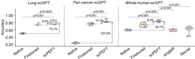
Extended Data Figure 1: Violin plots of benchmarking results using different scGPT pre-trained weights on the NSCLC dataset. The violin plots benchmark native, finetuned scGPT, and scPEFT using scGPT pre-trained weights from whole human data, pan-cancer data, and lung-specific data.
Extended Data Figure 1: Violin plots of benchmarking results using different scGPT pre-trained weights on the NSCLC dataset. The violin plots benchmark native, finetuned scGPT, and scPEFT using scGPT pre-trained weights from whole human data, pan-cancer data, and lung-specific data.CC BY · PubMed Central
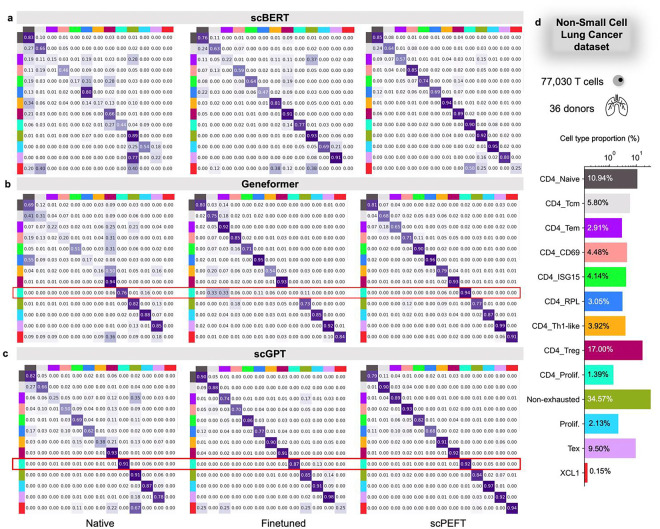
Extended Data Figure 2: Confusion matrices for benchmarking cell type identification on the NSCLC dataset. a-c, Confusion matrices for native, fine-tuned scLLMs, and scPEFT with (a) scBERT, (b) Geneformer, and (x) scGPT as respective backbones. Each cell reflects the percentage of instances from the row-defined cell type that are predicted as the column-defined cell type. High values along the diagonal indicate accurate predictions, while off-diagonal values represent misclassifications. The results exhibit the superior performance of scPEFT, particularly in identifying rare cell types (proportion < 5%). Notably, CD4+ enriched proliferation T cells were accurately identified by native scLLMs but misclassified by finetuned models, demonstrating an instance of catastrophic forgetting. This issue was not observed by scPEFT and was marked by the red boxes. d, Bar plot depicting the proportion of each cell type in the dataset.
Extended Data Figure 2: Confusion matrices for benchmarking cell type identification on the NSCLC dataset. a-c, Confusion matrices for native, fine-tuned scLLMs, and scPEFT with (a) scBERT, (b) Geneformer, and (x) scGPT as respective backbones. Each cell reflects the percentage of instances from the row-defined cell type that are predicted as the column-defined cell type. High values along the diagonal indicate accurate predictions, while off-diagonal values represent misclassifications. The results exhibit the superior performance of scPEFT, particularly in identifying rare cell types (proportion < 5%). Notably, CD4+ enriched proliferation T cells were accurately identified by native scLLMs but misclassified by finetuned models, demonstrating an instance of catastrophic forgetting. This issue was not observed by scPEFT and was marked by the red boxes. d, Bar plot depicting the proportion of each cell type in the dataset.CC BY · PubMed Central
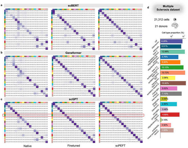
Extended Data Figure 3: Confusion matrices for benchmarking cell type identification on the MS dataset. a-c, Confusion matrices for native, fine-tuned scLLMs, and scPEFT with (a) scBERT, (b) Geneformer, and (c) scGPT as respective backbones. Notably, multiple cell types were accurately identified by native scLLMs but misclassified by their finetuned models, demonstrating clear instances of catastrophic forgetting. This issue was not observed by scPEFT and was marked by the red boxes. d, Bar plot depicting the proportion of each cell type in the dataset.
Extended Data Figure 3: Confusion matrices for benchmarking cell type identification on the MS dataset. a-c, Confusion matrices for native, fine-tuned scLLMs, and scPEFT with (a) scBERT, (b) Geneformer, and (c) scGPT as respective backbones. Notably, multiple cell types were accurately identified by native scLLMs but misclassified by their finetuned models, demonstrating clear instances of catastrophic forgetting. This issue was not observed by scPEFT and was marked by the red boxes. d, Bar plot depicting the proportion of each cell type in the dataset.CC BY · PubMed Central
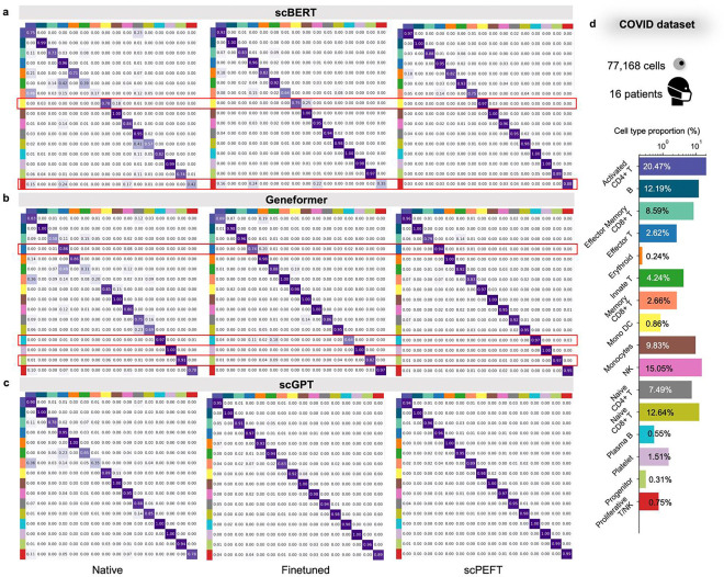
Extended Data Figure 4: Confusion matrices for benchmarking cell type identification on the COVID dataset. a-c, Confusion matrices for native, fine-tuned scLLMs, and scPEFT with (a) scBERT, (b) Geneformer, and (c) scGPT as respective backbones. Notably, the excitatory neuron cells at cortex layer 2–3 were accurately identified by native scGPT but misclassified by its finetuned model, demonstrating an instance of catastrophic forgetting. This issue was not observed by scPEFT and was marked by the red boxes. d, Bar plot depicting the proportion of each cell type in the dataset.
Extended Data Figure 4: Confusion matrices for benchmarking cell type identification on the COVID dataset. a-c, Confusion matrices for native, fine-tuned scLLMs, and scPEFT with (a) scBERT, (b) Geneformer, and (c) scGPT as respective backbones. Notably, the excitatory neuron cells at cortex layer 2–3 were accurately identified by native scGPT but misclassified by its finetuned model, demonstrating an instance of catastrophic forgetting. This issue was not observed by scPEFT and was marked by the red boxes. d, Bar plot depicting the proportion of each cell type in the dataset.CC BY · PubMed Central
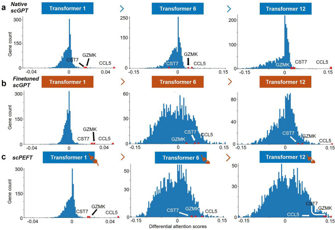
Extended Data Figure 5: Histograms of differential attention scores for COVID-related cell-state-specific genes. Histograms of differential attention scores were derived from (a) native, (b) fine-tuned scGPT, and (c) scPEFT models, respectively, in analysis of COVID-related cell-state-specific genes in Memory CD8+ T cells versus Naïve Memory CD8+ T cells. Histograms were generated for the top, middle, and last Transformer layers in these models. Red crosses mark the bins where genes of interest (CCL5, GZMK, and CST7) were located.
Extended Data Figure 5: Histograms of differential attention scores for COVID-related cell-state-specific genes. Histograms of differential attention scores were derived from (a) native, (b) fine-tuned scGPT, and (c) scPEFT models, respectively, in analysis of COVID-related cell-state-specific genes in Memory CD8+ T cells versus Naïve Memory CD8+ T cells. Histograms were generated for the top, middle, and last Transformer layers in these models. Red crosses mark the bins where genes of interest (CCL5, GZMK, and CST7) were located.CC BY · PubMed Central
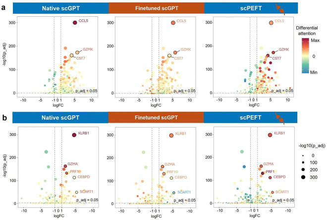
Extended Data Figure 6: Volcano plots of COVID-related cell-state-specific genes. The COVID-related cell-state-specific genes were analyzed in the comparisons of (a) Memory CD8+ T cells versus Naïve Memory CD8+ T cells, and (b) Effector Memory CD8+ T cells versus Memory CD8+ T cells, respectively. Volcano plots of Differential Gene Expression (DEG) analysis are colored by differential attention scores from native scGPT, finetuned scGPT, and scPEFT trained on the COVID dataset. The top attention genes of interest were highlighted with their names. Dot size reflects the genes’ adjusted p-values from DEG.
Extended Data Figure 6: Volcano plots of COVID-related cell-state-specific genes. The COVID-related cell-state-specific genes were analyzed in the comparisons of (a) Memory CD8+ T cells versus Naïve Memory CD8+ T cells, and (b) Effector Memory CD8+ T cells versus Memory CD8+ T cells, respectively. Volcano plots of Differential Gene Expression (DEG) analysis are colored by differential attention scores from native scGPT, finetuned scGPT, and scPEFT trained on the COVID dataset. The top attention genes of interest were highlighted with their names. Dot size reflects the genes’ adjusted p-values from DEG.CC BY · PubMed Central
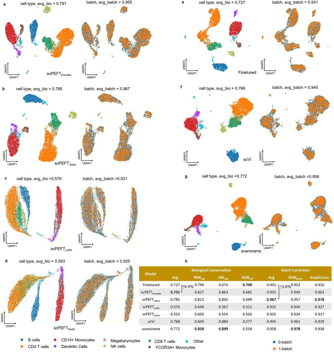
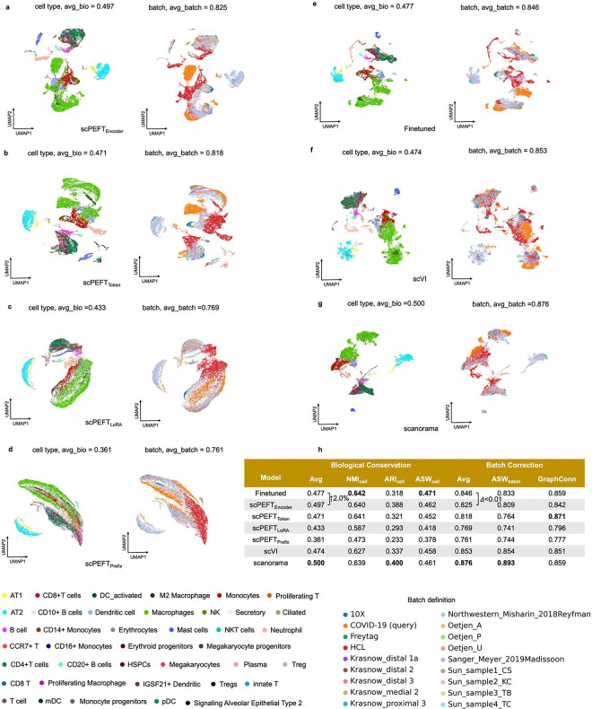
Extended Data Figure 7: Batch correction results on the PBMC 10K dataset. a-g, UMAP visualizations of corrected cell embeddings, color-coded by cell type and batch, for scPEFT (using Encoder adapter, Token adapter, LoRA, and Prefix adapter), fine-tuned scGPT, scVI, and Scanorama. h, Comparative table of batch correction performance across these methods.
Extended Data Figure 7: Batch correction results on the PBMC 10K dataset. a-g, UMAP visualizations of corrected cell embeddings, color-coded by cell type and batch, for scPEFT (using Encoder adapter, Token adapter, LoRA, and Prefix adapter), fine-tuned scGPT, scVI, and Scanorama. h, Comparative table of batch correction performance across these methods.CC BY · PubMed Central
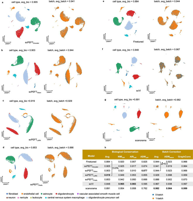
Extended Data Figure 8: Batch correction results on the Perirhinal cortex dataset. a-g, UMAP visualizations of corrected cell embeddings, color-coded by cell type and batch, for scPEFT (using Encoder adapter, Token adapter, LoRA, and Prefix adapter), fine-tuned scGPT, scVI, and Scanorama. h, Comparative table of batch correction performance across these methods.
Extended Data Figure 8: Batch correction results on the Perirhinal cortex dataset. a-g, UMAP visualizations of corrected cell embeddings, color-coded by cell type and batch, for scPEFT (using Encoder adapter, Token adapter, LoRA, and Prefix adapter), fine-tuned scGPT, scVI, and Scanorama. h, Comparative table of batch correction performance across these methods.CC BY · PubMed Central
Extended Data Figure 9: Batch correction results on the COVID-BATCH dataset. a-g, UMAP visualizations of corrected cell embeddings, color-coded by cell type and batch, for scPEFT (using Encoder adapter, Token adapter, LoRA, and Prefix adapter), fine-tuned scGPT, scVI, and Scanorama. h, Comparative table of batch correction performance across these methods.
Extended Data Figure 9: Batch correction results on the COVID-BATCH dataset. a-g, UMAP visualizations of corrected cell embeddings, color-coded by cell type and batch, for scPEFT (using Encoder adapter, Token adapter, LoRA, and Prefix adapter), fine-tuned scGPT, scVI, and Scanorama. h, Comparative table of batch correction performance across these methods.CC BY · PubMed Central
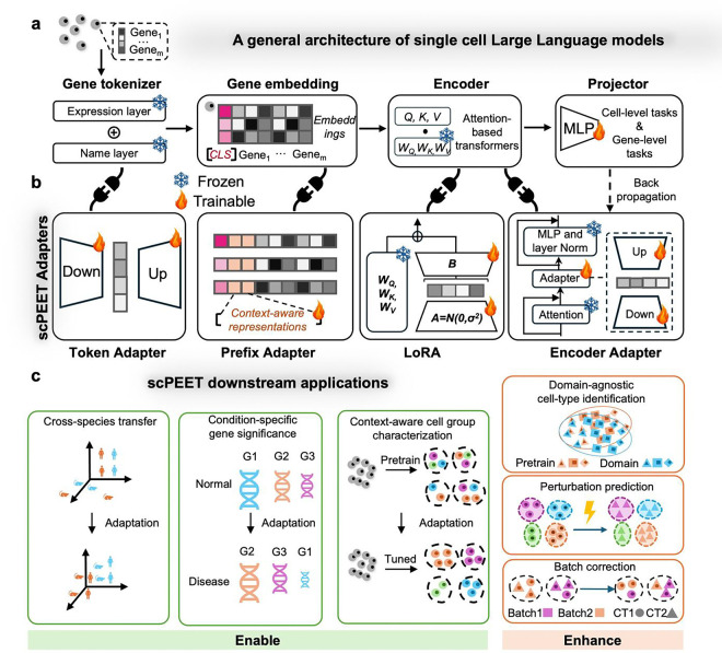
Figure 1: Overview of scPEFT. a, scLLM Architecture. A typical scLLM features a gene tokenizer that encodes gene identities and expression profiles into gene embeddings. This is followed by the Encoder, comprising multiple Transformer blocks that aggregate gene expression in cells into gene and cell representations. The final module, a projector, transforms these gene and cell embeddings into task-specific outputs. With the adapters from scPEFT, it can be adapted to various out-of-context applications without updating its original parameters, through task-specific objective functions and back-propagation. b, Adapters in scPEFT. Four types of adapters enhance scLLM’s domain adaptability: (i) Token Adapter, a compact autoencoder integrated into the gene tokenizer, refines gene token embeddings for specific tasks in reduced dimensional space. (ii) Prefix Adapter, which appends tunable tokens to gene tokens to incorporate task-specific information. (iii) RoLA (Rank-ordered Low-rank Approximation), which introduces low-rank matrices A and B into the Transformers to approximate model adjustments for the target domain. (iv) Encoder Adapter, another autoencoder attached to a Transformer block, customizes gene contextual embeddings to new biological contexts. These adapters can be used in combination. c, Downstream applications: scPEFT tailors scLLMs for a range of downstream applications in specific biological contexts, including domain-agnostic cell-type identification, condition-specific gene significance, context-aware cell group characterization, cross-species transfer, perturbation prediction, and so on.
Figure 1: Overview of scPEFT. a, scLLM Architecture. A typical scLLM features a gene tokenizer that encodes gene identities and expression profiles into gene embeddings. This is followed by the Encoder, comprising multiple Transformer blocks that aggregate gene expression in cells into gene and cell representations. The final module, a projector, transforms these gene and cell embeddings into task-specific outputs. With the adapters from scPEFT, it can be adapted to various out-of-context applications without updating its original parameters, through task-specific objective functions and back-propagation. b, Adapters in scPEFT. Four types of adapters enhance scLLM’s domain adaptability: (i) Token Adapter, a compact autoencoder integrated into the gene tokenizer, refines gene token embeddings for specific tasks in reduced dimensional space. (ii) Prefix Adapter, which appends tunable tokens to gene tokens to incorporate task-specific information. (iii) RoLA (Rank-ordered Low-rank Approximation), which introduces low-rank matrices A and B into the Transformers to approximate model adjustments for the target domain. (iv) Encoder Adapter, another autoencoder attached to a Transformer block, customizes gene contextual embeddings to new biological contexts. These adapters can be used in combination. c, Downstream applications: scPEFT tailors scLLMs for a range of downstream applications in specific biological contexts, including domain-agnostic cell-type identification, condition-specific gene significance, context-aware cell group characterization, cross-species transfer, perturbation prediction, and so on.CC BY · PubMed Central
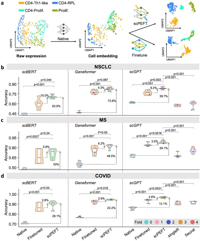
Figure 2: Cell type identification results of scPEFT under disease conditions. a, an illustration of how native scGPT, finetuned scGPT, and scPEFT models drive cell embeddings in their feature space. The data points represent a 10% random sample of cells from four annotated cell types in the query partition of the NSCLC dataset. Native scGPT appears to cluster cells according to their identities but misinterprets some CD4-Th1-like cells into CD4-proliferative cells, likely due to expression shifts in the tumor microenvironment compared to its training data of normal cells. Finetuned scGPT achieves better separation of CD4-Th1-like and CD4-RPL cells but performs worse in distinguishing CD4-proliferative cells from proliferation cells than the native scGPT, indicating catastrophic forgetting of pretrained knowledge. In contrast, scPEFT preserves the capability of native scGPT while benefiting from domain adaptation. b-d, Violin plots benchmarking native, finetuned, and scPEFT models using scBERT, Geneformer, and scGPT as backbones, along with SingleR and Seurat, under five-fold cross-validation on the NSCLC, MS and COVID datasets, respectively. Statistical significance between scPEFT and other models was assessed using a paired Student’s t-test across the five-fold validation results.
Figure 2: Cell type identification results of scPEFT under disease conditions. a, an illustration of how native scGPT, finetuned scGPT, and scPEFT models drive cell embeddings in their feature space. The data points represent a 10% random sample of cells from four annotated cell types in the query partition of the NSCLC dataset. Native scGPT appears to cluster cells according to their identities but misinterprets some CD4-Th1-like cells into CD4-proliferative cells, likely due to expression shifts in the tumor microenvironment compared to its training data of normal cells. Finetuned scGPT achieves better separation of CD4-Th1-like and CD4-RPL cells but performs worse in distinguishing CD4-proliferative cells from proliferation cells than the native scGPT, indicating catastrophic forgetting of pretrained knowledge. In contrast, scPEFT preserves the capability of native scGPT while benefiting from domain adaptation. b-d, Violin plots benchmarking native, finetuned, and scPEFT models using scBERT, Geneformer, and scGPT as backbones, along with SingleR and Seurat, under five-fold cross-validation on the NSCLC, MS and COVID datasets, respectively. Statistical significance between scPEFT and other models was assessed using a paired Student’s t-test across the five-fold validation results.CC BY · PubMed Central
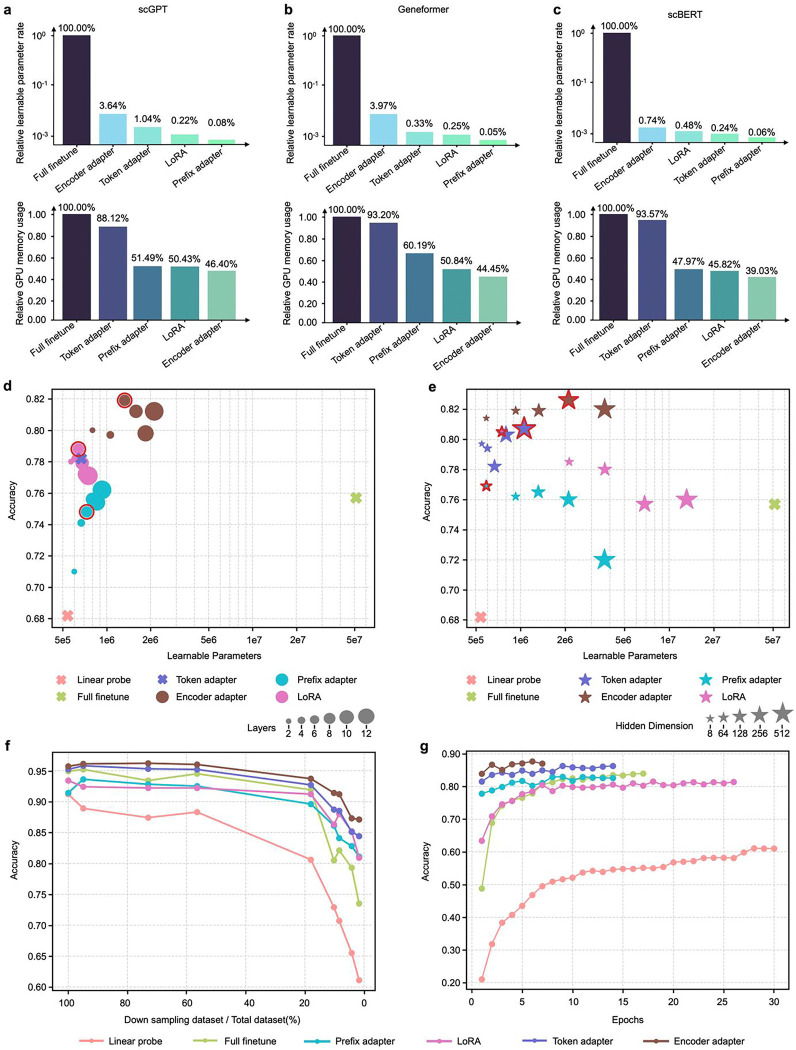
Figure 3: Efficiency analysis of scPEFT and related scLLMs. a-c, percentage of learnable parameters and GPU memory usage for fine-tuned scGPT, Geneformer, and scBERT, respectively, relative to scPEFT adapters. Evaluations were conducted using a batch size of 100 cells (the maximum setting for finetuning) on an Nvidia RTX A6000 GPU. GPU memory requirements depend not only on the learnable parameters but also on the gradient propagation path within the model. d, validation accuracies plotted against the number of learnable parameters for varying number of Transformer layers with adapters. Larger dots represent configurations with more layers, hence more parameters. Default scPEFT settings are highlighted. Notably, the highest parameter counts may not yield peak performance, suggesting that overparameterization may misalign with the model’s intrinsic dimensionality, thereby reducing generalization, as reflected in lower validation accuracies. e, Validation accuracies versus learnable parameters for adapters with different hidden representation dimensions. Larger stars indicate higher intermediate embedding dimensions, requiring more tunable parameters. Default scPEFT settings are highlighted. f, validation accuracies of fine-tuned scGPT versus scPEFT adapters using a progressively scaled-down reference dataset. Data points along each curve represent models trained on smaller subsets of the referenced set. g, validation accuracies of training checkpoints for fine-tuned scGPT versus scPEFT adapters. The final point on each curve marks the convergence epoch, defined by no improvement in validation loss over five consecutive epochs or reaching the maximum training limit of 50 epochs.
Figure 3: Efficiency analysis of scPEFT and related scLLMs. a-c, percentage of learnable parameters and GPU memory usage for fine-tuned scGPT, Geneformer, and scBERT, respectively, relative to scPEFT adapters. Evaluations were conducted using a batch size of 100 cells (the maximum setting for finetuning) on an Nvidia RTX A6000 GPU. GPU memory requirements depend not only on the learnable parameters but also on the gradient propagation path within the model. d, validation accuracies plotted against the number of learnable parameters for varying number of Transformer layers with adapters. Larger dots represent configurations with more layers, hence more parameters. Default scPEFT settings are highlighted. Notably, the highest parameter counts may not yield peak performance, suggesting that overparameterization may misalign with the model’s intrinsic dimensionality, thereby reducing generalization, as reflected in lower validation accuracies. e, Validation accuracies versus learnable parameters for adapters with different hidden representation dimensions. Larger stars indicate higher intermediate embedding dimensions, requiring more tunable parameters. Default scPEFT settings are highlighted. f, validation accuracies of fine-tuned scGPT versus scPEFT adapters using a progressively scaled-down reference dataset. Data points along each curve represent models trained on smaller subsets of the referenced set. g, validation accuracies of training checkpoints for fine-tuned scGPT versus scPEFT adapters. The final point on each curve marks the convergence epoch, defined by no improvement in validation loss over five consecutive epochs or reaching the maximum training limit of 50 epochs.CC BY · PubMed Central
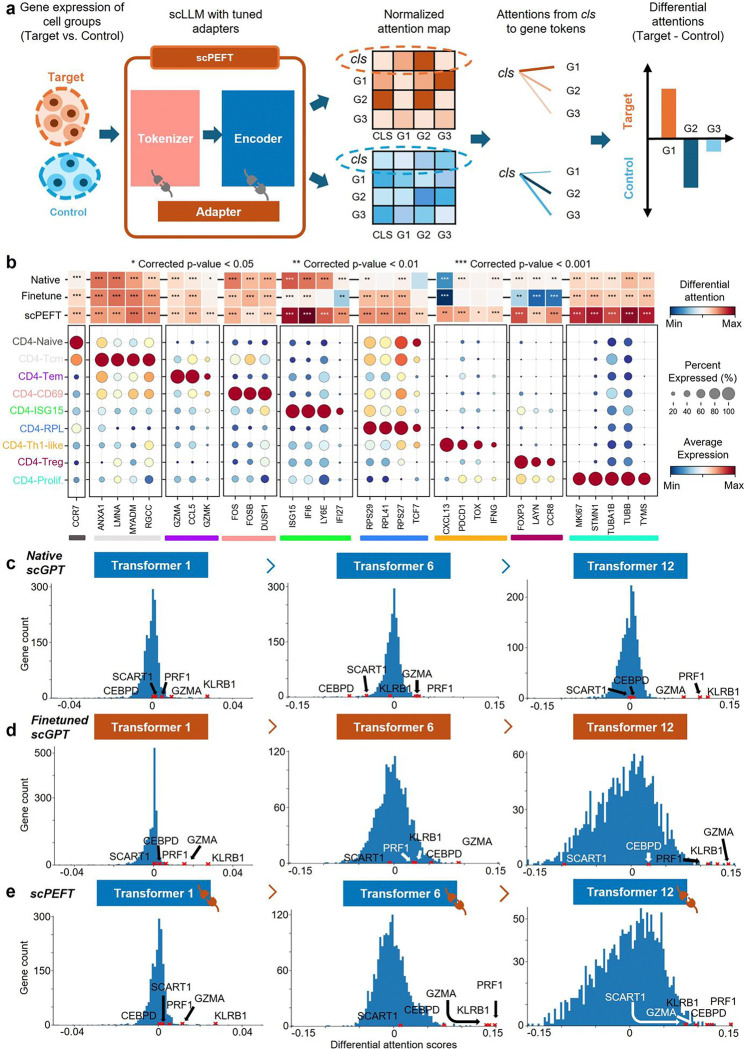
Figure 4: Condition-specific cell-state-associated gene analysis via attention mechanism. a, workflow for determining gene contributions to specific cell states under given conditions. Attention scores, describing the cell representation cls token’s attention on gene tokens, are extracted and normalized from scLLMs with tuned adapters to assess gene-cell associations. Differential attention scores between control and target cell states are calculated to reveal gene roles in cell-state differentiation under the specified condition. b, validation of differential attention values from native and fine-tuned scGPT, as well as scPEFT, in relation to cell-type-associated signature genes on the NSCLC dataset. The cell-type-associated signature genes were sourced from the original study [18]. A heatmap illustrates differential attention scores derived from native, fine-tuned scGPT, and scPEFT models for each signature gene. Differential attention scores were calculated between the target T cell subtype, defined by the corresponding signature genes, and other T cell subtypes as controls. Statistical significance is denoted by stars on the heatmap, based on corrected p-values obtained using the Wilcoxon rank-sum test and Benjamini-Yekutieli false discovery rate control [57]. Dot plots display the expression profiles of each signature gene across all T cell subtypes. Color bars beneath the signature genes indicate their associated target T cell subtypes, corresponding to the subtype labels on the y-axis. c-e, Histograms of differential attention scores from native, fine-tuned scGPT, and scPEFT models, respectively, in the analysis of COVID-related cell-state-specific genes in Effector Memory CD8+ T cells versus Memory CD8+ T cells. Histograms were generated for the top, middle, and last Transformer layers in these models. Red crosses mark the bins where key effector molecules (KLRB1, GZMA, PRF1) and two effector-function-associated genes (CEBPD and SCART1) were located.
Figure 4: Condition-specific cell-state-associated gene analysis via attention mechanism. a, workflow for determining gene contributions to specific cell states under given conditions. Attention scores, describing the cell representation cls token’s attention on gene tokens, are extracted and normalized from scLLMs with tuned adapters to assess gene-cell associations. Differential attention scores between control and target cell states are calculated to reveal gene roles in cell-state differentiation under the specified condition. b, validation of differential attention values from native and fine-tuned scGPT, as well as scPEFT, in relation to cell-type-associated signature genes on the NSCLC dataset. The cell-type-associated signature genes were sourced from the original study [18]. A heatmap illustrates differential attention scores derived from native, fine-tuned scGPT, and scPEFT models for each signature gene. Differential attention scores were calculated between the target T cell subtype, defined by the corresponding signature genes, and other T cell subtypes as controls. Statistical significance is denoted by stars on the heatmap, based on corrected p-values obtained using the Wilcoxon rank-sum test and Benjamini-Yekutieli false discovery rate control [57]. Dot plots display the expression profiles of each signature gene across all T cell subtypes. Color bars beneath the signature genes indicate their associated target T cell subtypes, corresponding to the subtype labels on the y-axis. c-e, Histograms of differential attention scores from native, fine-tuned scGPT, and scPEFT models, respectively, in the analysis of COVID-related cell-state-specific genes in Effector Memory CD8+ T cells versus Memory CD8+ T cells. Histograms were generated for the top, middle, and last Transformer layers in these models. Red crosses mark the bins where key effector molecules (KLRB1, GZMA, PRF1) and two effector-function-associated genes (CEBPD and SCART1) were located.CC BY · PubMed Central
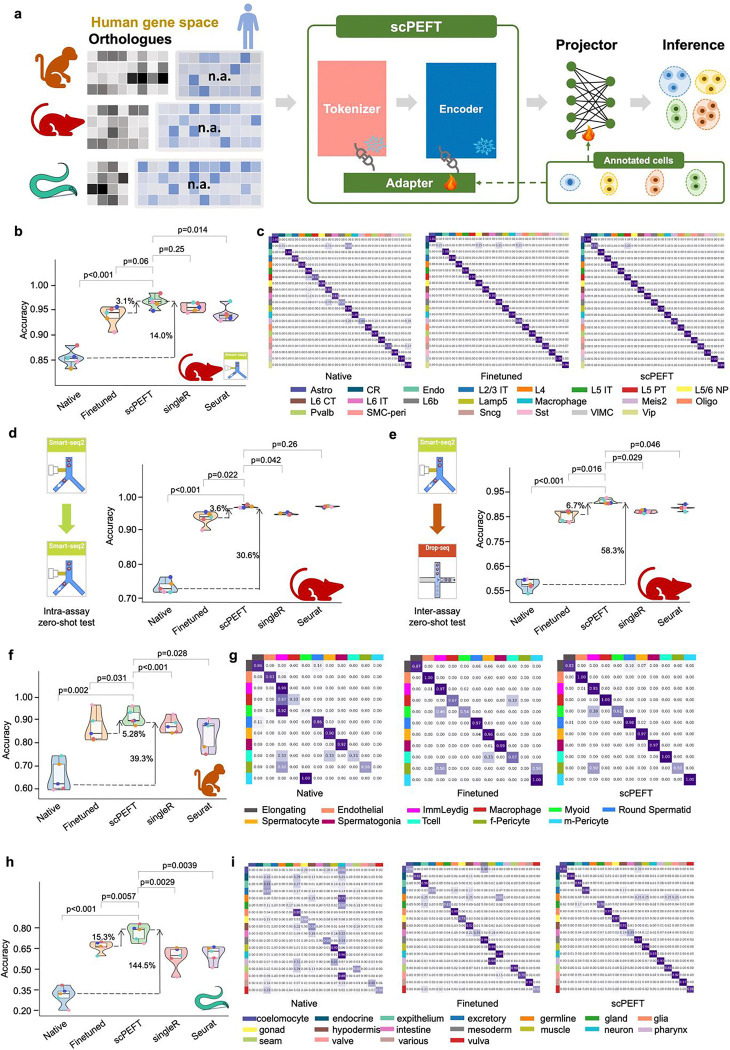
Figure 5: Cross-species transfer results of scPEFT. a, Schematic workflow for adapting the human-pretrained scGPT model to data from other species, including mice, macaque, and C. elegans. During adaptation, non-orthologous genes are masked, and adapters from scPEFT are trained using a small subset of annotated cells from the target species, enabling cross-species cell type contextualization. b, Benchmarking performance of native scGPT, fine-tuned scGPT, and scPEFT across five-fold cross-validation on a mice dataset, alongside comparisons to established cell-type identification tools SingleR and Seurat. Violin plots display performance metrics, with paired Student’s t-tests evaluating the statistical significance of differences between scPEFT and other methods across cross-validation results. c, Confusion matrices illustrate the alignment between annotated and predicted cell types for native scGPT, fine-tuned scGPT, and scPEFT on the mice dataset. d,e, Zero-shot benchmarking violin plots for native, finetune, and scPEFT under five-fold cross-validation on intra-assay and inter-assay interdependent test, respectively, revealing greater stability of scPEFT than finetune strategy in handling assay variance. f,h, Benchmarking violin plots for native, finetune, and scPEFT under five-fold cross-validation on a macaque and C. elegans dataset, respectively. g,i, Confusion matrices for native, finetune, and scPEFT showing the match between annotated and predicted cell types on a macaque and C. elegans dataset, respectively.
Figure 5: Cross-species transfer results of scPEFT. a, Schematic workflow for adapting the human-pretrained scGPT model to data from other species, including mice, macaque, and C. elegans. During adaptation, non-orthologous genes are masked, and adapters from scPEFT are trained using a small subset of annotated cells from the target species, enabling cross-species cell type contextualization. b, Benchmarking performance of native scGPT, fine-tuned scGPT, and scPEFT across five-fold cross-validation on a mice dataset, alongside comparisons to established cell-type identification tools SingleR and Seurat. Violin plots display performance metrics, with paired Student’s t-tests evaluating the statistical significance of differences between scPEFT and other methods across cross-validation results. c, Confusion matrices illustrate the alignment between annotated and predicted cell types for native scGPT, fine-tuned scGPT, and scPEFT on the mice dataset. d,e, Zero-shot benchmarking violin plots for native, finetune, and scPEFT under five-fold cross-validation on intra-assay and inter-assay interdependent test, respectively, revealing greater stability of scPEFT than finetune strategy in handling assay variance. f,h, Benchmarking violin plots for native, finetune, and scPEFT under five-fold cross-validation on a macaque and C. elegans dataset, respectively. g,i, Confusion matrices for native, finetune, and scPEFT showing the match between annotated and predicted cell types on a macaque and C. elegans dataset, respectively.CC BY · PubMed Central
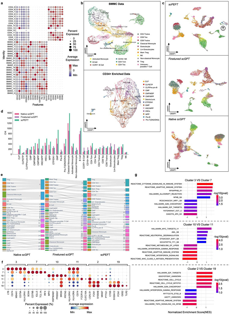
Figure 6: cPEFT identifies developmental cell populations in BMMC and CD34+ enriched CITE-seq data. s a, Protein expression patterns for annotated cell identities in BMMC and CD34+ enriched samples, depicted by circle sizes representing the fraction of cells expressing a protein and colors indicating average protein expression levels. b, UMAP visualizations of protein expression profile clusters from BMMC and CD34+ enriched cells. c, UMAP visualizations of cell representations from native, finetuned scGPT, and scPEFT, trained on scRNA-seq data from BMMC and CD34+ enriched cells, excluding cell identity annotations. Arrow points to a BMMC mature B cell subset which clusters closer to pre-proB, and PreB cells, annotated by protein. d, Evaluation of cell representations from native, fine-tuned scGPT, and scPEFT models using the Calinski–Harabasz index. This metric assesses the ability of generative embeddings from each model to effectively characterize protein-annotated cell groups. e, Sankey diagrams illustrating the assignment of cells from protein-annotated identities (left) to clustering results (right), obtained using the Leiden algorithm at a resolution of 1.5. Clustering was performed on embeddings generated by native, fine-tuned scGPT, and scPEFT models. Finetuned scGPT and scPEFT identified more distinct clusters than native scGPT. Notably, scPEFT was able to avoid some perplexing splitting/combining that confounds the interpretation of the finetuned model. For instance, the finetuned model nearly equally split the CD4 Tcm population between a group of memory Treg and exhausted CD4 T cells as well as two groups of Treg and CD4 Tnaive, combined, suggesting some confusion among memory programs. f, Expression profiles of genes receiving high differential attention scores in subgroups #3 vs. #7, #10 vs. #11, and #2 vs. #19 from panel e, with colors representing mean expression within each cluster and dot sizes indicating the fraction of cells expressing a gene. g, Gene Set Enrichment Analysis (GSEA) based on attention differential scores for these subgroups, showcasing Normalized Enrichment Scores with colors denoting the significance of enriched phenotypes.
Figure 6: cPEFT identifies developmental cell populations in BMMC and CD34+ enriched CITE-seq data. s a, Protein expression patterns for annotated cell identities in BMMC and CD34+ enriched samples, depicted by circle sizes representing the fraction of cells expressing a protein and colors indicating average protein expression levels. b, UMAP visualizations of protein expression profile clusters from BMMC and CD34+ enriched cells. c, UMAP visualizations of cell representations from native, finetuned scGPT, and scPEFT, trained on scRNA-seq data from BMMC and CD34+ enriched cells, excluding cell identity annotations. Arrow points to a BMMC mature B cell subset which clusters closer to pre-proB, and PreB cells, annotated by protein. d, Evaluation of cell representations from native, fine-tuned scGPT, and scPEFT models using the Calinski–Harabasz index. This metric assesses the ability of generative embeddings from each model to effectively characterize protein-annotated cell groups. e, Sankey diagrams illustrating the assignment of cells from protein-annotated identities (left) to clustering results (right), obtained using the Leiden algorithm at a resolution of 1.5. Clustering was performed on embeddings generated by native, fine-tuned scGPT, and scPEFT models. Finetuned scGPT and scPEFT identified more distinct clusters than native scGPT. Notably, scPEFT was able to avoid some perplexing splitting/combining that confounds the interpretation of the finetuned model. For instance, the finetuned model nearly equally split the CD4 Tcm population between a group of memory Treg and exhausted CD4 T cells as well as two groups of Treg and CD4 Tnaive, combined, suggesting some confusion among memory programs. f, Expression profiles of genes receiving high differential attention scores in subgroups #3 vs. #7, #10 vs. #11, and #2 vs. #19 from panel e, with colors representing mean expression within each cluster and dot sizes indicating the fraction of cells expressing a gene. g, Gene Set Enrichment Analysis (GSEA) based on attention differential scores for these subgroups, showcasing Normalized Enrichment Scores with colors denoting the significance of enriched phenotypes.CC BY · PubMed Central

Verification

Claims the paper makes

The claims and statistics of this paper haven’t been checked yet.

Run the check — free, no account needed. It reads the open-access text and checks the sources it cites.

A few free checks a day · papers already verified open instantly · results are public

Self-check

Percentages, sums and reported differences re-computed from the paper’s own numbers.

The self-check needs the paper’s full text.

Citations

Cites

Show all 62 references

Research works printed but not linked: 1

  1. Hu, E. J. et al. LoRA: Low-rank adaptation of large language models. In The 10th International Conference on Learning Representations (ICLR, 2022).

Cited by

Sorted by citations

See all 6

Metadata

Authors
He, F., Fei, R., Krull, J. E., Yu, Y., Zhang, X., Wang, X., Cheng, H., Gao, M., Su, L., Chen, Y., Li, J., Jin, B., Chang, Y., Ma, A., Ma, Q., & Xu, D.
Affiliations
  • University of Missouri (US)
  • The Ohio State University (US)
Corresponding author
  • Ma, Qin
  • Xu, Dong
Venue
Nature Machine Intelligence
Published
2025-12-31
Volume
8
Issue
1
Pages
118-133
ISSN
2522-5839 (ISSN-L)
OpenAlex
W7117735905
Cited by
5
References
62
Citations by year
2026: 4 · 2025: 1
FWCI
1.50
Type
journal article
Language
en
Open access
Locations
  • Nature Machine Intelligence (publishedVersion)
  • bioRxiv (Cold Spring Harbor Laboratory) (acceptedVersion, cc-by-nc)
  • Research Square (acceptedVersion, cc-by)
  • PubMed (publishedVersion)
  • PubMed Central (submittedVersion, cc-by)
Funders
Foundation for the National Institutes of Health (R35GM126985, R01GM152585, P01CA278732, P01AI177687, U54AG075931, R01DK138504)
Topics
  • Life Sciences › Biochemistry, Genetics and Molecular Biology › Molecular Biology › Single-cell and spatial transcriptomics
  • Physical Sciences › Computer Science › Artificial Intelligence › Domain Adaptation and Few-Shot Learning
  • Life Sciences › Biochemistry, Genetics and Molecular Biology › Molecular Biology › Gene Regulatory Network Analysis
Keywords
parameter-efficient fine-tuning, adapters, catastrophic forgetting, zero-shot prediction, single-cell atlas, interspecies transmission, cell population identification, GPU memory reduction

About these checks

Registry, retraction, Cites and statistics results are computed, not judged, with one exception: a printed reference that no index lists is read once by a model before it counts as a citation error. Claims are extracted by a model and kept only when their quote appears verbatim in the text; each cited source is then checked, with a model’s help, for whether it says what the claim relies on. Those verdicts can be wrong — read the claim’s quote and the cited source before relying on one.

Statistics and arithmetic are recomputed, not judged.

Confidence bands are shown only for fields calibrated on our annotated test set. None is calibrated yet, so no band is shown.

What counts as a claim ›How we measure confidence ›How we check this ›

Are you an author, or do you see a problem with this page? Request a review