

The DOI matches its Crossref record; 90 of the 90 references it cites were found in a registry or an index; 45 of the 61 sources its claims cite were checked for support.
Deviations in effective connectivity explain different hallucination subtypes in Parkinson's disease psychosis
Open accessPublished version of a preprint
Abstract
Abstract Psychosis and visual hallucinations (VH) in Parkinson’s disease (PD) substantially impact patient outcomes, yet the underlying neural mechanisms remain unclear, limiting effective treatments. Here we used dynamic causal modeling to leverage the fast temporal dynamics captured with electroencephalography data during a visual mismatch negativity task in people with PD with ( N = 20) and without ( N = 18) VH to examine effective connectivity. We found reduced top-down and enhanced bottom-up connectivity in ventral visual and prefrontal regions during task performance in PD-VH, suggesting deficits in sensory prediction updating and an overreliance on visual input. Connectivity patterns differed with hallucination complexity, with complex VH being associated with altered top-down and bottom-up right-hemisphere connectivity, and multimodal hallucinations to more widespread bilateral disruption. Increased task activity, as computed with source reconstruction, correlated positively with normative serotonergic 5-HT 2A receptor distribution. These findings highlight specific neural targets for early therapeutic interventions, supporting a transdiagnostic computational architecture of hallucinations.
Overview
CoChat Summary
## TL;DR
Using EEG during a visual mismatch negativity task, this study found that Parkinson’s disease psychosis with visual hallucinations (PD-VH; N = 20) showed reduced top-down and increased bottom-up effective connectivity in ventral visual and prefrontal regions versus Parkinson’s disease without hallucinations (N = 18). Connectivity patterns also varied by hallucination subtype and severity, with complex hallucinations linked to more right-hemisphere disruption and multimodal hallucinations to broader bilateral disruption.
## Key findings
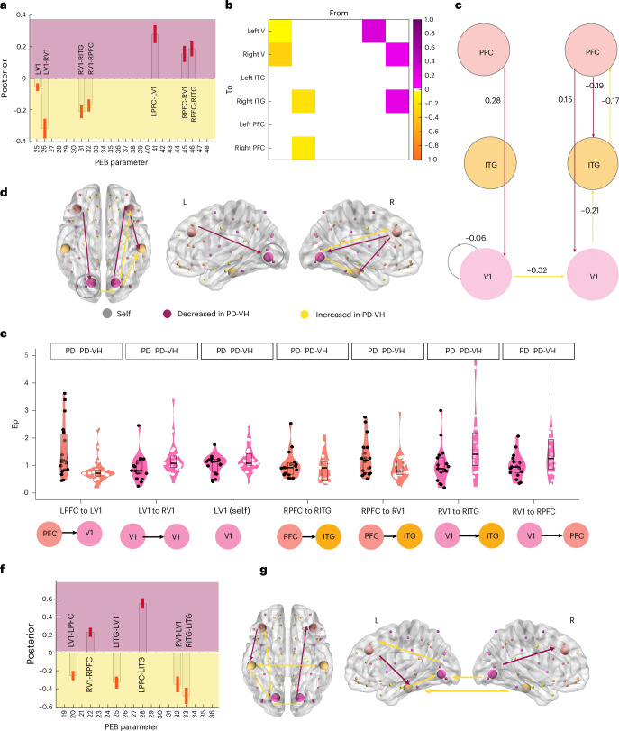
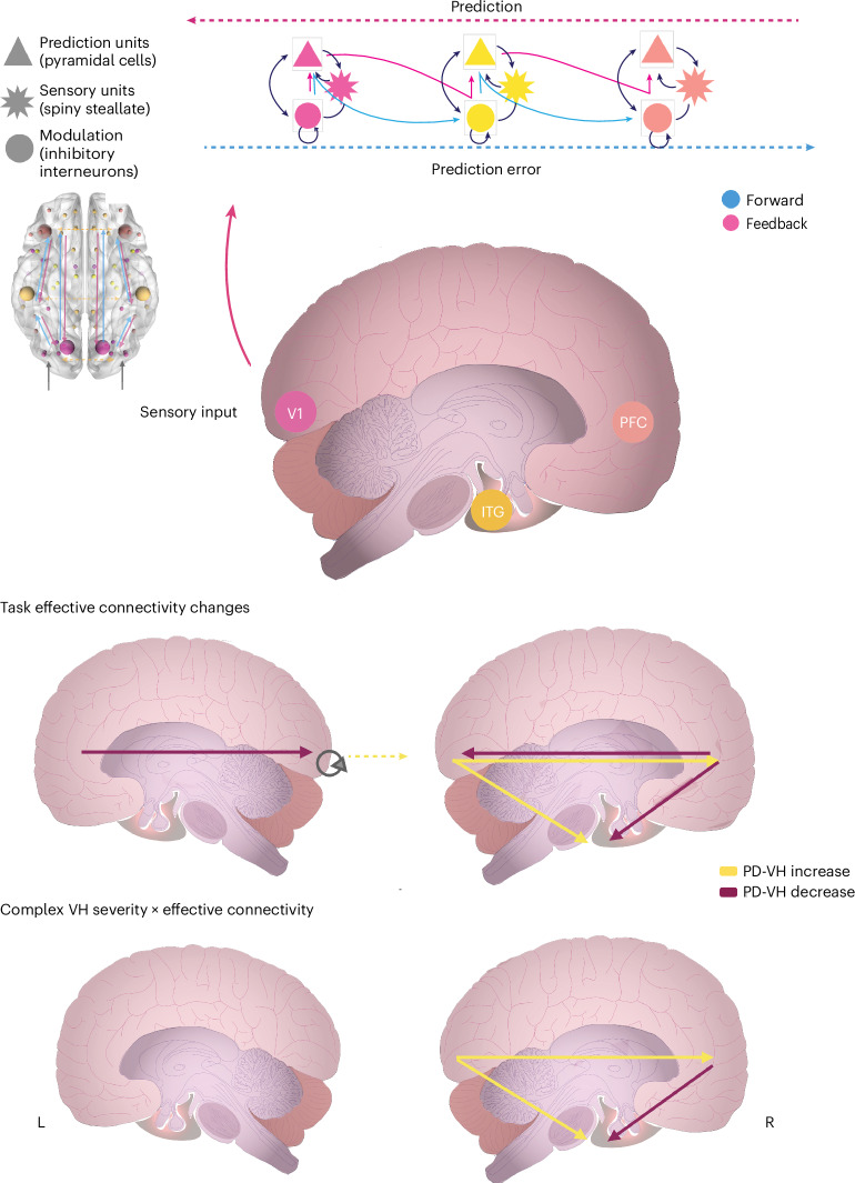
- In PD-VH, the task condition (rare vs standard) was associated with increased bottom-up connectivity and decreased self-inhibition for V1, plus decreased top-down connection strength; the top-down effect had posterior probability (pp) > 0.99.
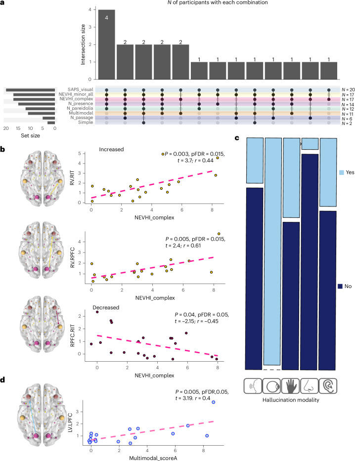
- Hallucination severity tracked connectivity patterns: complex visual hallucinations (CVH) were associated with reduced top-down connectivity from right PFC to right ITG and enhanced bottom-up connectivity from right V1 to right PFC and right ITG.
- In a regression/cross-validation model, right-hemisphere top-down and bottom-up connectivity predicted CVH severity with R2 = 0.49, adjusted R2 = 0.4, P = 0.014, RMSE = 2.26, and MAE = 177.
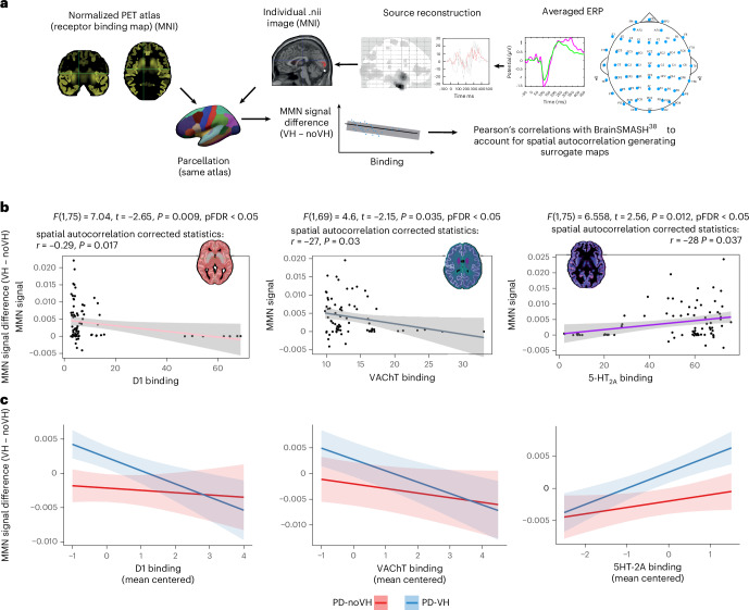
- Source-localized task activity correlated with normative receptor maps: 5-HT2A showed a positive correlation (r = 0.284, P = 0.037), while D1 showed a negative correlation (r = −0.29, P = 0.017); VAChT was negative in the exploratory analyses and remained directionally similar in sensitivity analyses.
## Methods
- Design and sample: Case-control comparison of 20 PD-VH participants and 18 PD-noVH participants; ethics-approved, clinicaltrials.gov registered (NCT03661125). Hallucination phenomenology and subtype were assessed with SAPS-PD and NEVHI.
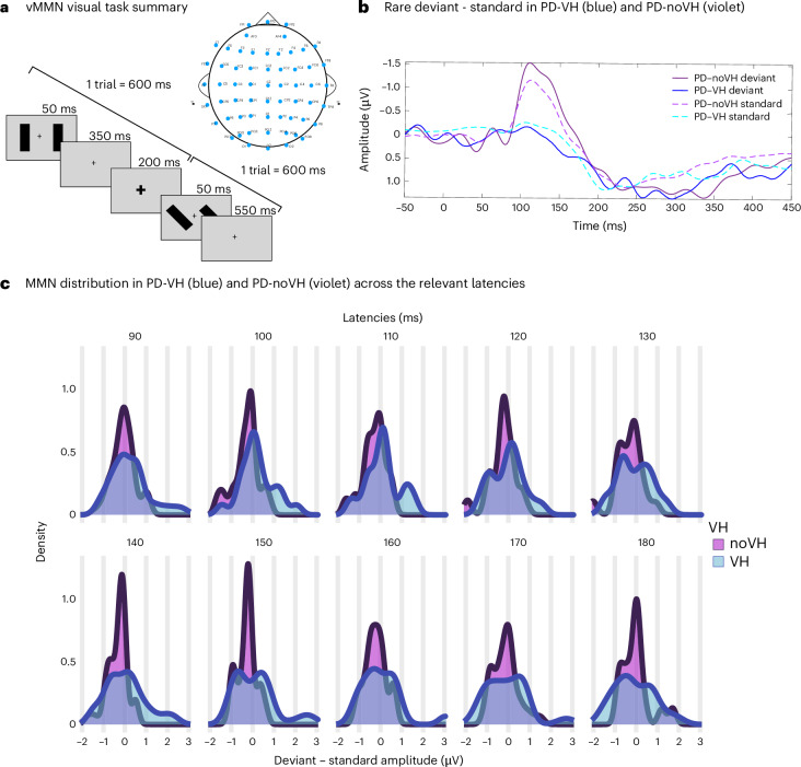
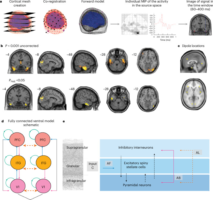
- Task and data: Participants completed a visual mismatch negativity (vMMN) EEG task with 1,000 trials; analyses focused on standard vs rare deviant trials. EEG was preprocessed in SPM12, with source reconstruction over 60–400 ms.
- Main analysis: Dynamic causal modeling (DCM) with parametric empirical Bayes (PEB) tested task-modulated effective connectivity in ventral and dorsal pathways; follow-up analyses included linear regression, leave-one-out cross-validation, neural mass model simulations, and exploratory receptor atlas correlations.
## Limitations
- The paper states that the study did not include healthy controls, so it cannot fully determine whether findings are specific to PD or disentangle aging-related effects.
- It also states that EEG cannot robustly assess subcortical sources, and the receptor-binding analyses are exploratory because both EEG source reconstruction and receptor-density maps have low spatial resolution.
- Evident from the text: the receptor-atlas/PET analyses rely on normative maps rather than individual PET data, so they are indirect and hypothesis-generating rather than participant-specific.
Model-written summary. Check it against the paper before relying on it.
Integrity signals
Registry
Cites
Statistics
Figures
Verification
Claims the paper makes
27 Valid · 4 Not valid · 8 Not checked · 86 Own results and interpretations
Every source the paper cites for this claim was checked and supports it.
In the paper
“Dynamic causal modeling (DCM), leveraging EEG’s temporal precision, enables exploration of these mechanisms21–23.”
§ Deviations in effective connectivity explain different hallucination subtypes in Parkinson’s disease psychosis > Main Parkinson’s disease (PD) is characterized by motor and nonmotor symptoms1, among which, psychosis and visual hallucinations (VH) can have a substantial impact on patient outcomes and are associated with cognitive decline2–4. Although morphological5–7 and functional7–9 alterations differentiate patients with PD with VH (PD-VH) from those without VH, standard cognitive assessments do not provide mechanistic insights into VH. A recent review of frameworks10 converges on the proposal that ‘ascending’ sensory disturbances together with ‘descending’ factors (for example, expectations, specific object features and attention) contribute to VH. Among the different proposals, hierarchical predictive coding, a cognitive model of psychosis, suggests that hallucinations arise from disrupted balance between top-down expectations and bottom-up sensory inputs, with a stronger influence of expectations observed in people with hallucinations11–13. Mismatch negativity (MMN) is an event-related potential computed by averaging the time-locked brain activity that can be observed when a rare stimulus is presented within a sequence of frequent stimuli in an electroencephalography (EEG) recording14. MMN is widely considered to be a robust marker of psychosis15, thought to reflect an attempt to minimize this prediction error and capture its disruption16–19. We recently demonstrated reduced visual MMN responses in PD-VH20, paralleling auditory MMN impairments observed in psychosis15. However, traditional event-related potential (ERP) analyses cannot uncover the directional neural mechanisms involved. Dynamic causal modeling (DCM), leveraging EEG’s temporal precision, enables exploration of these mechanisms21–23. DCM has proved instrumental in clarifying the disruption of MMN in people with schizophrenia-related psychosis24, and a spectral DCM study (using resting-state functional magnetic resonance imaging (fMRI)) found increased resting-state top-down and reduced bottom-up connectivity in the visual network in PD with VH, a pattern that was predictive of hallucination severity8. Here, we employ DCM with ERP neural mass models across dorsal (primary visual area, V1, and inferior parietal lobe (IPL)) and ventral (primary visual, V1 and inferior temporal gyrus (ITG)) visual pathways interacting with the prefrontal cortex (PFC) during a visual MMN task. The main aim of this study is to investigate the possible neural mechanisms and effective connectivity patterns underlying the reduced visual MMN observed in PD-VH, using DCM to test the hierarchical organization proposed by the predictive-coding framework11–13. Second, we aim to investigate the relation of changes in effective connectivity to hallucination complexity. We include the PFC to test prediction error impairment, because top-down connectivity is crucial for integrating sensory information25,26. We focus on the dorsal and ventral pathways on the basis of relevant literature20,27–33 and because changing the line orientation in our MMN task is a process known to involve specific parietal regions34. We hypothesize reduced top-down and enhanced bottom-up connectivity at task in PD-VH, reflecting impaired prediction updating and reliance on compromised sensory information, as visual input is known to be defective in PD-VH35. In addition, exploratory analyses relate MMN-derived source activity to cortical distributions of receptors, previously implicated in PD-VH.
What the sources say
- Supportedcites [21] Kiebel, S. J., Garrido, M. I., Moran, R. J. & Friston, K. J. Dynamic causal modelling for EEG and MEG. Cogn. Neurodyn. 2, 121–136 (2008). source
The Introduction describes DCM as a spatiotemporal model that uses M/EEG responses to infer neuronal dynamics and interactions. It also emphasizes the temporal detail in M/EEG data, supporting the claim that DCM can use EEG data to explore such mechanisms. No page numbers are supplied in our copy. (LLM, conf=0.87)
- Supportedcites [22] Kiebel, S. J., David, O. & Friston, K. J. Dynamic causal modelling of evoked responses in EEG/MEG with lead field parameterization. NeuroImage 30, 1273–1284 (2006). source
The Introduction describes DCM for EEG/MEG evoked responses as using temporal, physiologically informed constraints to infer coupling among cortical regions. The Theory section explains that the model estimates effective connectivity from measured EEG/MEG responses, supporting the claim that it can explore the underlying mechanisms. (LLM, conf=0.88)
- Supportedcites [23] Friston, K. A theory of cortical responses. Philos. Trans. R. Soc. B 360, 815–836 (2005). source
The section “Dynamic causal modelling with neural mass models” describes using DCM with EEG event-related responses to investigate their neurobiological mechanisms. “Perceptual learning and the MMN” shows the model using scalp time-series to examine changes in cortical connectivity. The paper does not use the phrase “EEG’s temporal precision,” but its analysis of time-resolved responses supports the paraphrase. (LLM, conf=0.87)
plainstates a cause- Asserted: passes — The authors put it forward as true.
- Checkable: passes — It can be checked against evidence.
- Load-bearing: passes — The paper’s conclusions depend on it.
- Based: passes — The paper gives a reason why it holds.
- Supported
Every source the paper cites for this claim was checked and supports it.
In the paper
“First, that of an E–I imbalance similar to what has been proposed in schizophrenia51.”
§ Deviations in effective connectivity explain different hallucination subtypes in Parkinson’s disease psychosis > Discussion > VH severity may be related to disinhibition
What the sources say
- Supportedcites [51] Liu, Y. et al. A selective review of the excitatory–inhibitory imbalance in schizophrenia: underlying biology, genetics, microcircuits, and symptoms. Front. Cell Dev. Biol. 9, 664535 (2021). source
The Summary and Future Outlook section (page not provided) states that E–I imbalance has been proposed as a core pathology of schizophrenia and describes clinical and animal-model evidence for it. The citation supports the schizophrenia analogy; the citing authors' explanation of their own observed pattern is not attributed to this source. (LLM, conf=0.94)
hedgedstates a possible cause- Asserted: passes — The authors put it forward as true.
- Checkable: passes — It can be checked against evidence.
- Load-bearing: passes — The paper’s conclusions depend on it.
- Based: passes — The paper gives a reason why it holds.
- Supported
The cited sources support only part of this claim — read each source’s verdict below.
In the paper
“The result of increased activity from rV1 being associated to VH and VH severity aligns with the finding in schizophrenia of disinhibition in sensory areas (A1) predicting abnormal auditory perception in the aMMN and positive symptoms24.”
§ Deviations in effective connectivity explain different hallucination subtypes in Parkinson’s disease psychosis > Discussion > Decreased top-down and increased bottom-up connectivity for vMMN in PD-VH are related to hallucination severity
What the sources say
- Partly supportedcites [24] Adams, R. A. et al. Computational modeling of electroencephalography and functional magnetic resonance imaging paradigms indicates a consistent loss of pyramidal cell synaptic gain in schizophrenia. Biol. Psychiatry 91, 202–215 (2022). sourceExperimental — accuracy not yet measured
The Results sections on resting-state fMRI and 40-Hz ASSR associate A1 disinhibition with abnormal auditory percepts, and the resting-state fMRI results also associate it with positive symptoms. But the MMN Results locate the association with abnormal auditory percepts in the left inferior frontal gyrus, not A1. The paper reports associations rather than showing that A1 disinhibition predicts these outcomes in the MMN paradigm. (LLM, conf=0.85)
plainstates an association- Asserted: passes — The authors put it forward as true.
- Checkable: passes — It can be checked against evidence.
- Load-bearing: unclear — It is unclear whether the paper’s conclusions depend on it.
- Based: passes — The paper gives a reason why it holds.
- Partly supported
The cited sources support only part of this claim — read each source’s verdict below.
In the paper
“Lower GABA+/creatine in the ITG in PD-VH33 supports this view.”
§ Deviations in effective connectivity explain different hallucination subtypes in Parkinson’s disease psychosis > Discussion > VH severity may be related to disinhibition
What the sources say
- Partly supportedcites [33] Firbank, M. J. et al. Reduced occipital GABA in Parkinson disease with visual hallucinations. Neurology 91, e675–e685 (2018). sourceExperimental — accuracy not yet measured
The Results report lower GABA+/creatine in PD with visual hallucinations, supporting the group comparison. But the Methods (MRI acquisition; page not available) place the spectroscopy voxel in the midline occipital lobe, not the ITG, so the claimed location is unsupported. (LLM, conf=0.90)
plainstates an association- Asserted: passes — The authors put it forward as true.
- Checkable: passes — It can be checked against evidence.
- Load-bearing: unclear — It is unclear whether the paper’s conclusions depend on it.
- Based: passes — The paper gives a reason why it holds.
- Partly supported
Every source the paper cites for this claim was checked and supports it.
In the paper
“suggesting that spontaneous feedforward activity in the visual cortex can also lead to hallucinations, rather than feedback activity alone41”
§ Deviations in effective connectivity explain different hallucination subtypes in Parkinson’s disease psychosis > Discussion > Decreased top-down and increased bottom-up activity underlies PD-VH reduction of vMMN We found that in PD-VH there was a significant reduction of top-down activity and an increase in bottom-up connectivity. Top-down connectivity is crucial for the integration of sensory information24–26, and this finding suggests that in PD-VH there is a failure to update predictions based on sensory evidence and an overreliance on or over-response to sensory information during the task. This can contribute to the difficulties in stimulus detection due to retinal degeneration29,33 and responding to the changes in the sensory environment. This interpretation resonates with the principles of a recently proposed framework on VH in PD10. When taking a hypothesis-free approach, the feedforward model, with a right-hemisphere pattern of increased V1-ITG and V1-PFC connectivity, emerged as the strongest contributor. In line with this result, recent work has shown that people are more likely to hallucinate when sensory information is noisy, especially when combined with strong expectations41. Interestingly, this was found with an orientation perception task, consistent with our stimuli, and in the input layers of V2, where orientation-specific activity takes place, lending further support to our interpretation and suggesting that spontaneous feedforward activity in the visual cortex can also lead to hallucinations, rather than feedback activity alone41. When further exploring our results using positron emission tomography (PET)-derived normative gradients as biologically informed spatial priors to test whether the pattern of MMN-related alterations aligns with known neurochemical distributions, we found that local source differences were positively correlated with 5-HT2A density. This finding may suggest that this receptor may be facilitating cortical disinhibition, consistent with previous proposals42,43 and studies showing 5-HT2A antagonists reducing psychosis-like symptoms20,42. Based on our results, we may speculate that this reflects an aberrant or a compensatory excitation. We also find a negative relationship with dopaminergic receptor D1; dopamine is proposed to encode precision weighting44. One possible interpretation is that this could bias the system toward assigning undue salience to bottom-up inputs, resulting in illusions and/or hallucinations, as well as disruption of normal mismatch detection. The analysis of the latent connectivity (A matrix) complements these results, revealing a similar pattern to that seen in the task-modulated model, suggesting that patients with PD-VH not only respond more strongly to sensory information during the visual mismatch negativity (vMMN) task but may do so more generally. The reduced connectivity from the left PFC to the ITG, with increased PFC-V1 connectivity, appears to support this interpretation, as well as previous models of VH in Lewy Body Diseases suggesting a disruption on the object-related semantic network45.
What the sources say
- Supportedcites [41] Haarsma, J., Deveci, N., Corbin, N., Callaghan, M. F. & Kok, P. Expectation cues and false percepts generate stimulus-specific activity in distinct layers of the early visual cortex. J. Neurosci. 43, 7946–7957 (2023). source
The Discussion reports that high-confidence false percepts were reflected in the middle input layers of early visual cortex, rather than in feedback-related expectation signals, and concludes that stimulus-like feedforward activity can generate false percepts. The Introduction also describes spontaneous early-visual-cortex activity generating hallucinatory perceptions in Charles Bonnet syndrome. The paper treats the link from its own false-percept experiment to hallucinations as a possibility, consistent with the citing sentence’s ‘suggesting.’ (LLM, conf=0.88)
hedgedstates a cause- Asserted: passes — The authors put it forward as true.
- Checkable: passes — It can be checked against evidence.
- Load-bearing: passes — The paper’s conclusions depend on it.
- Based: passes — The paper gives a reason why it holds.
- Supported
40 more claims
Create a free account to see all 125 claims and every source verdict.
- Probably not a substantial claim. 43
- It may state two separate claims as one. 46
Citations
Cites
Fitting Linear Mixed-Effects Models Using lme4
Bates, D., Mächler, M., Bolker, B., & Walker, S. (2015) · Journal of Statistical Software · doi:10.18637/jss.v067.i01 · Cited by 86,835
lmerTest Package: Tests in Linear Mixed Effects Models
Kuznetsova, A., Brockhoff, P. B., & Christensen, R. H. B. (2017) · Journal of Statistical Software · doi:10.18637/jss.v082.i13 · Cited by 24,346
Modern Applied Statistics with S
Venables, W. N., & Ripley, B. D. (2002) · Statistics and Computing · doi:10.1007/978-0-387-21706-2 · Cited by 24,305
Wickham, H., Averick, M., Bryan, J., Chang, W., McGowan, L., François, R., Grolemund, G., Hayes, A., Henry, L., Hester, J., et al. (2019) · Journal of Open Source Software · doi:10.21105/joss.01686 · Cited by 22,138 · cited 1×
“R packages Tidyverse 83 and dplyr 84 were used for these analyses (Supplementary Information 7).”
Desikan, R. S., Ségonne, F., Fischl, B., Quinn, B. T., Dickerson, B. C., Blacker, D., Buckner, R. L., Dale, A. M., Maguire, R. P., Hyman, B. T., et al. (2006) · NeuroImage · doi:10.1016/j.neuroimage.2006.01.021 · Cited by 13,324
Oostenveld, R., Fries, P., Maris, E., & Schoffelen, J.-M. (2011) · Computational Intelligence and Neuroscience · doi:10.1155/2011/156869 · Cited by 10,323
Building Predictive Models in R Using the caret Package
Kuhn, M. (2008) · Journal of Statistical Software · doi:10.18637/jss.v028.i05 · Cited by 8,863
Fischl, B., Salat, D. H., Busa, E., Albert, M., Dieterich, M., Haselgrove, C., van der Kouwe, A., Killiany, R., Kennedy, D., Klaveness, S., et al. (2002) · Neuron · doi:10.1016/s0896-6273(02)00569-x · Cited by 8,610
The fusiform face area: a module in human extrastriate cortex specialized for face perception
Kanwisher, N., McDermott, J., & Chun, M. M. (1997) · The Journal of Neuroscience · doi:10.1523/jneurosci.17-11-04302.1997 · Cited by 7,738 · cited 1×
“…of the effect of VH in response to perturbation of network-level excitatory/inhibitory dynamics recapitulate our results illustrating increased sensitivity in the PD-VH network, particularly in ITG regions, extensively involved in object recognition, particularly of human and animal stimuli 48,49 .”
Rao, R. P. N., & Ballard, D. H. (1999) · Nature Neuroscience · doi:10.1038/4580 · Cited by 5,483
Metadata
- Authors
- Vignando, M., Ffytche, D., Mazibuko, N., Palma, G., Bhat, A., Montagnese, M., Dave, S., Tai, Y. F., Batzu, L., Leta, V., Chaudhuri, K. R., Williams Gray, C. H., & Mehta, M. A.
- Affiliations
- King's College London (GB)
- University of Southampton (GB)
- University of Exeter (GB)
- University of Cambridge (GB)
- University of London (GB)
- Hammersmith Hospital (GB)
- King's College Hospital NHS Foundation Trust (GB)
- Corresponding author
- Vignando, Miriam
- ORCID
- Venue
- Nature Mental Health
- Published
- 2026-06-10
- Volume
- 4
- Issue
- 6
- Pages
- 994-1009
- ISSN
- 2731-6076 (ISSN-L)
- PMC
- PMC13259922
- OpenAlex
- W7164158954
- Cited by
- 0
- References
- 83
- FWCI
- 0.00
- Type
- journal article
- Language
- en
- Open access
- hybrid
- doi.org
- Licence
- cc-by
- creativecommons.org/licenses/by/4.0 (tdm)
- creativecommons.org/licenses/by/4.0 (vor)
- Locations
- Nature Mental Health (publishedVersion, cc-by)
- Research Square (submittedVersion, cc-by)
- PubMed (publishedVersion)
- PubMed Central (publishedVersion, cc-by)
- Apollo (University of Cambridge) (submittedVersion, cc-by)
- Funders
- Alzheimer's Research UK (ARUK-RF2022B-002)
- RCUK | Medical Research Council (MR/R005931/1)
- Topics
- Life Sciences › Neuroscience › Cognitive Neuroscience › Hallucinations in medical conditions
- Health Sciences › Medicine › Clinical Neurology › Parkinson's Disease Mechanisms and Treatments
- Life Sciences › Neuroscience › Cognitive Neuroscience › Functional Brain Connectivity Studies
- Keywords
- Parkinson's disease psychosis, visual hallucinations, effective connectivity, mismatch negativity, hemispheric lateralization, multimodal hallucination, prefrontal cortex, ventral visual cortex
About these checks
Registry, retraction, Cites and statistics results are computed, not judged, with one exception: a printed reference that no index lists is read once by a model before it counts as a citation error. Claims are extracted by a model and kept only when their quote appears verbatim in the text; each cited source is then checked, with a model’s help, for whether it says what the claim relies on. Those verdicts can be wrong — read the claim’s quote and the cited source before relying on one.
The claim verdicts this report counts are experimental: their accuracy is not yet measured.
A language model drew the claims from the paper on 2026-10-11, under version 3 of the claim definition (model deepseek-v4.1-flash, prompt extract-v3).
A second model labelled each claim on 2026-10-11 (model jev-1.13-20260917, definition revision gold-guideline-v0.2).
Cited sources are checked with a language model’s help (gpt-6-sol).
Statistics and arithmetic are recomputed, not judged.
Confidence bands are shown only for fields calibrated on our annotated test set. None is calibrated yet, so no band is shown.
What counts as a claim ›How we measure confidence ›How we check this ›
Are you an author, or do you see a problem with this page? Request a review
How to cite
Cite CoChat for the checks on this page, and the paper for its own findings.
- This page
- CoChat. (2026). Citation check: Deviations in effective connectivity explain different hallucination subtypes in Parkinson's disease psychosis. https://app.cochat.ai/papers/10.1038/s44220-026-00669-7
- The paper
- Vignando, M., Ffytche, D., Mazibuko, N., Palma, G., Bhat, A., Montagnese, M., Dave, S., Tai, Y. F., Batzu, L., Leta, V., Chaudhuri, K. R., Williams Gray, C. H., & Mehta, M. A. (2026). Deviations in effective connectivity explain different hallucination subtypes in Parkinson's disease psychosis. Nature Mental Health. https://doi.org/10.1038/s44220-026-00669-7
Data sources
Bibliographic data: Semantic Scholar, OpenAlex, Crossref.
Contains information from the Semantic Scholar Academic Graph Datasets, made available under the ODC Attribution License (ODC-BY 1.0).
Semantic Scholar’s data is described in: Kinney, R., et al. (2023). The Semantic Scholar Open Data Platform. arXiv:2301.10140.
