

The DOI matches its Crossref record; 2 of the 2 references it cites were found in a registry or an index.
PCR amplification of repetitive DNA: a limitation to genome editing technologies and many other applications
Overview
The claims and statistics of this paper haven’t been checked yet.
Run the check — free, no account needed. It reads the open-access text and checks the sources it cites.
Integrity signals
Registry
Cites
Statistics
Figures
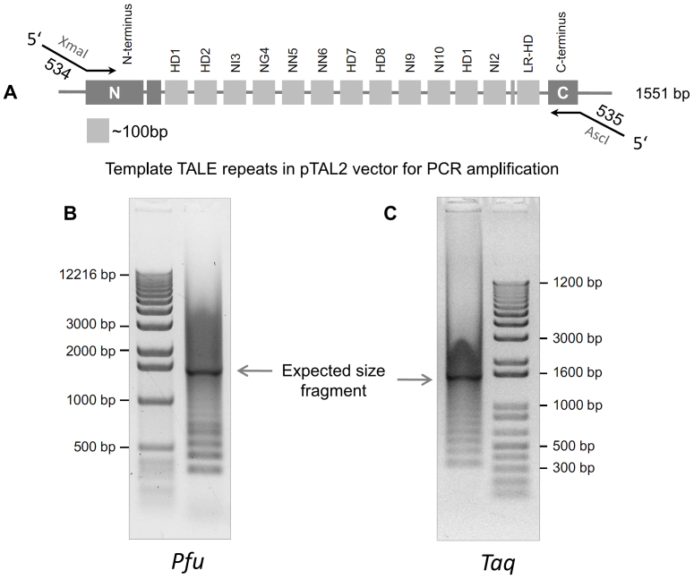
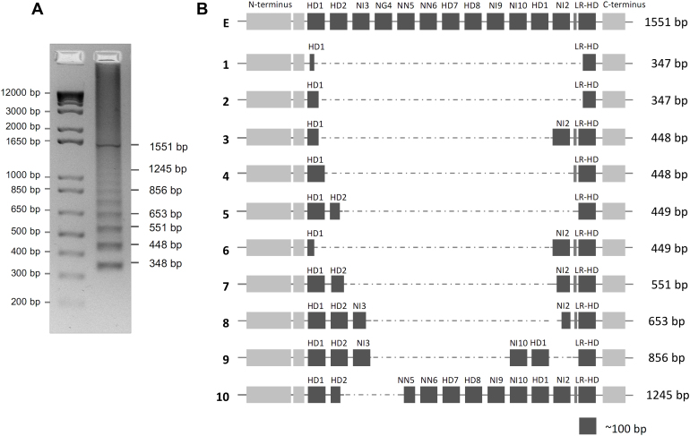
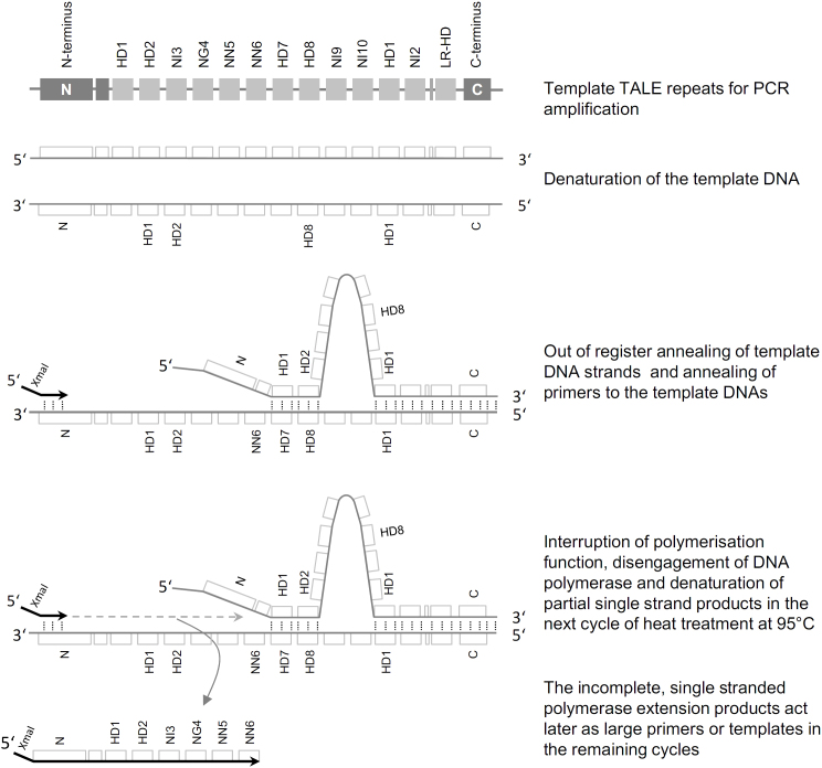
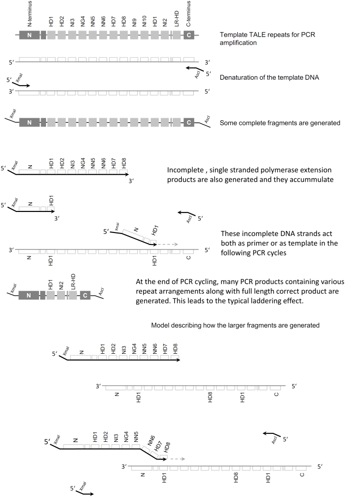
Verification
Claims the paper makes
The claims and statistics of this paper haven’t been checked yet.
Run the check — free, no account needed. It reads the open-access text and checks the sources it cites.
Claims other papers make about this one
Each is checked against this paper’s own text: does it say what the citing paper says it does?
In the citing paper
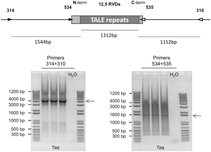
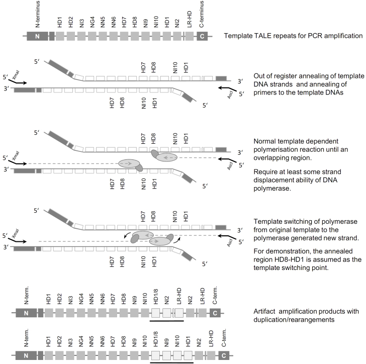
“this process also explains the typical PCR fragmentation patterns observed when using transcript-activator like effector (TALE) technology (69).”
From Tandem repeats lead to sequence assembly errors and impose multi-level challenges for genome and protein databases (2019) · cites this paper as [69]
Self-check
Percentages, sums and reported differences re-computed from the paper’s own numbers.
The self-check needs the paper’s full text.
Citations
Cites
Genome structural variation discovery and genotyping
Alkan, C., Coe, B. P., & Eichler, E. E. (2011) · Nature Reviews Genetics · doi:10.1038/nrg2958 · Cited by 1,630
Scherzinger, E., Lurz, R., Turmaine, M., Mangiarini, L., Hollenbach, B., Hasenbank, R., Bates, G. P., Davies, S. W., Lehrach, H., & Wanker, E. E. (1997) · Cell · doi:10.1016/s0092-8674(00)80514-0 · Cited by 1,291
Research works printed but not linked: 37
- Moscou, M. J. & Bogdanove, A. J. A simple cipher governs DNA recognition by TAL effectors. Science 326, 1501 (2009). · DOI
- Filipovska, A., Razif, M. F., Nygard, K. K. & Rackham, O. A universal code for RNA recognition by PUF proteins. Nat Chem Biol 7, 425–427 (2011). · DOI
- Lu, G., Dolgner, S. J. & Hall, T. M. Understanding and engineering RNA sequence specificity of PUF proteins. Curr Opin Struct Biol 19, 110–115 (2009). · DOI
- Barkan, A. et al. A combinatorial amino Acid code for RNA recognition by pentatricopeptide repeat proteins. PLoS Genet 8, e1002910 (2012). · DOI
- Yagi, Y., Hayashi, S., Kobayashi, K., Hirayama, T. & Nakamura, T. Elucidation of the RNA recognition code for pentatricopeptide repeat proteins involved in organelle RNA editing in plants. PLoS ONE 8, e57286 (2013). · DOI
- Yin, P. et al. Structural basis for the modular recognition of single-stranded RNA by PPR proteins. Nature 504, 168–171 (2013). · DOI
- Filipovska, A. & Rackham, O. Modular recognition of nucleic acids by PUF, TALE and PPR proteins. Mol Biosyst 8, 699–708 (2012). · DOI
- Fichtner, F., Castellanos, R. U. & Ülker, B. Precision genetic modifications: a new era in molecular biology and crop improvement. Planta 239, 921 (2014). · DOI
- Hopkins, C. M., White, F. F., Choi, S. H., Guo, A. & Leach, J. E. A family of avirulence genes from Xanthomonas oryzae pv.oryzae. Mol. Plant-Microbe Interact 5, 451–459 (1992). · DOI
- Boch, J. et al. Breaking the code of DNA binding specificity of TAL-type III effectors. Science 326, 1509–1512 (2009). · DOI · Paper page
- Cermak, T. et al. Efficient design and assembly of custom TALEN and other TAL effector-based constructs for DNA targeting. Nucleic Acids Res 39, e82 (2011). · DOI
- Miller, J. C. et al. A TALE nuclease architecture for efficient genome editing. Nature Biotechnol 29, 143–148 (2011). · DOI · Paper page
- Viguera, E., Canceill, D. & Ehrlich, S. D. In vitro replication slippage by DNA polymerases from thermophilic organisms. J Mol Biol 312, 323–333 (2001). · DOI
- Canceill, D., Viguera, E. & Ehrlich, S. D. Replication slippage of different DNA polymerases is inversely related to their strand displacement efficiency. J Biol Chem 274, 27481–27490 (1999). · DOI
- Rapley, R. Enhancing PCR amplification and sequencing using DNA-binding proteins. Mol Biotechnol 2, 295–298 (1994). · DOI
- Hartl, D. L. Molecular melodies in high and low C. Nat Rev Genet 1, 145–149 (2000). · DOI
- Treangen, T. J. & Salzberg, S. L. Repetitive DNA and next-generation sequencing: computational challenges and solutions. Nat Rev Genet 13, 36–46 (2012). · DOI · Paper page
- Korbel, J. O. et al. Paired-end mapping reveals extensive structural variation in the human genome. Science 318, 420–426 (2007). · DOI · Paper page
- Jurka, J., Kapitonov, V. V., Kohany, O. & Jurka, M. V. Repetitive sequences in complex genomes: structure and evolution. Annu Rev Genomics Hum Genet 8, 241–259 (2007). · DOI · Paper page
- Shapiro, J. A. & von Sternberg, R. Why repetitive DNA is essential to genome function. Biol Rev Camb Philos Soc 80, 227–250 (2005). · DOI
- Li, Y.-C., Korol, A. B., Fahima, T. & Nevo, E. Microsatellites Within Genes: Structure, Function and Evolution. Mol Biol Evol 21, 991–1007 (2004). · DOI
- Jin, P., Alisch, R. S. & Warren, S. T. RNA and microRNAs in fragile X mental retardation. Nat Cell Biol 6, 1048–1053 (2004). · DOI
- Muragaki, Y., Mundlos, S., Upton, J. & Olsen, B. R. Altered growth and branching patterns in synpolydactyly caused by mutations in HOXD13. Science 272, 548–551 (1996). · DOI
- Lee, J. E. & Cooper, T. A. Pathogenic mechanisms of myotonic dystrophy. Biochem Soc Trans 37, 1281–1286 (2009). · DOI
- Kunkel, T. A. Nucleotide repeats. Slippery DNA and diseases. Nature 365, 207–208 (1993). · DOI
- Sahdev, S., Saini, S., Tiwari, P., Saxena, S. & Singh Saini, K. Amplification of GC-rich genes by following a combination strategy of primer design, enhancers and modified PCR cycle conditions. Mol Cell Probes 21, 303–307 (2007). · DOI
- Brakenhoff, R. H., Schoenmakers, J. G. & Lubsen, N. H. Chimeric cDNA clones: a novel PCR artifact. Nucleic Acids Res 19, 1949 (1991). · DOI
- Meyerhans, A., Vartanian, J. P. & Wain-Hobson, S. DNA recombination during PCR. Nucleic Acids Res 18, 1687–1691 (1990). · DOI
- Dover, G. Slippery DNA runs on and on and on. Nat Genet 10, 254–256 (1995). · DOI
- Briggs, A. W. et al. Iterative capped assembly: rapid and scalable synthesis of repeat-module DNA such as TAL effectors from individual monomers. Nucleic Acids Res (2012). · DOI
- Dean, F. B., Nelson, J. R., Giesler, T. L. & Lasken, R. S. Rapid amplification of plasmid and phage DNA using Phi 29 DNA polymerase and multiply-primed rolling circle amplification. Genome Res 11, 1095–1099 (2001). · DOI
- Blanco, L. et al. Highly efficient DNA synthesis by the phage phi 29 DNA polymerase. Symmetrical mode of DNA replication. J Biol Chem 264, 8935–8940 (1989). · DOI
- de Vega, M., Lazaro, J. M., Mencia, M., Blanco, L. & Salas, M. Improvement of phi29 DNA polymerase amplification performance by fusion of DNA binding motifs. P Nat Acad Sci USA 107, 16506–16511 (2010). · DOI
- Odelberg, S. J., Weiss, R. B., Hata, A. & White, R. Template-switching during DNA synthesis by Thermus aquaticus DNA polymerase I. Nucleic Acids Res 23, 2049–2057 (1995). · DOI
- Hugenholtz, P. & Huber, T. Chimeric 16S rDNA sequences of diverse origin are accumulating in the public databases. Int J Syst Evol Microbiol 53, 289–293 (2003). · DOI
- Berney, C., Fahrni, J. & Pawlowski, J. How many novel eukaryotic ‘kingdoms’? Pitfalls and limitations of environmental DNA surveys. BMC Biol 2, 13 (2004). · DOI
- Wintzingerode, V. F., Göbel, U. B. & Stackebrandt, E. Determination of microbial diversity in environmental samples: pitfalls of PCR-based rRNA analysis. FEMS Microbiol Rev 21, 213–229 (1997). · DOI
Cited by
Sorted by citations
Biallelic expansion of an intronic repeat in RFC1 is a common cause of late-onset ataxia
Cortese, A., Simone, R., Sullivan, R., Vandrovcova, J., Tariq, H., Yau, W. Y., Humphrey, J., Jaunmuktane, Z., Sivakumar, P., Polke, J., et al. (2019) · Nature Genetics · doi:10.1038/s41588-019-0372-4 · Cited by 575
Tørresen, O. K., Star, B., Mier, P., Andrade-Navarro, M. A., Bateman, A., Jarnot, P., Gruca, A., Grynberg, M., Kajava, A. V., Promponas, V. J., et al. (2019) · Nucleic Acids Research · doi:10.1093/nar/gkz841 · Cited by 347
DNA Structural Barcode Copying and Random Access
Bošković, F., Ohmann, A., Keyser, U. F., & Chen, K. (2021) · Small Structures · doi:10.1002/sstr.202000144 · Cited by 24
Monomer‐scale design of functional protein polymers using consensus repeat sequences
Chang, M. P., Huang, W., & Mai, D. J. (2021) · Journal of Polymer Science · doi:10.1002/pol.20210506 · Cited by 20
Metadata
- Authors
- Hommelsheim, C. M., Frantzeskakis, L., Huang, M., & Ülker, B.
- Venue
- Scientific Reports
- Published
- 2014-05-23
- Volume
- 4
- Issue
- 1
- ISSN
- 2045-2322
- PMC
- PMC4031481
- Cited by
- 121
- References
- 2
- Language
- en
About these checks
Registry, retraction, Cites and statistics results are computed, not judged, with one exception: a printed reference that no index lists is read once by a model before it counts as a citation error. Claims are extracted by a model and kept only when their quote appears verbatim in the text; each cited source is then checked, with a model’s help, for whether it says what the claim relies on. Those verdicts can be wrong — read the claim’s quote and the cited source before relying on one.
Statistics and arithmetic are recomputed, not judged.
Confidence bands are shown only for fields calibrated on our annotated test set. None is calibrated yet, so no band is shown.
What counts as a claim ›How we measure confidence ›How we check this ›
Are you an author, or do you see a problem with this page? Request a review
How to cite
Cite CoChat for the checks on this page, and the paper for its own findings.
- This page
- CoChat. (2026). Citation check: PCR amplification of repetitive DNA: a limitation to genome editing technologies and many other applications. https://app.cochat.ai/papers/10.1038/srep05052
- The paper
- Hommelsheim, C. M., Frantzeskakis, L., Huang, M., & Ülker, B. (2014). PCR amplification of repetitive DNA: a limitation to genome editing technologies and many other applications. Scientific Reports. https://doi.org/10.1038/srep05052
