

The DOI matches its Crossref record.
In situ formation of magnetopolymersomes via electroporation for MRI
Abstract
As the development of diagnostic/therapeutic (and combined: theranostic) nanomedicine grows, smart drug-delivery vehicles become ever more critical. Currently therapies consist of drugs tethered to, or encapsulated within nanoparticles or vesicles. There is growing interest in functionalising them with magnetic nanoparticles (MNPs) to target the therapeutics by localising them using magnetic fields. An alternating magnetic field induces remote heating of the particles (hyperthermia) triggering drug release or cell death. Furthermore, MNPs are diagnostic MRI contrast agents. There is considerable interest in MNP embedded vehicles for nanomedicine, but their development is hindered by difficulties producing consistently monodisperse MNPs and their reliable loading into vesicles. Furthermore, it is highly advantageous to "trigger" MNP production and to tune the MNP's size and magnetic response. Here we present the first example of a tuneable, switchable magnetic delivery vehicle for nanomedical application. These are comprised of robust, tailored polymer vesicles (polymersomes) embedded with superparamagnetic magnetite MNPs (magnetopolymersomes) which show good MRI contrast (R2* = 148.8 s−1) and have a vacant core for loading of therapeutics. Critically, the magnetopolymersomes are produced by a pioneering nanoreactor method whereby electroporation triggers the in situ formation of MNPs within the vesicle membrane, offering a switchable, tuneable magnetic responsive theranostic delivery vehicle.
Overview
The claims and statistics of this paper haven’t been checked yet.
Run the check — free, no account needed. It reads the open-access text and checks the sources it cites.
Integrity signals
Registry
Cites
Statistics
Figures
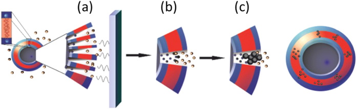
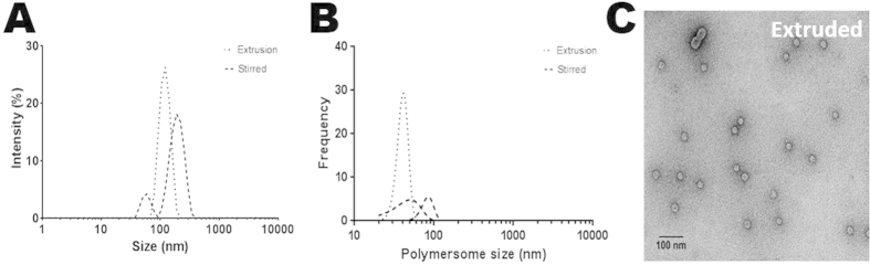
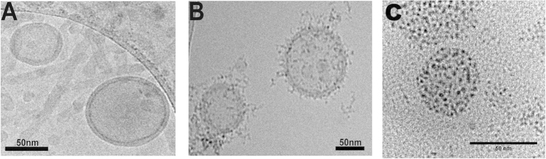
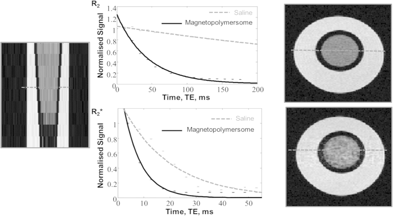
Verification
Claims the paper makes
The claims and statistics of this paper haven’t been checked yet.
Run the check — free, no account needed. It reads the open-access text and checks the sources it cites.
Claims other papers make about this one
Each is checked against this paper’s own text: does it say what the citing paper says it does?
In the citing paper
“In fact, only magnetoliposomes containing Mms6-mediated BMNPs have been described so far, and their production is intended for magnetic resonance imaging [28,29,30].”
From Biomimetic Magnetoliposomes as Oxaliplatin Nanocarriers: In Vitro Study for Potential Application in Colon Cancer (2020) · cites this paper as [29]
Self-check
Percentages, sums and reported differences re-computed from the paper’s own numbers.
The self-check needs the paper’s full text.
Citations
Cites
No references could be resolved yet.
Research works printed but not linked: 30
- Walde, P., Cosentino, K., Engel, H. & Stano, P. Giant vesicles: Preparations and applications. Chem Bio Chem 11, 848–865, 10.1002/cbic.201000010 (2010). · DOI
- Discher, B. M. et al. Polymersomes: Tough Vesicles Made from Diblock Copolymers. Science 284, 1143–1146, 10.1126/science.284.5417.1143 (1999). · DOI
- Discher, D. E. & Eisenberg, A. Polymer vesicles. Science 297, 967–973, 10.1126/science.1074972 (2002). · DOI
- Al-Jamal, W. T. & Kostarelos, K. Liposomes: From a Clinically Established Drug Delivery System to a Nanoparticle Platform for Theranostic Nanomedicine. Accounts Chem. Res. 44, 1094–1104, 10.1021/ar200105p (2011). · DOI
- Fang, C. et al. Fabrication of magnetic nanoparticles with controllable drug loading and release through a simple assembly approach. J. Control. Release 162, 233–241, 10.1016/j.jconrel.2012.06.028 (2012). · DOI
- Janib, S. M., Moses, A. S. & MacKay, J. A. Imaging and drug delivery using theranostic nanoparticles. Adv. Drug Deliver. Rev. 62, 1052–1063, 10.1016/j.addr.2010.08.004 (2010). · DOI
- Qiu, D., An, X., Chen, Z. & Ma, X. Microstructure study of liposomes decorated by hydrophobic magnetic nanoparticles. Chem. Phys. Lipids 165, 563–570, 10.1016/j.chemphyslip.2012.06.004 (2012). · DOI
- Ren, T. et al. Multifunctional polymer vesicles for ultrasensitive magnetic resonance imaging and drug delivery. J. Mater. Chem. 22, 12329–12338, 10.1039/C2JM31891A (2012). · DOI
- Sanson, C. et al. Doxorubicin loaded magnetic polymersomes: theranostic nanocarriers for MR imaging and magneto-chemotherapy. ACS Nano 5, 1122–1140, 10.1021/nn102762f (2011). · DOI
- Pankhurst, Q. A., Connolly, J., Jones, S. & Dobson, J. Applications of magnetic nanoparticles in biomedicine. J. Phys. D: Appl. Phys. 36, R167–R181, 10.1088/0022-3727/36/13/201 (2003). · DOI
- Drummond, D. C., Zignani, M. & Leroux, J. Current status of pH-sensitive liposomes in drug delivery. Prog. Lipid Res. 39, 409–460, 10.1016/S0163-7827(00)00011-4 (2000). · DOI
- Simões, S., Moreira, J. N., Fonseca, C., Düzgüneş, N. & Pedroso de Lima, M. C. On the formulation of pH-sensitive liposomes with long circulation times. Adv. Drug Deliver. Rev. 56, 947–965, 10.1016/j.addr.2003.10.038 (2004). · DOI
- Li, M. H. & Keller, P. Stimuli-responsive polymer vesicles. Soft Matter 5, 927–937, 10.1039/B815725A (2009). · DOI
- Schmaljohann, D. Thermo-and pH-responsive polymers in drug delivery. Adv. Drug Deliver. Rev. 58, 1655–1670 (2006). · DOI · Paper page
- Immordino, M. L., Dosio, F. & Cattel, L. Stealth liposomes: review of the basic science, rationale and clinical applications, existing and potential. Int. J. Nanomedicine 1, 297–315 (2006). · DOI · Paper page
- Dobson, J. Magnetic nanoparticles for drug delivery. Drug Develop. Res. 67, 55–60, 10.1002/ddr.20067 (2006). · DOI
- Dobson, J. Magnetic micro-and nano-particle-based targeting for drug and gene delivery. Nanomedicine 1, 31–37 (2006). · DOI
- Laurent, S. et al. Magnetic Iron Oxide Nanoparticles: Synthesis, Stabilization, Vectorization, Physicochemical Characterizations and Biological Applications. Chem. Rev. 108, 2064–2110, 10.1021/cr068445e (2008). · DOI · Paper page
- Deok Kong, S. et al. Magnetic field activated lipid–polymer hybrid nanoparticles for stimuli-responsive drug release. Acta Biomater. 9, 5447–5452, 10.1016/j.actbio.2012.11.006 (2013). · DOI
- Escoffre, J. M. et al. What is (still not) known of the mechanism by which electroporation mediates gene transfer and expression in cells and tissues. Mol. Biotechnol. 41, 286–295, 10.1007/s12033-008-9121-0 (2009). · DOI
- Weaver, J. C. Electroporation: a general phenomenon for manipulating cells and tissues. J. Cell. Biochem. 51, 426–435, 10.1002/jcb.2400510407 (1993). · DOI
- Wang, L. et al. Encapsulation of Biomacromolecules within Polymersomes by Electroporation. Angew. Chem. Int. Ed. 51, 11122–11125, 10.1002/anie.201204169 (2012). · DOI
- Schoenbach, K. H., Beebe, S. J. & Buescher, E. S. Intracellular effect of ultrashort electrical pulses. Bioelectromagnetics 22, 440–448 (2001). · DOI
- Hillmyer, M. A. & Bates, F. S. Synthesis and Characterization of Model Polyalkane−Poly(ethylene oxide) Block Copolymers. Macromolecules 29, 6994–7002, 10.1021/ma960774t (1996). · DOI
- Blanazs, A., Armes, S. P. & Ryan, A. J. Self-Assembled Block Copolymer Aggregates: From Micelles to Vesicles and their Biological Applications. Macromol. Rapid Comm. 30, 267–277, 10.1002/marc.200800713 (2009). · DOI · Paper page
- Lee, J. C. M. et al. Preparation, stability and in vitro performance of vesicles made with diblock copolymers. Biotechnol. Bioeng. 73, 135–145 (2001). · DOI
- Danino, D. Cryo-TEM of Molecular Assemblies. Curr. Opin. Colloid Interface Sci. 17, 316–329, 10.1016/j.cocis.2012.10.003 (2012). · DOI
- Peters, A. M. et al. T2* measurements in human brain at 1.5, 3 and 7 T. Magn. Reson. Imaging 25, 748–753, 10.1016/j.mri.2007.02.014 (2007). · DOI
- Mastronarde, D. N. Automated electron microscope tomography using robust prediction of specimen movements. J. Struct. Biol. 152, 36–51, 10.1016/j.jsb.2005.07.007 (2005). · DOI
- Kremer, J. R., Mastronarde, D. N. & McIntosh, J. R. Computer visualization of three-dimensional image data using IMOD. J. Struct. Biol. 116, 71–76, 10.1006/jsbi.1996.0013 (1996). · DOI
Cited by
Sorted by citations
Garcia-Pinel, B., Jabalera, Y., Ortiz, R., Cabeza, L., Jimenez-Lopez, C., Melguizo, C., & Prados, J. (2020) · Pharmaceutics · doi:10.3390/pharmaceutics12060589 · Cited by 40
Metadata
- Authors
- Bain, J., Ruiz-Pérez, L., Kennerley, A. J., Muench, S. P., Thompson, R., Battaglia, G., & Staniland, S. S.
- Venue
- Scientific Reports
- Published
- 2015-09-22
- Volume
- 5
- Issue
- 1
- ISSN
- 2045-2322
- PMC
- PMC4585761
- Cited by
- 24
- References
- 0
- Language
- en
About these checks
Registry, retraction, Cites and statistics results are computed, not judged, with one exception: a printed reference that no index lists is read once by a model before it counts as a citation error. Claims are extracted by a model and kept only when their quote appears verbatim in the text; each cited source is then checked, with a model’s help, for whether it says what the claim relies on. Those verdicts can be wrong — read the claim’s quote and the cited source before relying on one.
Statistics and arithmetic are recomputed, not judged.
Confidence bands are shown only for fields calibrated on our annotated test set. None is calibrated yet, so no band is shown.
What counts as a claim ›How we measure confidence ›How we check this ›
Are you an author, or do you see a problem with this page? Request a review
