

The DOI matches its Crossref record; 2 of the 2 references it cites were found in a registry or an index.
Inhibition of HSV-1 Replication by Gene Editing Strategy
Abstract
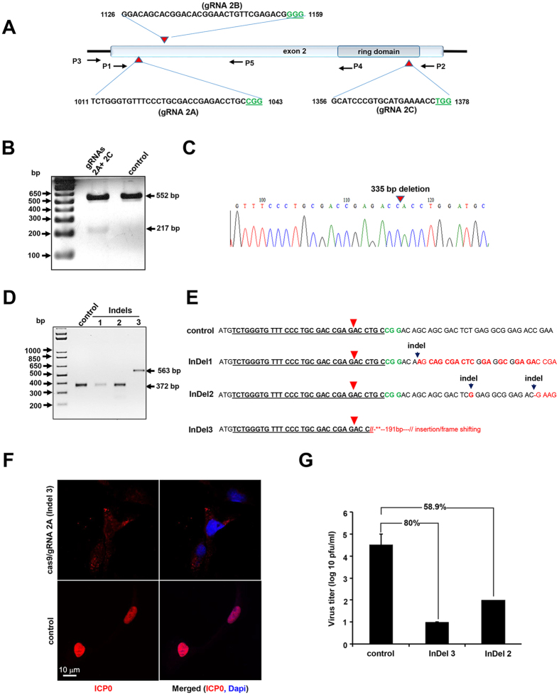
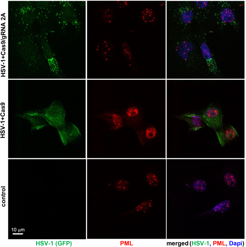
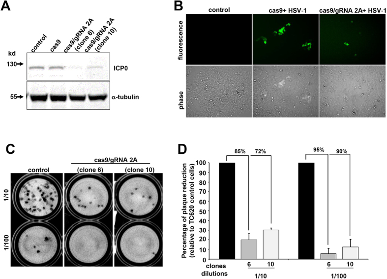
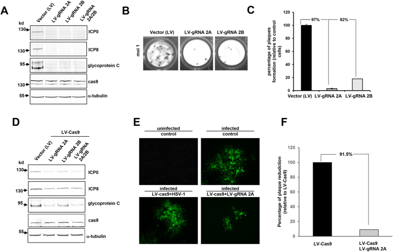
HSV-1 induced illness affects greater than 85% of adults worldwide with no permanent curative therapy. We used RNA-guided CRISPR/Cas9 gene editing to specifically target for deletion of DNA sequences of the HSV-1 genome that span the region directing expression of ICP0, a key viral protein that stimulates HSV-1 gene expression and replication. We found that CRISPR/Cas9 introduced InDel mutations into exon 2 of the ICP0 gene profoundly reduced HSV-1 infectivity in permissive human cell culture models and protected permissive cells against HSV-1 infection. CRISPR/Cas9 mediated targeting ICP0 prevented HSV-1-induced disintegration of promonocytic leukemia (PML) nuclear bodies, an intracellular event critical to productive HSV-1 infection that is initiated by interaction of the ICP0 N-terminus with PML. Combined treatment of cells with CRISPR targeting ICP0 plus the immediate early viral proteins, ICP4 or ICP27, completely abrogated HSV-1 infection. We conclude that RNA-guided CRISPR/Cas9 can be used to develop a novel, specific and efficacious therapeutic and prophylactic platform for targeted viral genomic ablation to treat HSV-1 diseases.
Overview
The claims and statistics of this paper haven’t been checked yet.
Run the check — free, no account needed. It reads the open-access text and checks the sources it cites.
Integrity signals
Registry
Cites
Statistics
Figures
Verification
Claims the paper makes
The claims and statistics of this paper haven’t been checked yet.
Run the check — free, no account needed. It reads the open-access text and checks the sources it cites.
Claims other papers make about this one
Each is checked against this paper’s own text: does it say what the citing paper says it does?
The cited source says what the claim attributes to it.
In the citing paper
“target of herpes simplex virus-1 genome regions essential to viral protein expression during early and late phases of viral infection/reinfection to suppress infection and prevent new infection (Roehm et al., 2016)”
From Delivering CRISPR: a review of the challenges and approaches (2018) · cites this paper as Roehm et al., 2016
Self-check
Percentages, sums and reported differences re-computed from the paper’s own numbers.
The self-check needs the paper’s full text.
Citations
Cites
Harnessing the CRISPR/Cas9 system to disrupt latent HIV-1 provirus
Ebina, H., Misawa, N., Kanemura, Y., & Koyanagi, Y. (2013) · Scientific Reports · doi:10.1038/srep02510 · Cited by 565
RNA-guided endonuclease provides a therapeutic strategy to cure latent herpesviridae infection
Wang, J., & Quake, S. R. (2014) · Proceedings of the National Academy of Sciences · doi:10.1073/pnas.1410785111 · Cited by 220
Research works printed but not linked: 42
- Whitley, R. J. & Roizman B. Herpes simplex virus infections. Lancet 357, 1513–1518. · DOI
- Xu, F. et al. Trends in herpes simplex virus type 1 and type 2 seroprevalence in the United States. JAMA 296, 964–973. · DOI
- Baringer, J. R. Herpes Virus Encephalitis. In: Davis, L. E., Kennedy, P. G. E. (eds) Infectious diseases of the nervous system. Butterworth-Heinemann. Oxford pp. 139–164 (2000).
- Kennedy, P. G. E. & Chaudhuri. A. Herpes simplex encephalitis. J. Neurol. Neurosurg. Psychiatry 73, 237–238 (2002). · DOI
- Kennedy, P. G. E. & Steiner, I. Recent issues in herpes simplex encephalitis. J. Neurovirol. 19, 346–350 (2013). · DOI
- Westerberg, B. D., Atashband, S. & Kozak, F. K. A systematic review of the incidence of sensorineural hearing loss in neonates exposed to herpes simplex virus (HSV). Int. J. Pediatr. Otorhinolaryngol. 72, 931–937 (2008). · DOI
- Gilden, D. H., Mahalingam, R., Cohrs, R. J. & Tyler, K. L. Herpesvirus infections of the nervous system. Nature Clin. Practice 3, 82–94 (2007). · DOI
- Fraser, N. W., Lawrence, W. C., Wroblewska, Z., Gilden, D. H. & Koprowski, H. Herpes simplex type 1 DNA in human brain tissue. Proc Natl Acad Sci USA 78, 6461–6465 (1981) · DOI
- Mahalingam, R. Wellish, M. C., Dueland, A. N., Cohrs, R. J. & Gilden, D. H. Localization of herpes simplex virus and varicella zoster virus DNA in human ganglia. Ann. Neurol. 31, 444–448 (1992). · DOI
- Izumi, K. M., McKelvey, A. M., Devi-Rao, G., Wagner, E. K. & Stevens, J. G. Molecular and biological characterization of a type 1 herpes simplex virus (HSV-1) specifically deleted for expression of the latency-associated transcript (LAT). Microb. Pathog. 7, 121–134. · DOI
- Kang, W., Mukerjee, R. & Fraser, N. W. Establishment and maintenance of HSV latent infection is mediated through correct splicing of the LAT primary transcript. Virology 312, 233–244 (2003). · DOI
- Perng, G. C. et al. Virus-induced neuronal apoptosis blocked by the herpes simplex virus latency-associated transcript. Science 287, 1500–1503. · DOI
- Stevens, J. G., Wagner, E. K., Devi-Rao, G. B., Cook, M. L. & Feldman, L. T. RNA complemenatary to a herpesvirus alpha gene mRNA is prominent in latenly infected neurons. Science 235, 1056–1059 (1987). · DOI
- Belshe, R. B. et al. Efficacy results of a trial of a herpes simplex vaccine. N Engl J Med 366, 34–43 (2012). · DOI
- Garneau, J. E. et al. The CRISPR/Cas bacterial immune system cleaves bacteriophage and plasmid DNA. Nature 468, 67–71 (2010). · DOI
- Gasiunas, G., Barrangou, R., Horvath, P. & Siksnys, V. Cas9-crRNA ribonucleoprotein complex mediates specific DNA cleavage for adaptive immunity in bacteria. Proc Natl Acad Sci USA 109, 15539–15540 (2012). · DOI · Paper page
- Mali, P. et al. RNA-guided human genome engineering via Cas9. Science 339, 823–826 (2013). · DOI · Paper page
- Cong, L. et al. Multiplex genome engineering using CRISPR/Cas systems. Science 339, 819–823 (2013). · DOI · Paper page
- Hu, W. et al. RNA-directed gene editing specifically eradicates latent and prevents new HIV-1 infection. Proc. Natl. Acad. Sci. USA 111, 11461–11466 (2014). · DOI · Paper page
- Wollebo, H. S. et al. CRISPR/Cas9 system as an agent for eliminating polyomavirus JC infection. PLoS One 10, e0136046 (2015) · DOI
- Yuen, K. S. et al. CRISPR/Cas9-mediated genome editing of Epstein-Barr virus in human cells. J. Gen. Virol. 96, 626–636 (2015). · DOI
- Delboy, M. G. & Nicola, A. V. A pre-immediate early role for tegument ICP0 in the proteasome-dependent entry of herpes simplex virus. J. Virol. 85, 5910–5918 (2011). · DOI
- Boutell, C. & Everett, R. D. Regulation of alphaherpesvirus infections by the ICP0 family of proteins. J. Gen. Virol. 94, 465–481 (2013). · DOI
- Hagglund, R. & Roizman, B. Role of ICP0 in the strategy of conquest of the host cell by herpes simplex virus 1. J. Virol. 78, 2169–2178 (2004). · DOI
- Samaniego, L. A., Wu, N. & DeLuca, N. A. The herpes simplex virus immediate-early protein ICP0 affects transcription from the viral genome and infected-cell survival in the absence of ICP4 and ICP27. J. Virol. 71, 4614–4625 (1997). · DOI
- Halford, W. P. et al. ICP0 antagonizes Stat 1-dependent repression of herpes simples virus: implications for the regulation of viral latency. Virol. J. 3, 44 (2006). · DOI
- Halford, W. P., Püschel, R. & Rakowski, B. Herpes simplex virus 2 ICP0- mutant viruses are avirulent and immunogenic: implications for a genital herpes virus vaccine. PloS One 5, e12251 (2010). · DOI
- Sahin, U., Lallemand-Breitenbach, V. & de Thé, H. PML nuclear bodies: regulation, function and therapeutic perspectives. J. Pathol. 234, 289–291 (2014). · DOI
- Wang, S., Long, J. & Zheng, C. F. The potential link between PML NBs and ICP0 in regulating lytic and latent infection of HSV-1. Protein Cell 3, 372–382 (2012). · DOI
- Cuchet-Lourenco, D., Vanni, E., Glass, M., Orr, A. & Everett, R. D. Herpes simplex virus 1 ubiquitin ligase ICP0 interacts with PML isoform I and induces its SUMO-dependent degradation. J. Virol. 86, 11209–11222 (2012). · DOI
- Everett, R. D. et al. PML contributes to a cellular mechanism of repression of herpes simplex virus type 1 infection that is inactivated by ICP0. J. Virol. 80, 7995–8005 (2006). · DOI
- Merrill, J. E. & Matsushima, K. Production of and response to interleukin 1 by cloned human oligodendroglioma cell lines. J. Biol. Regul. Homeost. Agents 2, 77–86 (1988).
- Challberg, M. D. A method for identifying the viral genes required for herpesvirus DNA replication. Proc. Natl. Acad. Sci. USA 83, 9094–9098 (1986). · DOI
- Gao, M. & Knipe, D. M. Potential role for herpes simplex virus ICP8 DNA replication protein in stimulation of late gene expression. J. Virol. 65, 2666–2675 (1991). · DOI
- Tanguy Le Gac, N., Villani, G. & Boehmer, P. E. Herpes simplex virus type-1 single-strand DNA-binding protein (ICP8) enhances the ability of the viral DNA helicase-primase to unwind cisplatin-modified DNA. J. Biol. Chem. 273, 13801–13807 (1998). · DOI
- Friedman, H. M., Cohen, G. H., Eisenberg, R. J., Seidel, C. A. & Cines, D. B. Glycoprotein C of herpes simplex virus 1 acts as a receptor for the C3b complement component on infected cells. Nature 309, 633–635 (1984). · DOI
- Chen, J. & Silverstein, S. Herpes simplex viruses with mutations in the gene encoding ICP0 are defective in gene expression. J. Virol. 66, 2916–2927 (1992). · DOI
- Knipe, D. M. & Cliffe, A. Chromatin control of herpes simplex virus lytic and latent infection. Nature Rev. 6, 211–221 (2008).
- Horvath, P. & Barrangou, R. CRISPR/Cas9, the immune system of bacteria and archaea. Science 327, 167–170 (2010). · DOI
- Shekarabi, M. et al. Mutations in the nervous system—specific HSN2 exon of WNK1 cause heredity sensory neuropathy type II. J. Clin. Invest. 118, 2496–2505 (2008).
- Shekarabi, M. & Kennedy, T. E. The netrin-1 receptor DCC promotes filopodia formation and cell spreading by activating Cdc42 and Rac1. Mol. Cell. Neurosci. 19, 1–17 (2002). · DOI
- Cai, W. & Schaffer, P. A. Herpes simplex virus type 1 ICP0 regulates expression of immediate-early, early and late genes in productively infected cells. J. Virol. 66, 2904–2915 (1992). · DOI
Cited by
Sorted by citations
Delivering CRISPR: a review of the challenges and approaches
Lino, C. A., Harper, J. C., Carney, J. P., & Timlin, J. A. (2018) · Drug Delivery · doi:10.1080/10717544.2018.1474964 · Cited by 1,185
Wang, H.-X., Li, M., Lee, C. M., Chakraborty, S., Kim, H.-W., Bao, G., & Leong, K. W. (2017) · Chemical Reviews · doi:10.1021/acs.chemrev.6b00799 · Cited by 560
Ortinski, P. I., O’Donovan, B., Dong, X., & Kantor, B. (2017) · Molecular Therapy - Methods & Clinical Development · doi:10.1016/j.omtm.2017.04.002 · Cited by 146
Pandey, M., Ojha, D., Bansal, S., Rode, A. B., & Chawla, G. (2021) · Molecular Aspects of Medicine · doi:10.1016/j.mam.2021.101003 · Cited by 23
Vijayraghavan, S., & Kantor, B. (2017) · Journal of Visualized Experiments · doi:10.3791/56915 · Cited by 20
Metadata
- Authors
- Roehm, P. C., Shekarabi, M., Wollebo, H. S., Bellizzi, A., He, L., Salkind, J., & Khalili, K.
- Venue
- Scientific Reports
- Published
- 2016-04-11
- Volume
- 6
- Issue
- 1
- ISSN
- 2045-2322
- PMC
- PMC4827394
- Cited by
- 128
- References
- 2
- Language
- en
About these checks
Registry, retraction, Cites and statistics results are computed, not judged, with one exception: a printed reference that no index lists is read once by a model before it counts as a citation error. Claims are extracted by a model and kept only when their quote appears verbatim in the text; each cited source is then checked, with a model’s help, for whether it says what the claim relies on. Those verdicts can be wrong — read the claim’s quote and the cited source before relying on one.
Statistics and arithmetic are recomputed, not judged.
Confidence bands are shown only for fields calibrated on our annotated test set. None is calibrated yet, so no band is shown.
What counts as a claim ›How we measure confidence ›How we check this ›
Are you an author, or do you see a problem with this page? Request a review
How to cite
Cite CoChat for the checks on this page, and the paper for its own findings.
- This page
- CoChat. (2026). Citation check: Inhibition of HSV-1 Replication by Gene Editing Strategy. https://app.cochat.ai/papers/10.1038/srep23146
- The paper
- Roehm, P. C., Shekarabi, M., Wollebo, H. S., Bellizzi, A., He, L., Salkind, J., & Khalili, K. (2016). Inhibition of HSV-1 Replication by Gene Editing Strategy. Scientific Reports. https://doi.org/10.1038/srep23146
