

The DOI matches its Crossref record.
A quantitative method to measure biofilm removal efficiency from complex biomaterial surfaces using SEM and image analysis
Abstract
Biofilm accumulation on biomaterial surfaces is a major health concern and significant research efforts are directed towards producing biofilm resistant surfaces and developing biofilm removal techniques. To accurately evaluate biofilm growth and disruption on surfaces, accurate methods which give quantitative information on biofilm area are needed, as current methods are indirect and inaccurate. We demonstrate the use of machine learning algorithms to segment biofilm from scanning electron microscopy images. A case study showing disruption of biofilm from rough dental implant surfaces using cavitation bubbles from an ultrasonic scaler is used to validate the imaging and analysis protocol developed. Streptococcus mutans biofilm was disrupted from sandblasted, acid etched (SLA) Ti discs and polished Ti discs. Significant biofilm removal occurred due to cavitation from ultrasonic scaling (p < 0.001). The mean sensitivity and specificity values for segmentation of the SLA surface images were 0.80 ± 0.18 and 0.62 ± 0.20 respectively and 0.74 ± 0.13 and 0.86 ± 0.09 respectively for polished surfaces. Cavitation has potential to be used as a novel way to clean dental implants. This imaging and analysis method will be of value to other researchers and manufacturers wishing to study biofilm growth and removal.
Overview
The claims and statistics of this paper haven’t been checked yet.
Run the check — free, no account needed. It reads the open-access text and checks the sources it cites.
Integrity signals
Registry
Cites
Statistics
Figures
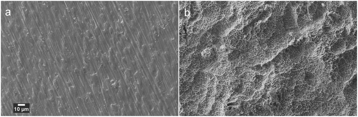
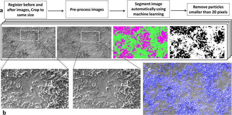
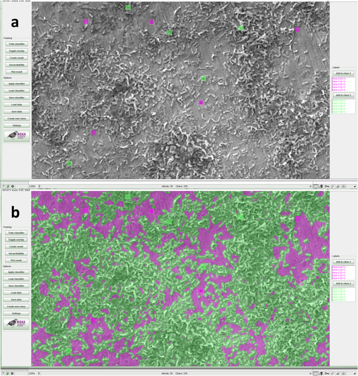
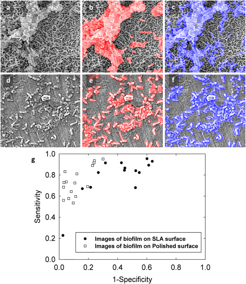
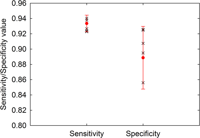
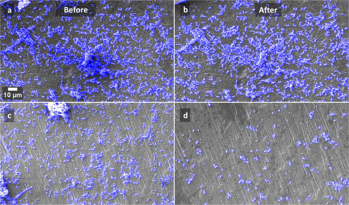
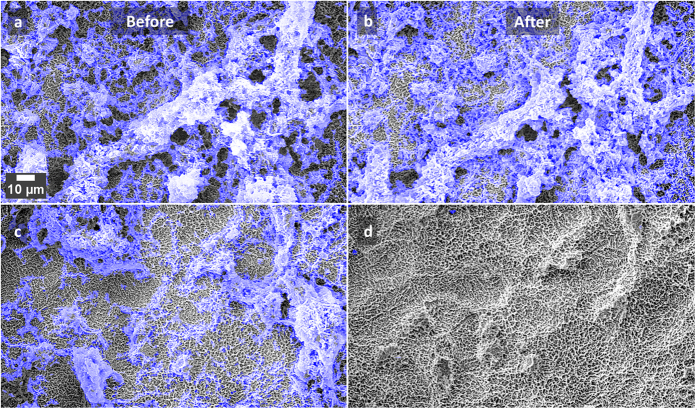
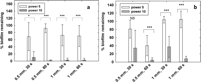
Verification
Claims the paper makes
The claims and statistics of this paper haven’t been checked yet.
Run the check — free, no account needed. It reads the open-access text and checks the sources it cites.
Claims other papers make about this one
Each is checked against this paper’s own text: does it say what the citing paper says it does?
In the citing paper
“A few studies have utilized TWIST-based biofilm segmentation using SEM images but primarily for the abiotic surfaces.38,65”
From SEMTWIST Quantification of Biofilm Infection in Human Chronic Wound Using Scanning Electron Microscopy and Machine Learning (2025) · cites this paper as [38]
In the citing paper
“Weka, an open-source software tool that enables users to perform ML-based image segmentation, object classification, and feature extraction with minimal programming experience,33-35 has been widely used in biological and clinical research to quantitatively analyze various types of microorganisms, cells, and tissues.36-40”
From SEMTWIST Quantification of Biofilm Infection in Human Chronic Wound Using Scanning Electron Microscopy and Machine Learning (2025) · cites this paper as [38]
Self-check
Percentages, sums and reported differences re-computed from the paper’s own numbers.
The self-check needs the paper’s full text.
Citations
Cites
No references could be resolved yet.
Research works printed but not linked: 39
- Costerton, J. W., Geesey, G. G. & Cheng, K. J. How Bacteria Stick. Sci Am 238, 86-& (1978). · DOI
- Costerton, J. W., Stewart, P. S. & Greenberg, E. P. Bacterial biofilms: a common cause of persistent infections. Science 284, 1318–1322 (1999). · DOI · Paper page
- Donlan, R. M. & Costerton, J. W. Biofilms: Survival mechanisms of clinically relevant microorganisms. Clin Microbiol Rev 15, 167-+, doi: 10.1128/Cmr.15.2.167-193.2002 (2002). · DOI · Paper page
- Bjarnsholt, T. The role of bacterial biofilms in chronic infections. APMIS 121, 1–58, doi: 10.1111/apm.12099 (2013). · DOI · Paper page
- Clegg, M. S., Vertucci, F. J., Walker, C., Belanger, M. & Britto, L. R. The effect of exposure to irrigant solutions on apical dentin biofilms in vitro. Journal of Endodontics 32, 434–437 (2006). · DOI
- Hadi, R., Vickery, K., Deva, A. & Charlton, T. Biofilm removal by medical device cleaners: comparison of two bioreactor detection assays. Journal of Hospital Infection 74, 160–167, doi: 10.1016/j.jhin.2009.10.023 (2010). · DOI
- Schwarz, F. et al. Influence of plaque biofilm removal on reestablishment of the biocompatibility of contaminated titanium surfaces. Journal of Biomedical Materials Research Part A 77, 437–444 (2006). · DOI
- Vickery, K., Pajkos, A. & Cossart, Y. Removal of biofilm from endoscopes: evaluation of detergent efficiency. American Journal of Infection Control 32, 170–176, doi: 10.1016/j.ajic.2003.10.009 (2004). · DOI
- Wu, Y. T., Zhu, H., Willcox, M. & Stapleton, F. The Effectiveness of Various Cleaning Regimens and Current Guidelines in Contact Lens Case Biofilm Removal. Investigative Opthalmology & Visual Science 52, 5287, doi: 10.1167/iovs.10-6785 (2011). · DOI
- Vertes, A., Hitchins, V. & Phillips, K. S. Analytical challenges of microbial biofilms on medical devices. Analytical Chemistry 84, 3858–3866 (2012). · DOI
- Park, J.-B., Jang, Y. J., Choi, B.-K., Kim, K.-K. & Ko, Y. Treatment With Various Ultrasonic Scaler Tips Affects Efficiency of Brushing of SLA Titanium Discs. Journal of Craniofacial Surgery 24, e119–e123, doi: 10.1097/SCS.0b013e3182668a1a (2013). · DOI
- John, G., Becker, J. & Schwarz, F. Rotating titanium brush for plaque removal from rough titanium surfaces–an in vitro study. Clinical Oral Implants Research 25, 838–842 (2014). · DOI
- Kite, P., Eastwood, K., Sugden, S. & Percival, S. L. Use of In Vivo-Generated Biofilms from Hemodialysis Catheters To Test the Efficacy of a Novel Antimicrobial Catheter Lock for Biofilm Eradication In Vitro. J. Clin. Microbiol. 42, 3073–3076, doi: 10.1128/JCM.42.7.3073-3076.2004 (2004). · DOI
- Qian, Z., Stoodley, P. & Pitt, W. G. Effect of low-intensity ultrasound upon biofilm structure from confocal scanning laser microscopy observation. Biomaterials 17, 1975–1980 (1996). · DOI
- Agarwal, A., Jern Ng, W. & Liu, Y. Removal of biofilms by intermittent low-intensity ultrasonication triggered bursting of microbubbles. Biofouling 30, 359–365 (2014). · DOI
- Cruz, P. C. et al. The effectiveness of chemical denture cleansers and ultrasonic device in biofilm removal from complete dentures. Journal of Applied Oral Science 19, 668–673, doi: 10.1590/S1678-77572011000600021 (2011). · DOI
- Fricke, K. et al. Atmospheric Pressure Plasma: A High-Performance Tool for the Efficient Removal of Biofilms. PLoS ONE 7, e42539, doi: 10.1371/journal.pone.0042539 (2012). · DOI
- Hägi, T. T. et al. A Biofilm Pocket Model to Evaluate Different Non-Surgical Periodontal Treatment Modalities in Terms of Biofilm Removal and Reformation, Surface Alterations and Attachment of Periodontal Ligament Fibroblasts. PLoS ONE 10, e0131056, doi: 10.1371/journal.pone.0131056 (2015). · DOI
- Li, J. et al. Biofilm formation of Candida albicans on implant overdenture materials and its removal. Journal of Dentistry 40, 686–692, doi: 10.1016/j.jdent.2012.04.026 (2012). · DOI
- Nance, W. C. et al. A high-throughput microfluidic dental plaque biofilm system to visualize and quantify the effect of antimicrobials. Journal of Antimicrobial Chemotherapy 68, 2550–2560 (2013). · DOI
- Salles, A. E. S., Macedo, L. D., Fernandes, R. A. G., Silva‐Lovato, C. H. & Paranhos, H. d. F. O. Comparative analysis of biofilm levels in complete upper and lower dentures after brushing associated with specific denture paste and neutral soap. Gerodontology 24, 217–223 (2007). · DOI
- Sedgley, C., Applegate, B. & Nagel, A. & Hall, D. Real-Time Imaging and Quantification of Bioluminescent Bacteria in Root Canals In Vitro. Journal of Endodontics 30, 893–898, doi: 10.1097/01.DON.0000132299.02265.6C (2004). · DOI
- Tawakoli, P. N., Sauer, B., Becker, K., Buchalla, W. & Attin, T. Interproximal biofilm removal by intervallic use of a sonic toothbrush compared to an oral irrigation system. BMC oral health 15, 91 (2015). · DOI
- Whittaker, C., Ridgway, H. & Olson, B. H. Evaluation of Cleaning Strategies for Removal of Biofilms from Reverse-Osmosis Membranes. Applied and Environmental Microbiology 48, 395–403 (1984). · DOI
- Zhang, Y. & Hu, Z. Combined treatment of Pseudomonas aeruginosa biofilms with bacteriophages and chlorine. Biotechnology and Bioengineering 110, 286–295, doi: 10.1002/bit.24630 (2013). · DOI
- Murphy, J., Ali, M. J. & Psaltis, A. J. Biofilm Quantification on Nasolacrimal Silastic Stents After Dacryocystorhinostomy. Ophthalmic Plastic and Reconstructive Surgery 31, 396–400, doi: 10.1097/IOP.0000000000000361 (2015). · DOI
- Chan, T. F., Esedoglu, S. & Nikolova, M. Algorithms for finding global minimizers of image segmentation and denoising models. Siam J Appl Math 66, 1632–1648, doi: 10.1137/040615286 (2006). · DOI
- Le Guéhennec, L., Soueidan, A., Layrolle, P. & Amouriq, Y. Surface treatments of titanium dental implants for rapid osseointegration. Dental Materials 23, 844–854 (2007). · DOI · Paper page
- Rimondini, L., Fare, S., Brambilla, E. & Felloni, A. The effect of surface roughness on early in vivo plaque colonization on titanium. Journal of Periodontology 68, 556–562 (1997). · DOI
- Rosen, P. et al. Peri-implant mucositis and peri-implantitis: a current understanding of their diagnoses and clinical implications. J Periodontol 84, 436–443 (2013). · DOI
- Walmsley, A. D., Lea, S. C., Landini, G. & Moses, A. J. Advances in power driven pocket/root instrumentation. Journal of Clinical Periodontology 35, 22–28, doi: 10.1111/j.1600-051X.2008.01258.x (2008). · DOI
- Wang, C. & Cheng, J. Cavitation microstreaming generated by a bubble pair in an ultrasound field. The Journal of the Acoustical Society of America 134, 1675–1682, doi: 10.1121/1.4812896 (2013). · DOI
- Felver, B., King, D. C., Lea, S. C., Price, G. J. & Damien Walmsley, A. Cavitation occurrence around ultrasonic dental scalers. Ultrasonics Sonochemistry 16, 692–697, doi: 10.1016/j.ultsonch.2008.11.002 (2009). · DOI
- Brennen, C. E. Cavitation and Bubble Dynamics. (Cambridge University Press, 2013). · DOI
- Blake, J. R. & Gibson, D. C. Cavitation Bubbles Near Boundaries. Annual Review of Fluid Mechanics 19, 99–123, doi: 10.1146/annurev.fl.19.010187.000531 (1987). · DOI
- Lauterborn, W., Kurz, T., Geisler, R., Schanz, D. & Lindau, O. Acoustic cavitation, bubble dynamics and sonoluminescence. Ultrasonics Sonochemistry 14, 484–491, doi: 10.1016/j.ultsonch.2006.09.017 (2007). · DOI
- Macaskie, L. E. et al. A novel non line-of-sight method for coating hydroxyapatite onto the surfaces of support materials by biomineralization. Journal of Biotechnology 118, 187–200, doi: 10.1016/j.jbiotec.2005.03.006 (2005). · DOI
- Schindelin, J. et al. Fiji: an open-source platform for biological-image analysis. Nature Methods 9, 676–682 (2012). · DOI · Paper page
- Arganda-Carreras, I. et al. Consistent and elastic registration of histological sections using vector-spline regularization. Lect Notes Comput Sc 4241, 85–95 (2006). · DOI
Cited by
Sorted by citations
SuRVoS: Super-Region Volume Segmentation workbench
Luengo, I., Darrow, M. C., Spink, M. C., Sun, Y., Dai, W., He, C. Y., Chiu, W., Pridmore, T., Ashton, A. W., Duke, E. M. H., et al. (2017) · Journal of Structural Biology · doi:10.1016/j.jsb.2017.02.007 · Cited by 89
Singh, S., Muniz De Oliveira, F., Wang, C., Kumar, M., Xuan, Y., DeMazumder, D., Sen, C. K., & Roy, S. (2025) · Advances in Wound Care · doi:10.1089/wound.2024.0291 · Cited by 6
Metadata
- Authors
- Vyas, N., Sammons, R. L., Addison, O., Dehghani, H., & Walmsley, A. D.
- Venue
- Scientific Reports
- Published
- 2016-09-07
- Volume
- 6
- Issue
- 1
- ISSN
- 2045-2322
- PMC
- PMC5013386
- Cited by
- 98
- References
- 0
- Language
- en
About these checks
Registry, retraction, Cites and statistics results are computed, not judged, with one exception: a printed reference that no index lists is read once by a model before it counts as a citation error. Claims are extracted by a model and kept only when their quote appears verbatim in the text; each cited source is then checked, with a model’s help, for whether it says what the claim relies on. Those verdicts can be wrong — read the claim’s quote and the cited source before relying on one.
Statistics and arithmetic are recomputed, not judged.
Confidence bands are shown only for fields calibrated on our annotated test set. None is calibrated yet, so no band is shown.
What counts as a claim ›How we measure confidence ›How we check this ›
Are you an author, or do you see a problem with this page? Request a review
