

The DOI matches its Crossref record; 6 of the 6 references it cites were found in a registry or an index.
The toxicity of nanoparticles and their interaction with cells: an in vitro metabolomic perspective
Abstract
The interaction of nanoparticles (NPs) with cells depends on their physicochemical properties and can lead to cytotoxic events. Metabolomics reveals the molecular mechanisms of this nanotoxicity and avoids the misleading interferences with NPs.
Overview
The claims and statistics of this paper haven’t been checked yet.
Run the check — free, no account needed. It reads the open-access text and checks the sources it cites.
Integrity signals
Registry
Cites
Statistics
Figures
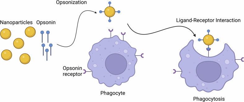
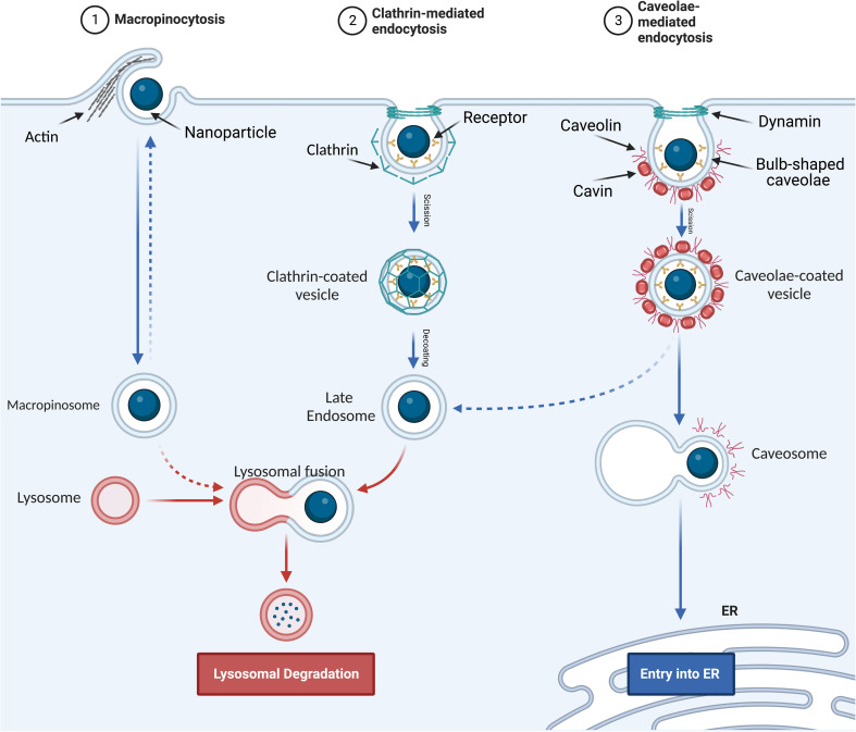
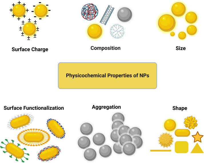
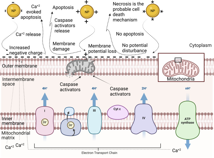
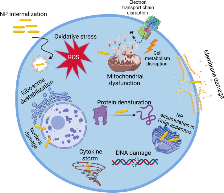
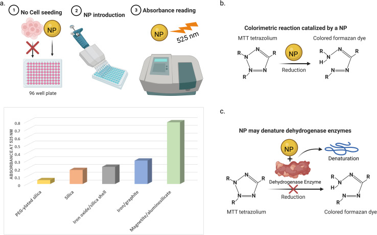
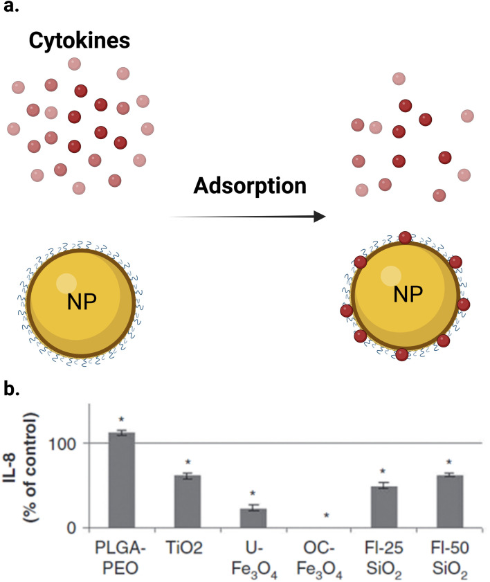
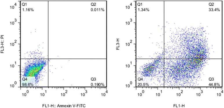
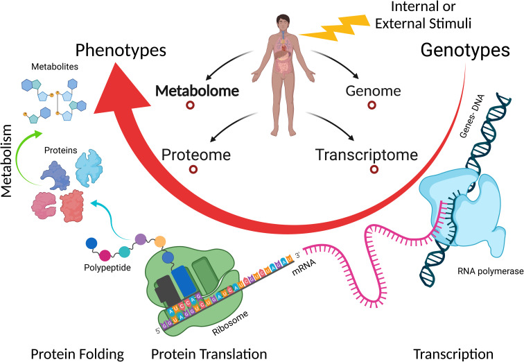
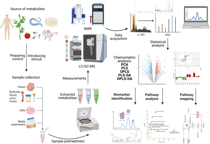
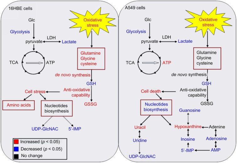
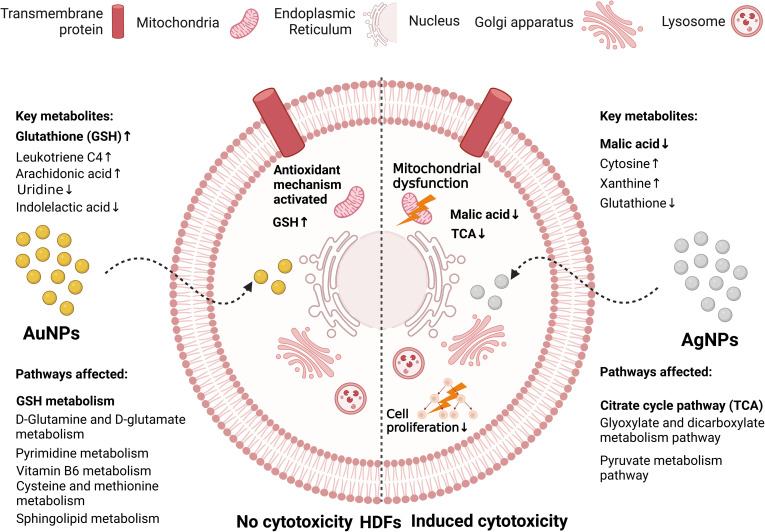


Verification
Claims the paper makes
The claims and statistics of this paper haven’t been checked yet.
Run the check — free, no account needed. It reads the open-access text and checks the sources it cites.
Claims other papers make about this one
Each is checked against this paper’s own text: does it say what the citing paper says it does?
In the citing paper
“Cytotoxicity is often evaluated through in vitro analyses using human and animal cell lines. These tests consider viability, oxidative stress, apoptosis, and membrane integrity after exposure to nanoparticles [72].”
From Advances in green-synthesized magnetic nanoparticles for targeted cancer therapy: mechanisms, applications, and future perspectives (2026) · cites this paper as [72]
Self-check
Percentages, sums and reported differences re-computed from the paper’s own numbers.
The self-check needs the paper’s full text.
Citations
Cites
Apoptosis: A Review of Programmed Cell Death
Elmore, S. (2007) · Toxicologic Pathology · doi:10.1080/01926230701320337 · Cited by 13,853
Cellular uptake of nanoparticles: Journey inside the cell
Behzadi, S., Serpooshan, V., Tao, W., Hamaly, M. A., Alkawareek, M. Y., Dreaden, E. C., Brown, D., Alkilany, A. M., Farokhzad, O. C., & Mahmoudi, M. (2017) · Chemical Society Reviews · doi:10.1039/c6cs00636a · Cited by 2,859
Phagocytosis: An Immunobiologic Process
Gordon, S. (2016) · Immunity · doi:10.1016/j.immuni.2016.02.026 · Cited by 902
Nanotechnology in cosmetics: Opportunities and challenges
Raj, S., Jose, S., Sumod, U., & Sabitha, M. (2012) · Journal of Pharmacy And Bioallied Sciences · doi:10.4103/0975-7406.99016 · Cited by 545
Kinoshita, S., Akira, S., & Kishimoto, T. (1992) · Proceedings of the National Academy of Sciences · doi:10.1073/pnas.89.4.1473 · Cited by 327
Guo, C., Weber, R. J. M., Buckley, A., Mazzolini, J., Robertson, S., Delgado-Saborit, J. M., Rappoport, J. Z., Warren, J., Hodgson, A., Sanderson, P., et al. (2021) · International Journal of Molecular Sciences · doi:10.3390/ijms22020556 · Cited by 25
Research works printed but not linked: 308
- X.Cheng , in Nanolithography , Elsevier , 2014 , pp. 348–375 · DOI
- Hulla, 2015, Hum. Exp. Toxicol., 34, 1318, 10.1177/0960327115603588 · DOI
- Wright, 2016, Med. J. Aust., 204, 369, 10.5694/mja15.01128 · DOI
- Santos, 2015, Mater. Today: Proc., 2, 456
- He, 2019, J. Food Drug Anal., 27, 1, 10.1016/j.jfda.2018.12.002 · DOI
- Fraceto, 2016, Front. Environ. Sci., 4, 10.3389/fenvs.2016.00020 · DOI
- Chellaram, 2014, APCBEE Proc., 8, 109, 10.1016/j.apcbee.2014.03.010 · DOI
- Mitchell, 2021, Nat. Rev. Drug Discovery, 20, 101, 10.1038/s41573-020-0090-8 · DOI · Paper page
- Javed, 2021, Fibers Polym., 22, 77, 10.1007/s12221-021-0051-9 · DOI
- Mathew, 2019, J. King Saud Univ., Sci., 31, 586, 10.1016/j.jksus.2018.03.015 · DOI
- Sekoai, 2019, Fuel, 237, 380, 10.1016/j.fuel.2018.10.030 · DOI
- Zhang, 2009, Sensors, 9, 1033, 10.3390/s90201033 · DOI
- Borm, 2006, Part. Fibre Toxicol., 3, 11, 10.1186/1743-8977-3-11 · DOI
- A. B.Asha and R.Narain , in Polymer Science and Nanotechnology , Elsevier , 2020 , pp. 343–359 · DOI
- Ray, 2009, J. Environ. Sci. Health, Part C: Environ. Carcinog. Ecotoxicol. Rev., 27, 1, 10.1080/10590500802708267 · DOI
- Saleh, 2020, Environ. Technol. Innovation, 20, 101067, 10.1016/j.eti.2020.101067 · DOI
- Seaton, 2010, J. R. Soc., Interface, 7, 10.1098/rsif.2009.0252.focus · DOI
- Kan, 2018, Inhalation Toxicol., 30, 335, 10.1080/08958378.2018.1535634 · DOI
- Chrishtop, 2021, Toxicology, 462, 10.1016/j.tox.2021.152952 · DOI
- Hallock, 2009, J. Chem. Health Saf., 16, 16, 10.1016/j.jchas.2008.04.001 · DOI
- Sohaebuddin, 2010, Part. Fibre Toxicol., 7, 22, 10.1186/1743-8977-7-22 · DOI
- Alshatwi, 2013, Food Addit. Contam., Part A, 30, 1, 10.1080/19440049.2012.729160 · DOI
- Senapati, 2015, Food Chem. Toxicol., 85, 61, 10.1016/j.fct.2015.06.018 · DOI
- Yuan, 2020, Theranostics, 10, 4589, 10.7150/thno.34065 · DOI
- Nogueira, 2014, Asian Pac. J. Cancer Prev., 15, 4739, 10.7314/APJCP.2014.15.12.4739 · DOI
- Ramalingam, 2016, RSC Adv., 6, 20598, 10.1039/C5RA26781A · DOI
- Fu, 2014, J. Food Drug Anal., 22, 64, 10.1016/j.jfda.2014.01.005 · DOI
- Zhang, 2019, Bull. Environ. Contam. Toxicol., 103, 181, 10.1007/s00128-019-02622-0 · DOI
- Shin, 2019, Arch. Toxicol., 93, 1201, 10.1007/s00204-019-02402-z · DOI
- Zhao, 2019, J. Nanomater., 2019, 1
- Duan, 2013, PLoS One, 8, e62087, 10.1371/journal.pone.0062087 · DOI
- Shvedova, 2003, J. Toxicol. Environ. Health, Part A, 66, 1909, 10.1080/713853956 · DOI
- Magrez, 2006, Nano Lett., 6, 1121, 10.1021/nl060162e · DOI
- Jeng, 2006, J. Environ. Sci. Health, Part A: Toxic/Hazard. Subst. Environ. Eng., 41, 2699, 10.1080/10934520600966177 · DOI
- Blacklock, 2010, Biomaterials, 31, 7167, 10.1016/j.biomaterials.2010.06.002 · DOI
- Bahadar, 2016, Iran. Biomed. J., 20, 1
- Wei, 2014, J. Exp. Nanosci., 9, 625, 10.1080/17458080.2012.683534 · DOI
- Fröhlich, 2017, J. Nanobiotechnol., 15, 84, 10.1186/s12951-017-0320-3 · DOI
- Clish, 2015, Molecular Case Studies, 1, a000588, 10.1101/mcs.a000588 · DOI
- A.Klassen , A. T.Faccio , G. A. B.Canuto , P. L. R.da Cruz , H. C.Ribeiro , M. F. M.Tavares and A.Sussulini , in Advances in Experimental Medicine and Biology , 2017 , pp. 3–17
- Peng, 2020, Environ. Pollut., 267, 115434, 10.1016/j.envpol.2020.115434 · DOI
- Ž.Krpetić , S.Anguissola , D.Garry , P. M.Kelly and K. A.Dawson , in Advances in Experimental Medicine and Biology , 2014 , pp. 135–156
- Nierenberg, 2018, Rep. Practical Oncol. Radiother., 23, 300, 10.1016/j.rpor.2018.05.005 · DOI
- Manzanares, 2020, Pharmaceutics, 12, 371, 10.3390/pharmaceutics12040371 · DOI
- Colucci-Guyon, 2005, Curr. Biol., 15, 2007, 10.1016/j.cub.2005.09.051 · DOI
- Mahmoudi, 2011, Chem. Rev., 111, 3407, 10.1021/cr1003166 · DOI
- Kou, 2013, Asian J. Pharm. Sci., 8, 1, 10.1016/j.ajps.2013.07.001 · DOI
- K.Hirota and H.Ter , in Molecular Regulation of Endocytosis , 2012 , 10.5772/45820 · DOI
- Lim, 2011, Immunol. Cell Biol., 89, 836, 10.1038/icb.2011.20 · DOI
- Pastan, 1981, Annu. Rev. Physiol., 43, 239, 10.1146/annurev.ph.43.030181.001323 · DOI
- Kovtun, 2015, J. Cell Sci., 128, 1269, 10.1242/jcs.167866 · DOI · Paper page
- Lajoie, 2010, Int. Rev. Cell Mol. Biol., 282, 135, 10.1016/S1937-6448(10)82003-9 · DOI
- Jones, 2009, Adv. Drug Delivery Rev., 61, 438, 10.1016/j.addr.2009.03.005 · DOI
- Zhu, 2013, Acc. Chem. Res., 46, 622, 10.1021/ar300031y · DOI
- Huang, 2020, Sci. Adv., 6, eaba1321, 10.1126/sciadv.aba1321 · DOI
- Li, 2019, Small, 15, 1901687, 10.1002/smll.201901687 · DOI
- Shrestha, 2020, Adv. Colloid Interface Sci., 279, 102162, 10.1016/j.cis.2020.102162 · DOI
- Hui, 2008, J. Phys. Chem. C, 112, 11336, 10.1021/jp801632p · DOI
- Sabourian, 2020, Int. J. Mol. Sci., 21, 8019, 10.3390/ijms21218019 · DOI
- Kettler, 2014, Environ. Toxicol. Chem., 33, 481, 10.1002/etc.2470 · DOI
- Dong, 2020, Int. J. Nanomed., 15, 9089, 10.2147/IJN.S276105 · DOI
- Chithrani, 2007, Nano Lett., 7, 1542, 10.1021/nl070363y · DOI
- Chithrani, 2006, Nano Lett., 6, 662, 10.1021/nl052396o · DOI
- Sun, 2019, Front. Bioeng. Biotechnol., 7, 414, 10.3389/fbioe.2019.00414 · DOI
- Gliga, 2014, Part. Fibre Toxicol., 11, 11, 10.1186/1743-8977-11-11 · DOI
- Wu, 2016, Nanoscale, 8, 1237, 10.1039/c5nr07681a · DOI
- Woźniak, 2017, J. Mater. Sci.: Mater. Med., 28, 92, 10.1007/s10856-017-5902-y · DOI
- Asati, 2010, ACS Nano, 4, 5321, 10.1021/nn100816s · DOI
- Jeon, 2018, Nanomaterials, 8, 1028, 10.3390/NANO8121028 · DOI
- Schauer, 2009, Curr. Opin. Struct. Biol., 19, 507, 10.1016/j.sbi.2009.06.003 · DOI
- Fröhlich, 2012, Int. J. Nanomed., 7, 5577, 10.2147/IJN.S36111 · DOI
- He, 2010, Biomaterials, 31, 3657, 10.1016/j.biomaterials.2010.01.065 · DOI · Paper page
- Untener, 2013, ACS Appl. Mater. Interfaces, 5, 8366, 10.1021/am402848q · DOI
- Schaeublin, 2011, Nanoscale, 3, 410, 10.1039/c0nr00478b · DOI
- Li, 2021, Chem. Phys. Lett., 766, 138336, 10.1016/j.cplett.2021.138336 · DOI
- Bai, 2020, ACS Nano, 14, 289, 10.1021/acsnano.9b04407 · DOI
- Li, 2008, J. Phys. Chem. B, 112, 16647, 10.1021/jp8051906 · DOI
- Muthukumarasamyvel, 2017, ACS Omega, 2, 3527, 10.1021/acsomega.7b00353 · DOI
- Sun, 2018, J. Mater. Chem. B, 6, 1633, 10.1039/c7tb03153j · DOI
- Jambhrunkar, 2014, Mol. Pharm., 11, 3642, 10.1021/mp500385n · DOI
- Shao, 2015, Cell Proliferation, 48, 465, 10.1111/cpr.12192 · DOI
- Tao, 2015, Adv. Healthcare Mater., 4, 1203, 10.1002/adhm.201400751 · DOI
- Lund, 2011, Biomaterials, 32, 9776, 10.1016/j.biomaterials.2011.09.018 · DOI
- Yeh, 2013, Nanoscale, 5, 12140, 10.1039/c3nr04037b · DOI
- Ding, 2018, Small, 14, 1801451, 10.1002/smll.201801451 · DOI
- Carnovale, 2019, ACS Omega, 4, 242, 10.1021/acsomega.8b03227 · DOI
- Steckiewicz, 2019, J. Mater. Sci.: Mater. Med., 30, 22, 10.1007/s10856-019-6221-2 · DOI
- Zhang, 2017, Sci. Rep., 7, 7315, 10.1038/s41598-017-07588-9 · DOI
- Dong, 2019, Sci. Total Environ., 690, 158, 10.1016/j.scitotenv.2019.06.532 · DOI
- Savage, 2019, Methods Mol. Biol., 1894, 1, 10.1007/978-1-4939-8916-4_1 · DOI
- Kumar, 2017, Int. Nano Lett., 7, 243, 10.1007/s40089-017-0221-3 · DOI
- J. M.Veranth , in Nanoscience and Nanotechnology , John Wiley & Sons, Inc. , Hoboken, NJ, USA , 2008 , pp. 261–286 · DOI
- Sayes, 2007, Toxicol. Sci., 97, 163, 10.1093/toxsci/kfm018 · DOI
- Han, 2011, Toxicology, 287, 99, 10.1016/j.tox.2011.06.011 · DOI
- Stueker, 2014, Small, 10, 2006, 10.1002/smll.201303639 · DOI
- Drasler, 2017, NanoImpact, 8, 99, 10.1016/j.impact.2017.08.002 · DOI
- Díaz, 2008, Small, 4, 2025, 10.1002/smll.200800199 · DOI
- Hoshino, 2004, Nano Lett., 4, 2163, 10.1021/nl048715d · DOI
- Ray, 2012, Cell. Signalling, 24, 981, 10.1016/j.cellsig.2012.01.008 · DOI · Paper page
- He, 2017, Cell. Physiol. Biochem., 44, 532, 10.1159/000485089 · DOI
- Nita, 2016, Oxid. Med. Cell. Longevity, 2016, 3164734, 10.1155/2016/3164734 · DOI
- Higashi, 2018, Anal. Chem., 90, 5773, 10.1021/acs.analchem.8b00118 · DOI
- Katerji, 2019, Oxid. Med. Cell. Longevity, 2019, 1279250, 10.1155/2019/1279250 · DOI
- Kalyanaraman, 2012, Free Radical Biol. Med., 52, 1, 10.1016/j.freeradbiomed.2011.09.030 · DOI
- Whongsiri, 2018, Methods Mol. Biol., 1655, 109, 10.1007/978-1-4939-7234-0_9 · DOI
- M.Jeszka-Skowron , T.Podgórski and B.Czarczyńska-Goślińska , Determination of Antioxidant Biomarkers in Biological Fluids , Springer , Cham , 2021 , 10.1007/978-3-030-61879-7_11 · DOI
- Fantel, 1996, Teratology, 53, 196, 10.1002/(SICI)1096-9926(199603)53:3<196::AID-TERA7>3.0.CO;2-2 · DOI
- Ohkawa, 1979, Anal. Biochem., 95, 351, 10.1016/0003-2697(79)90738-3 · DOI
- Pylväs, 2011, Anticancer Res., 31, 1411
- Toyokuni, 1997, Lab. Invest., 76, 365
- Chiou, 2003, Clin. Chim. Acta, 334, 87, 10.1016/S0009-8981(03)00191-8 · DOI
- Golbamaki, 2015, Nanoscale, 7, 2154, 10.1039/c4nr06670g · DOI
- Aranda, 2013, Toxicol. in Vitro, 27, 954, 10.1016/j.tiv.2013.01.016 · DOI
- Pfaller, 2010, Nanotoxicology, 4, 52, 10.3109/17435390903374001 · DOI
- Yu, 2021, Ecotoxicol. Environ. Saf., 208, 111609, 10.1016/j.ecoenv.2020.111609 · DOI
- Tada-Oikawa, 2020, Chem. Res. Toxicol., 33, 1226, 10.1021/acs.chemrestox.9b00478 · DOI
- Lin, 2020, Toxicol. Ind. Health, 36, 759, 10.1177/0748233720947214 · DOI
- Guadagnini, 2015, Nanotoxicology, 9, 13, 10.3109/17435390.2013.829590 · DOI
- Piret, 2017, Arch. Toxicol., 91, 2315, 10.1007/s00204-016-1897-2 · DOI
- Ruffolo, 1964, Am. J. Pathol., 45, 741
- Ahamed, 2011, Toxicology, 283, 101, 10.1016/j.tox.2011.02.010 · DOI
- Park, 2008, Toxicol. Lett., 180, 222, 10.1016/j.toxlet.2008.06.869 · DOI
- Zhu, 2010, J. Nanosci. Nanotechnol., 10, 8584, 10.1166/jnn.2010.2488 · DOI
- Xu, 2012, J. Biomed. Mater. Res., 100 A, 738, 10.1002/jbm.a.33270 · DOI
- Xue, 2016, J. Appl. Toxicol., 36, 352, 10.1002/jat.3199 · DOI
- Reus, 2020, Toxicol. in Vitro, 63, 104723, 10.1016/j.tiv.2019.104723 · DOI
- Fiers, 1999, Oncogene, 18, 7719, 10.1038/sj.onc.1203249 · DOI
- Kumar, 2015, Food Chem. Toxicol., 85, 45, 10.1016/j.fct.2015.06.012 · DOI
- Vermes, 1995, J. Immunol. Methods, 184, 39, 10.1016/0022-1759(95)00072-I · DOI
- Vafaei, 2020, Biol. Trace Elem. Res., 198, 109, 10.1007/s12011-020-02054-6 · DOI
- Bancos, 2012, ISRN Nanotechnol., 2012, 454072, 10.5402/2012/454072 · DOI
- Ong, 2014, PLoS One, 9, e90650, 10.1371/journal.pone.0090650 · DOI
- Naseer, 2018, Nanotechnol. Rev., 7, 623, 10.1515/ntrev-2018-0076 · DOI
- Ramirez, 2013, ALTEX, 30, 209, 10.14573/altex.2013.2.209 · DOI
- Pang, 2021, Nucleic Acids Res., 49, W388, 10.1093/nar/gkab382 · DOI
- Idle, 2007, Cell Metab., 6, 348, 10.1016/j.cmet.2007.10.005 · DOI
- Gomez-Casati, 2013, BioMed Res. Int., 2013, 792527, 10.1155/2013/792527 · DOI
- Masutin, 2022, Exp. Dermatol., 700, 10.1111/exd.14529 · DOI
- Deja, 2014, J. Pharm. Biomed. Anal., 100, 369, 10.1016/j.jpba.2014.08.020 · DOI
- Deja, 2013, J. Pharm. Biomed. Anal., 83, 43, 10.1016/j.jpba.2013.04.017 · DOI
- Mika, 2018, J. Pharm. Biomed. Anal., 149, 1, 10.1016/j.jpba.2017.10.037 · DOI
- Bu, 2010, Nanotechnology, 21, 125105, 10.1088/0957-4484/21/12/125105 · DOI
- Grintzalis, 2019, Nanotoxicology, 13, 783, 10.1080/17435390.2019.1577510 · DOI
- Lanekoff, 2022, Curr. Opin. Biotechnol., 75, 102693, 10.1016/j.copbio.2022.102693 · DOI
- Liu, 2012, Chromatographia, 75, 1301, 10.1007/s10337-012-2315-4 · DOI
- Steuer, 2019, Front. Chem., 7, 10.3389/fchem.2019.00319 · DOI
- Deng, 2022, Front. Environ. Sci. Eng., 16, 134, 10.1007/s11783-022-1569-9 · DOI
- Bouyahya, 2022, Environ. Sci. Pollut. Res., 29, 22913, 10.1007/s11356-022-18659-4 · DOI
- Chen, 2022, Chemosphere, 290, 133368, 10.1016/j.chemosphere.2021.133368 · DOI
- Kumar, 2017, Nanotechnology, 28, 475101, 10.1088/1361-6528/aa935e · DOI
- Shim, 2012, ACS Nano, 6, 7665, 10.1021/nn301113f · DOI
- Kou, 2022, Bioact. Mater., 9, 15, 10.1016/j.bioactmat.2021.07.009 · DOI
- Shin, 2018, BMB Rep., 51, 14, 10.5483/BMBRep.2018.51.1.237 · DOI
- Machuca, 2021, Pharmaceutics, 13, 1629, 10.3390/pharmaceutics13101629 · DOI
- Shin, 2021, Nanomaterials, 11, 2385, 10.3390/nano11092385 · DOI
- Lv, 2015, Bioanalysis, 7, 1527, 10.4155/bio.15.83 · DOI
- Ahmad, 2019, Adv. Funct. Mater., 29, 1904268, 10.1002/adfm.201904268 · DOI
- Feng, 2016, Toxicol. Nanomater., 10.1002/9783527689125.ch12 · DOI
- Schnackenberg, 2012, Methods Mol. Biol., 926, 141, 10.1007/978-1-62703-2-1_10 · DOI
- M.Farré and A. N.Jha , in Environmental Metabolomics , Elsevier , 2020 , pp. 259–281 · DOI
- Mortimer, 2021, Front. Bioeng. Biotechnol., 9, 10.3389/fbioe.2021.683520 · DOI
- Boisselier, 2009, Chem. Soc. Rev., 38, 1759, 10.1039/b806051g · DOI
- Wang, 2011, Nano Lett., 11, 772, 10.1021/nl103992v · DOI
- Zhang, 2013, Biomaterials, 34, 7117, 10.1016/j.biomaterials.2013.05.043 · DOI
- J. J.Ramsden , Metabolomics and Metabonomics , in Computational Biology , 2009 , pp. 1–6
- Xu, 2014, Nanoscale, 6, 8265, 10.1039/c4nr01035c · DOI
- Huang, 2020, Regener. Biomater., 7, 221, 10.1093/RB/RBZ051 · DOI
- Lindeque, 2018, Nanotoxicology, 12, 251, 10.1080/17435390.2018.1432779 · DOI
- Reinecke, 2012, Metabolomics, 8, 264, 10.1007/s11306-011-0309-0 · DOI
- Gioria, 2016, Nanotoxicology, 10, 736, 10.3109/17435390.2015.1121412 · DOI
- Ali, 2016, J. Am. Chem. Soc., 138, 15434, 10.1021/jacs.6b08787 · DOI
- Liu, 2016, Adv. Healthcare Mater., 5, 2363, 10.1002/adhm.201600164 · DOI
- Dahabiyeh, 2021, Biomolecules, 11, 364, 10.3390/biom11030364 · DOI
- Herance, 2019, Analyst, 144, 1265, 10.1039/c8an01827h · DOI
- Schätzlein, 2018, Anal. Bioanal. Chem., 410, 2793, 10.1007/s00216-018-0961-6 · DOI
- Palomino-Schätzlein, 2017, PLoS One, 12, e0189748, 10.1371/journal.pone.0189748 · DOI
- Paris, 2016, Biomed. Phys. Eng. Express, 2, 045003, 10.1088/2057-1976/2/4/045003 · DOI
- Rai, 2012, J. Appl. Microbiol., 112, 841, 10.1111/j.1365-2672.2012.05253.x · DOI
- Galdiero, 2011, Molecules, 16, 8894, 10.3390/molecules16108894 · DOI
- Wei, 2015, Drug Discovery Today, 20, 595, 10.1016/j.drudis.2014.11.014 · DOI
- Raja, 2020, Cancers, 12, 664, 10.3390/cancers12030664 · DOI
- Krzyzanowski, 2021, Materials, 14, 3383, 10.3390/ma14123383 · DOI
- Paciorek, 2020, Materials, 13, 2460, 10.3390/MA13112460 · DOI
- Sikorska, 2020, Materials, 13, 3227, 10.3390/ma13143227 · DOI
- Rajan, 2022, Chemosphere, 292, 133397, 10.1016/j.chemosphere.2021.133397 · DOI
- Carrola, 2016, Arch. Biochem. Biophys., 589, 53, 10.1016/j.abb.2015.08.022 · DOI
- Carrola, 2018, J. Proteome Res., 17, 1636, 10.1021/acs.jproteome.7b00905 · DOI
- Carrola, 2016, Nanotoxicology, 10, 1105, 10.1080/17435390.2016.1177744 · DOI
- Carrola, 2020, Eur. J. Inorg. Chem., 2020, 1867, 10.1002/ejic.202000095 · DOI
- Kim, 2011, Bull. Korean Chem. Soc., 32, 2021, 10.5012/bkcs.2011.32.6.2021 · DOI
- Babele, 2019, Front. Pharmacol., 10, 10.3389/fphar.2019.01016 · DOI
- Qu, 2021, Sci. Total Environ., 770, 145362, 10.1016/j.scitotenv.2021.145362 · DOI
- Cao, 2021, Environ. Sci. Technol., 55, 15776, 10.1021/acs.est.1c04059 · DOI
- Dekkers, 2018, Environ. Sci.: Nano, 5, 1506, 10.1039/c8en00071a · DOI
- Cao, 2022, J. Proteome Res., 21, 172, 10.1021/acs.jproteome.1c00668 · DOI
- Wang, 2019, J. Environ. Sci., 76, 310, 10.1016/j.jes.2018.05.017 · DOI
- Liu, 2020, Sci. Rep., 10, 20563, 10.1038/s41598-020-77521-0 · DOI
- Zhang, 2018, Environ. Sci.: Nano, 5, 2519, 10.1039/c8en00738a · DOI
- Hart, 1998, J. Occup. Environ. Med., 40, 29, 10.1097/00043764-199801000-00008 · DOI
- Bernard, 1990, J. Toxicol. Environ. Health, 29, 417, 10.1080/15287399009531402 · DOI
200 of 308 shown
Cited by
Sorted by citations
El-khawaga, A. M., Youssef, O. I., El-dydamoni, O. A., Ragab, W. M., & Mohammed, R. A. E. (2026) · European Journal of Medical Research · doi:10.1186/s40001-025-03804-9 · Cited by 14
Modulation of the miR-485-3p/PGC-1α Pathway by ASO-Loaded Nanoparticles Attenuates ALS Pathogenesis
Ryu, I. S., Ha, D.-I., Jung, Y.-J., Lee, H. J., Kim, I., Lim, Y. N., Min, H. S., Kim, S. H., Yoon, I., Cho, H.-J., et al. (2026) · International Journal of Molecular Sciences · doi:10.3390/ijms27020615 · Cited by 2
Metadata
- Authors
- Awashra, M., & Młynarz, P.
- Venue
- Nanoscale Advances
- Publisher
- Royal Society of Chemistry (RSC)
- Published
- 2023
- Volume
- 5
- Issue
- 10
- Pages
- 2674-2723
- ISSN
- 2516-0230
- PMC
- PMC10186990
- Cited by
- 319
- References
- 6
- Language
- en
- Funders
- Politechnika Wrocławska ((Wrocław University of Science and Technology), Wydział Chemiczny (Faculty of Chemistry))
About these checks
Registry, retraction, Cites and statistics results are computed, not judged, with one exception: a printed reference that no index lists is read once by a model before it counts as a citation error. Claims are extracted by a model and kept only when their quote appears verbatim in the text; each cited source is then checked, with a model’s help, for whether it says what the claim relies on. Those verdicts can be wrong — read the claim’s quote and the cited source before relying on one.
Statistics and arithmetic are recomputed, not judged.
Confidence bands are shown only for fields calibrated on our annotated test set. None is calibrated yet, so no band is shown.
What counts as a claim ›How we measure confidence ›How we check this ›
Are you an author, or do you see a problem with this page? Request a review
