

The DOI matches its Crossref record; 38 of the 38 references it cites were found in a registry or an index.
Synthetic bovine lactoferrin peptide Lfampin kills Entamoeba histolytica trophozoites by necrosis and resolves amoebic intracecal infection in mice
Open accessAbstract
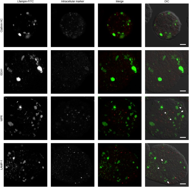
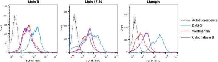
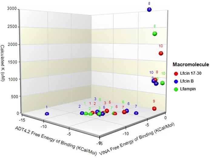
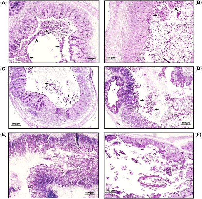
Abstract Amoebiasis caused by the protozoan parasite Entamoeba histolytica remains a public health problem in developing countries, making the identification of new anti-amoebic compounds a continuing priority. Previously, we have shown that lactoferrin (Lf) and several Lf-derived peptides exhibit in vitro anti-amoebic activity independently of their iron-binding activity. Here, we evaluated the amoebicidal effect of synthetic Lf-derived peptides Lfcin-B, Lfcin 17-30, and Lfampin, analyzed the mechanism of death induced by the peptides and determined their therapeutic effects on murine intestinal amoebiasis. MTT assays in trophozoite cultures of E. histolytica exposed to each peptide (1–1000 μM) showed that Lfampin is far more amoebicidal than Lfcins. Lfampin killed 80% of trophozoites at doses higher than 100 μM in 24 h, and FACs analysis using Annexin V/propidium iodide showed that death occurred mainly by necrosis. In contrast, Lfcin-B and Lfcin 17-30 appeared to have no significant effect on amoebic viability. FACs and confocal microscopy analysis using FITC-labeled peptides showed that all three peptides are internalized by the amoeba mainly using receptor (PI3K signaling) and actin-dependent pathways but independent of clathrin. Docking studies identified cholesterol in the amoeba’s plasma membrane as a possible target of Lfampin. Oral treatment of intracecally infected mice with the abovementioned peptides at 10 mg/kg for 4 days showed that Lfampin resolved 100% of the cases of intestinal amoebiasis, whereas Lfcin 17-30 and Lfcin-B were effective in resolving infection in 80 and 70% of cases, respectively. These data show that although synthetic bovine Lf-derived peptides exhibit varying amoebicidal potentials in vitro, they do resolve murine intestinal amoebiasis efficiently, suggesting that they may be useful as a therapeutic treatment.
Overview
The claims and statistics of this paper haven’t been checked yet.
Run the check — free, no account needed. It reads the open-access text and checks the sources it cites.
Integrity signals
Registry
Cites
Statistics
Figures
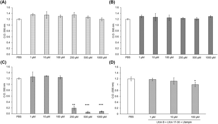
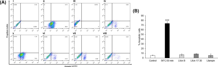
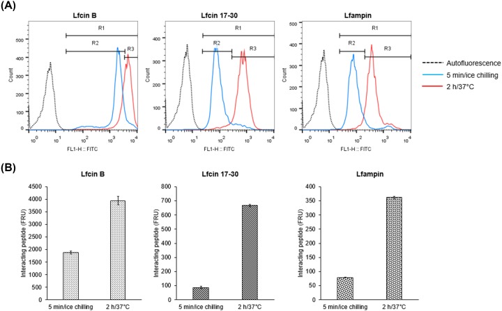
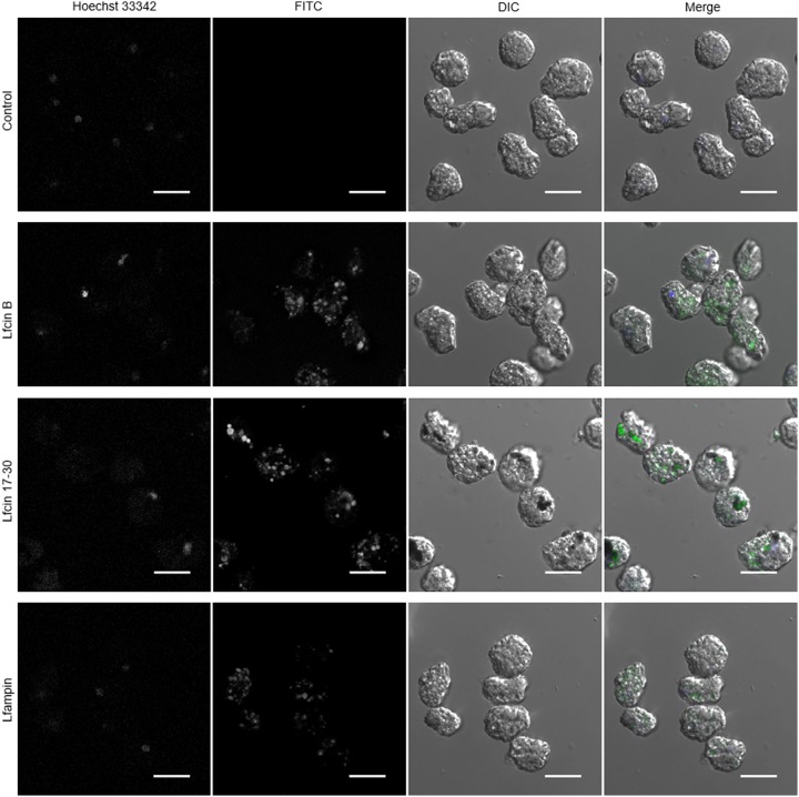
Verification
Claims the paper makes
The claims and statistics of this paper haven’t been checked yet.
Run the check — free, no account needed. It reads the open-access text and checks the sources it cites.
Claims other papers make about this one
Each is checked against this paper’s own text: does it say what the citing paper says it does?
The source backs part of the claim, not all of it — or backs it for a different population, condition or comparison.
In the citing paper
“In addition, LF is active against the intestinal protozoa Entamoeba histolytica and Giardia lamblia following receptor-mediated endocytosis of its antimicrobial component lactoferricin, that is generated by pepsin-mediated digestion (Aguilar-Diaz et al., 2017;Diaz-Godinez et al., 2019).”
From Iron in infection and immunity (2020) · cites this paper as Diaz-Godinez et al., 2019
Experimental — accuracy not yet measured
Self-check
Percentages, sums and reported differences re-computed from the paper’s own numbers.
The self-check needs the paper’s full text.
Citations
Cites
Trott, O., & Olson, A. J. (2010) · Journal of Computational Chemistry · doi:10.1002/jcc.21334 · Cited by 38,412
AutoDock4 and AutoDockTools4: Automated docking with selective receptor flexibility
Morris, G. M., Huey, R., Lindstrom, W., Sanner, M. F., Belew, R. K., Goodsell, D. S., & Olson, A. J. (2009) · Journal of Computational Chemistry · doi:10.1002/jcc.21256 · Cited by 26,294
Lozano, R., Naghavi, M., Foreman, K., Lim, S., Shibuya, K., Aboyans, V., Abraham, J., Adair, T., Aggarwal, R., Ahn, S. Y., et al. (2012) · The Lancet · doi:10.1016/s0140-6736(12)61728-0 · Cited by 14,070
Cell‐penetrating peptides: 20 years later, where do we stand?
Bechara, C., & Sagan, S. (2013) · FEBS Letters · doi:10.1016/j.febslet.2013.04.031 · Cited by 906
Identification of the bactericidal domain of lactoferrin
Bellamy, W., Takase, M., Yamauchi, K., Wakabayashi, H., Kawase, K., & TOMITA, M. (1992) · Biochimica et Biophysica Acta (BBA) - Protein Structure and Molecular Enzymology · doi:10.1016/0167-4838(92)90346-f · Cited by 890
Walsh, J. (1986) · Clinical Infectious Diseases · doi:10.1093/clinids/8.2.228 · Cited by 659
Antimicrobial properties of lactoferrin
Jenssen, H., & Hancock, R. E. W. (2008) · Biochimie · doi:10.1016/j.biochi.2008.05.015 · Cited by 521
Antibacterial activity of lactoferrin and a pepsin-derived lactoferrin peptide fragment
Yamauchi, K., Tomita, M., Giehl, T. J., & Ellison, R. T. (1993) · Infection and Immunity · doi:10.1128/iai.61.2.719-728.1993 · Cited by 500
Sequence specificity and transcriptional activation in the binding of lactoferrin to DNA
He, J., & Furmanski, P. (1995) · Nature · doi:10.1038/373721a0 · Cited by 356
Overview of Lactoferrin as a Natural Immune Modulator.
Legrand, D. (2016) · Jornal de Pediatria · doi:10.1016/j.jpeds.2016.02.071 · Cited by 274
Wassmann, C., Hellberg, A., Tannich, E., & Bruchhaus, I. (1999) · Journal of Biological Chemistry · doi:10.1074/jbc.274.37.26051 · Cited by 260
Lactoferrampin: a novel antimicrobial peptide in the N1-domain of bovine lactoferrin
van der Kraan, M. I. A., Groenink, J., Nazmi, K., Veerman, E. C. I., Bolscher, J. G. M., & Amerongen, A. V. N. (2004) · Peptides · doi:10.1016/j.peptides.2003.12.006 · Cited by 254
Lactoferrin: a multifunctional glycoprotein.
Vorland, L. H. (1999) · APMIS · doi:10.1111/j.1699-0463.1999.tb01499.x · Cited by 252
Bruni, N., Capucchio, M. T., Biasibetti, E., Pessione, E., Cirrincione, S., Giraudo, L., Corona, A., & Dosio, F. (2016) · Molecules · doi:10.3390/molecules21060752 · Cited by 240
Mechanisms of Carrier Formation during Clathrin-Independent Endocytosis
Ferreira, A. P. A., & Boucrot, E. (2017) · Trends in Cell Biology · doi:10.1016/j.tcb.2017.11.004 · Cited by 175
Samarawickrema, N. A. (1997) · Journal of Antimicrobial Chemotherapy · doi:10.1093/jac/40.6.833 · Cited by 155
Immunomodulatory effects of lactoferrin on antigen presenting cells
Puddu, P. E., Valenti, P., & Gessani, S. (2008) · Biochimie · doi:10.1016/j.biochi.2008.05.005 · Cited by 141
Flores-Villaseñor, H. M., Canizalez‐Román, A., Reyes‐López, M., Nazmi, K., de la Garza, M., Zazueta-Beltrán, J., León-Sicairos, N. M., & Bolscher, J. G. M. (2010) · BioMetals · doi:10.1007/s10534-010-9306-4 · Cited by 121
Self-citationGenotoxicity and carcinogenicity of metronidazole
Dobiáš, L., Černá, M., Rössner, P., & Šrám, R. J. (1994) · Mutation Research/Reviews in Genetic Toxicology · doi:10.1016/0165-1110(94)90001-9 · Cited by 111
Haney, E. F., Lau, F., & Vogel, H. J. (2007) · Biochimica et Biophysica Acta (BBA) - Biomembranes · doi:10.1016/j.bbamem.2007.04.018 · Cited by 87
van der Kraan, M. I. A., Nazmi, K., Teeken, A., Groenink, J., Hof, W. V. ‘., Veerman, E. C. I., Bolscher, J. G. M., & Amerongen, A. V. N. (2005) · Biological Chemistry · doi:10.1515/bc.2005.017 · Cited by 76
Laughlin, R. C., McGugan, G. C., Powell, R. R., et al. (2004) · Infection and Immunity · doi:10.1128/iai.72.9.5349-5357.2004 · Cited by 68
León-Sicairos, N. M., Reyes‐López, M., Ordaz‐Pichardo, C., & de la Garza, M. (2006) · Biochemistry and Cell Biology · doi:10.1139/o06-060 · Cited by 57
Self-citationLeón-Sicairos, N. M., Canizalez‐Román, A., Delagarza, M., et al. (2008) · Biochimie · doi:10.1016/j.biochi.2008.06.009 · Cited by 56
Self-citationLópez-Soto, L. F., León-Sicairos, N. M., Nazmi, K., Bolscher, J. G. M., & de la Garza, M. (2010) · BioMetals · doi:10.1007/s10534-010-9295-3 · Cited by 55
Self-citationMorphodynamics of the Actin-Rich Cytoskeleton in Entamoeba histolytica
Manich, M., Hernández‐Cuevas, N. A., Ospina‐Villa, J. D., Syan, S., Marchat, L. A., Olivo‐Marín, J., & Guillén, N. (2018) · Frontiers in Cellular and Infection Microbiology · doi:10.3389/fcimb.2018.00179 · Cited by 52
Drago-Serrano, M. E., Campos‐Rodríguez, R., Carrero, J. C., & de la Garza, M. (2018) · Current Pharmaceutical Design · doi:10.2174/1381612824666180327155929 · Cited by 45
Self-citationProgrammed cell death in Entamoeba histolytica induced by the aminoglycoside G418
Villalba, J. D., Gómez, C., Medel, O., et al. (2007) · Microbiology · doi:10.1099/mic.0.2007/008599-0 · Cited by 44
Penuliar, G. M., Nakada‐Tsukui, K., & Nozaki, T. (2015) · Frontiers in Microbiology · doi:10.3389/fmicb.2015.00354 · Cited by 42
Novel antibacterial lactoferrin peptides generated by rennet digestion and autofocusing technique
Elbarbary, H. A., Abdou, A. M., Park, E. Y., et al. (2010) · International Dairy Journal · doi:10.1016/j.idairyj.2009.12.019 · Cited by 39
Aguilar-Diaz, H., Canizalez-Roman, A., Nepomuceno-Mejia, T., Gallardo-Vera, F., Hornelas-Orozco, Y., Nazmi, K., Bolscher, J. G. M., Carrero, J. C., Leon-Sicairos, C., & Leon-Sicairos, N. (2017) · Biochemistry and Cell Biology · doi:10.1139/bcb-2016-0079 · Cited by 35
Self-citationPais-Morales, J., Betanzos, A., García‐Rivera, G., Chávez‐Munguía, B., Shibayama, M., & Orozco, E. (2016) · PLoS ONE · doi:10.1371/journal.pone.0146287 · Cited by 33
Use and endocytosis of iron-containing proteins by Entamoeba histolytica trophozoites
López-Soto, L. F., León-Sicairos, N. M., Reyes‐López, M., Serrano‐Luna, J., Ordaz‐Pichardo, C., Piña‐Vázquez, C., Ortíz-Estrada, G., & de la Garza, M. (2009) · Infection Genetics and Evolution · doi:10.1016/j.meegid.2009.05.018 · Cited by 31
Self-citationHeterotrimeric G-protein Signaling Is Critical to Pathogenic Processes in Entamoeba histolytica
Bosch, D. E., Kimple, A. J., Muller, R. E., et al. (2012) · PLoS Pathogens · doi:10.1371/journal.ppat.1003040 · Cited by 30
Intestinal amebiasis: histopathologic features in experimentally infected mice.
Pk, G., Mancilla, R., & Ortíz-Ortíz, L. (1994) · PubMed · Cited by 27
Silva, T., Abengózar, M. Á., Fernández‐Reyes, M., Andreu, D., Nazmi, K., Bolscher, J. G. M., Bastos, M., & Rivas, L. (2012) · Amino Acids · doi:10.1007/s00726-012-1304-0 · Cited by 26
León-Sicairos, N. M., Martínez-Pardo, L., Sánchez-Hernández, B. E., de la Garza, M., & Carrero, J. C. (2012) · Biochemistry and Cell Biology · doi:10.1139/o2012-008 · Cited by 19
Self-citationBinding and Endocytosis of Bovine Hololactoferrin by the ParasiteEntamoeba histolytica
Ortíz-Estrada, G., Calderón‐Salinas, V., Shibayama-Salas, M., León-Sicairos, N. M., & de la Garza, M. (2015) · BioMed Research International · doi:10.1155/2015/375836 · Cited by 6
Self-citation
Research works printed but not linked: 2
- Leon-Sicairos, 2006, Microbicidal action of lactoferrin and lactoferricin and their synergistic effect with metronidazole in Entamoeba histolytica, Biochem. Cell Biol., 84, 327
- Samarawickrema, 1997, Involvement of superoxide dismutase and pyruvate:ferredoxin oxidoreductase in mechanisms of metronidazole resistance in Entamoeba histolytica, J. Antimicrob. Chemother., 40, 833, 10.1093/jac/40.6.833 · DOI
Cited by
Sorted by citations
Iron in infection and immunity
Nairz, M., & Weiss, G. (2020) · Molecular Aspects of Medicine · doi:10.1016/j.mam.2020.100864 · Cited by 359
Metadata
- Authors
- Díaz-Godínez, C., González-Galindo, X., Meza-Menchaca, T., Bobes, R. J., de la Garza, M., León-Sicairos, N., Laclette, J. P., & Carrero, J. C.
- Affiliations
- Instituto de Investigaciones Biomédicas, Universidad Nacional Autónoma de México (MX)
- Laboratorio de Genómica Humana, Facultad de Medicina, Universidad Veracruzana, Calle Médicos y Odontólogos S/N., Col. Unidad del Bosque, 91010, Xalapa, Veracruz, México
- Department of Cellular Biology, Centro de Investigación y de Estudios Avanzados del IPN, Av. IPN 2508, San Pedro Zacatenco, 07360 México, D.F., México
- Universidad Autónoma de Sinaloa (MX)
- Corresponding author
- Carrero, Julio C.
- Venue
- Bioscience Reports
- Publisher
- Portland Press Ltd.
- Published
- 2019-01
- Volume
- 39
- Issue
- 1
- ISSN
- 0144-8463 (ISSN-L)
- 1573-4935
- PMC
- PMC6328891
- OpenAlex
- W2900943109
- Cited by
- 23
- References
- 38
- Citations by year
- 2026: 1 · 2025: 3 · 2024: 4 · 2023: 6 · 2022: 3 · 2020: 6
- FWCI
- 1.13
- Type
- journal article
- Language
- en
- Open access
- gold
- doi.org
- Locations
- Bioscience Reports (publishedVersion, cc-by)
- Europe PMC (PubMed Central) (publishedVersion, other-oa)
- PubMed Central (publishedVersion, cc-by)
- PubMed (publishedVersion)
- Greater South Information System
- Funders
- Consejo Nacional de Ciencia y Tecnología (284830)
- Topics
- Health Sciences › Medicine › Infectious Diseases › Amoebic Infections and Treatments
- Life Sciences › Immunology and Microbiology › Microbiology › Antimicrobial Peptides and Activities
- Life Sciences › Biochemistry, Genetics and Molecular Biology › Endocrinology › Legionella and Acanthamoeba research
- Keywords
- Entamoeba histolytica, amoebiasis, lactoferrin-derived peptides, amoebicidal activity, necrosis, peptide internalization, PI3K signaling, cholesterol, plasma membrane
- MeSH terms
- Animals, Antiprotozoal Agents, Cattle, Entamoeba histolytica, Entamoebiasis, Lactoferrin, Male, Mice, Inbred C3H, Necrosis, Peptides, Phosphatidylinositol 3-Kinases, Mice, Trophozoites
About these checks
Registry, retraction, Cites and statistics results are computed, not judged, with one exception: a printed reference that no index lists is read once by a model before it counts as a citation error. Claims are extracted by a model and kept only when their quote appears verbatim in the text; each cited source is then checked, with a model’s help, for whether it says what the claim relies on. Those verdicts can be wrong — read the claim’s quote and the cited source before relying on one.
Statistics and arithmetic are recomputed, not judged.
Confidence bands are shown only for fields calibrated on our annotated test set. None is calibrated yet, so no band is shown.
What counts as a claim ›How we measure confidence ›How we check this ›
Are you an author, or do you see a problem with this page? Request a review
