

The DOI matches its Crossref record; 41 of the 41 references it cites were found in a registry or an index.
NOX4-dependent neuronal autotoxicity and BBB breakdown explain the superior sensitivity of the brain to ischemic damage
Open accessAbstract
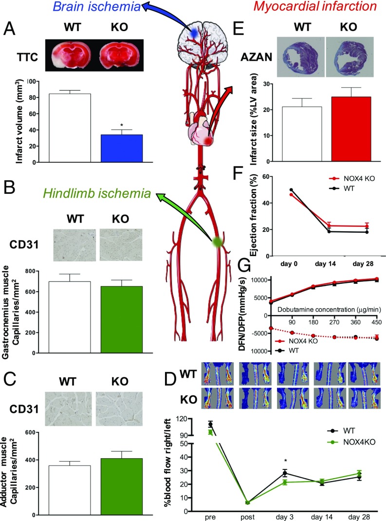
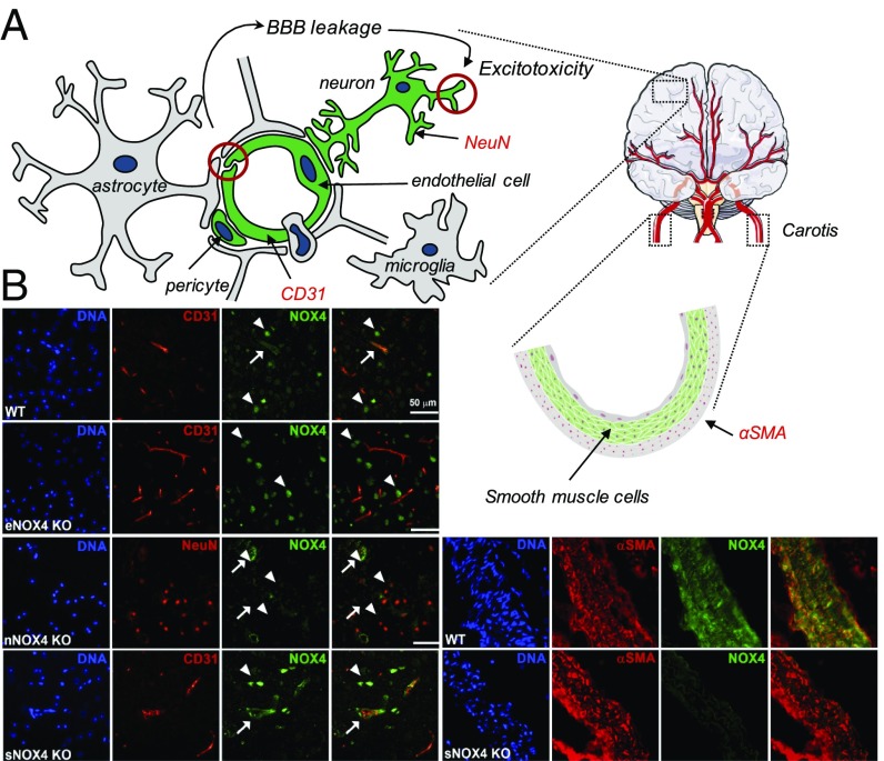
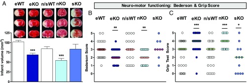
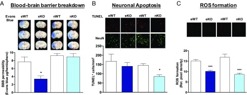
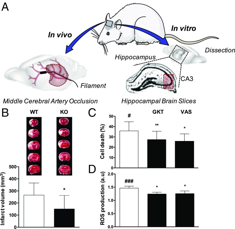
Ischemic injury represents the most frequent cause of death and disability, and it remains unclear why, of all body organs, the brain is most sensitive to hypoxia. In many tissues, type 4 NADPH oxidase is induced upon ischemia or hypoxia, converting oxygen to reactive oxygen species. Here, we show in mouse models of ischemia in the heart, brain, and hindlimb that only in the brain does NADPH oxidase 4 (NOX4) lead to ischemic damage. We explain this distinct cellular distribution pattern through cell-specific knockouts. Endothelial NOX4 breaks down the BBB, while neuronal NOX4 leads to neuronal autotoxicity. Vascular smooth muscle NOX4, the common denominator of ischemia within all ischemic organs, played no apparent role. The direct neuroprotective potential of pharmacological NOX4 inhibition was confirmed in an ex vivo model, free of vascular and BBB components. Our results demonstrate that the heightened sensitivity of the brain to ischemic damage is due to an organ-specific role of NOX4 in blood-brain-barrier endothelial cells and neurons. This mechanism is conserved in at least two rodents and humans, making NOX4 a prime target for a first-in-class mechanism-based, cytoprotective therapy in the unmet high medical need indication of ischemic stroke.
Overview
The claims and statistics of this paper haven’t been checked yet.
Run the check — free, no account needed. It reads the open-access text and checks the sources it cites.
Integrity signals
Registry
Cites
Statistics
Figures
Verification
Claims the paper makes
The claims and statistics of this paper haven’t been checked yet.
Run the check — free, no account needed. It reads the open-access text and checks the sources it cites.
Claims other papers make about this one
Each is checked against this paper’s own text: does it say what the citing paper says it does?
In the citing paper
“NOX4 is linked to oxidative stress and ROS production in brain injury [42,43].”
Self-check
Percentages, sums and reported differences re-computed from the paper’s own numbers.
The self-check needs the paper’s full text.
Citations
Cites
Zhang, Q., Wang, C., Zheng, M., Li, Y., Li, J., Zhang, L., Shang, X., & Yan, C. (2014) · Cerebrovascular Diseases · doi:10.1159/000369778 · Cited by 11,609
Projections of Global Mortality and Burden of Disease from 2002 to 2030
Mathers, C. D., & Loncar, D. (2006) · PLoS Medicine · doi:10.1371/journal.pmed.0030442 · Cited by 11,386
Meijer, L., Nelson, D. J., Riazanski, V., Gabdoulkhakova, A. G., Héry-Arnaud, G., Berre, R. L., Loaëc, N., Oumata, N., Galons, H., Nowak, E., et al. (2016) · Journal of Innate Immunity · doi:10.1159/000444256 · Cited by 5,008
Bederson, J. B., Pitts, L. H., Germano, I. M., Nishimura, M. C., Davis, R. L., & Bartkowski, H. M. (1986) · Stroke · doi:10.1161/01.str.17.6.1304 · Cited by 1,277
Tie2-Cre Transgenic Mice: A New Model for Endothelial Cell-Lineage Analysis in Vivo
Kisanuki, Y. Y., Hammer, R. E., Miyazaki, J.-I., Williams, S. C., Richardson, J. A., & Yanagisawa, M. (2001) · Developmental Biology · doi:10.1006/dbio.2000.0106 · Cited by 1,241
Chen, H., Yoshioka, H., Kim, G. S., Jung, J. E., Okami, N., Sakata, H., Maier, C. M., Narasimhan, P., Goeders, C. E., & Chan, P. H. (2010) · Antioxidants and Redox Signaling · doi:10.1089/ars.2010.3576 · Cited by 791
NADPH oxidase 4 (Nox4) is a major source of oxidative stress in the failing heart
Kuroda, J., Ago, T., Matsushima, S., Zhai, P., Schneider, M. D., & Sadoshima, J. (2010) · Proceedings of the National Academy of Sciences · doi:10.1073/pnas.1002178107 · Cited by 772
G12-G13–LARG–mediated signaling in vascular smooth muscle is required for salt-induced hypertension
Wirth, A., Benyó, Z., Lukasova, M., Leutgeb, B., Wettschureck, N., Gorbey, S., Örsy, P., Horváth, B., Maser‐Gluth, C., Greiner, E. F., et al. (2007) · Nature Medicine · doi:10.1038/nm1666 · Cited by 692
Couffinhal, T., Silver, M., Zheng, L., Kearney, M., Witzenbichler, B., & Isner, J. M. (1998) · Europe PMC (PubMed Central) · Cited by 672
Evolution of nadph oxidase inhibitors: Selectivity and mechanisms for target engagement
Altenhöfer, S., Radermacher, K. A., Kleikers, P. W. M., Wingler, K., & Schmidt, H. H. H. W. (2015) · Antioxidants & Redox Signaling · doi:10.1089/ars.2013.5814 · Cited by 521
Self-citation
Cited by
Sorted by citations
Reactive oxygen species (ROS) as pleiotropic physiological signalling agents
Sies, H., & Jones, D. P. (2020) · Nature Reviews Molecular Cell Biology · doi:10.1038/s41580-020-0230-3 · Cited by 5,335
Reactive Oxygen Species in Metabolic and Inflammatory Signaling
Forrester, S. J., Kikuchi, D. S., Hernandes, M. S., Xu, Q., & Griendling, K. K. (2018) · Circulation Research · doi:10.1161/circresaha.117.311401 · Cited by 2,081
Network pharmacology: curing causal mechanisms instead of treating symptoms
Nogales, C., Mamdouh, Z. M., List, M., Kiel, C., Casas, A. I., & Schmidt, H. H. H. W. (2021) · Trends in Pharmacological Sciences · doi:10.1016/j.tips.2021.11.004 · Cited by 1,251
Self-citationA randomized trial of epinephrine in out-of-hospital cardiac arrest
Perkins, G. D., Ji, C., Deakin, C. D., Quinn, T., Nolan, J. P., Scomparin, C., Regan, S., Long, J., Slowther, A., Pocock, H., et al. (2018) · New England Journal of Medicine · doi:10.1056/nejmoa1806842 · Cited by 794
Park, M. W., Cha, H. W., Kim, J., Kim, J. H., Yang, H., Yoon, S., Boonpraman, N., Yi, S. S., Yoo, I. D., & Moon, J.-S. (2021) · Redox Biology · doi:10.1016/j.redox.2021.101947 · Cited by 683
“NOX4 is linked to oxidative stress and ROS production in brain injury [42,43].”
Background
Metadata
- Authors
- Casas, A. I., Geuss, E., Kleikers, P. W. M., Mencl, S., Herrmann, A. M., Buendia, I., Egea, J., Meuth, S. G., Lopez, M. G., Kleinschnitz, C., & Schmidt, H. H. H. W.
- Affiliations
- Maastricht University (NL)
- Universitätsklinikum Würzburg (DE)
- Essen University Hospital (DE)
- University of Münster (DE)
- Instituto Teofilo Hernando, Department of Pharmacology, School of Medicine, Universidad Autónoma de Madrid, 28029 Madrid, Spain;
- Hospital Universitario de La Princesa (ES)
- Corresponding author
- Kleinschnitz, Christoph
- Schmidt, Harald H. H. W.
- ORCID
- Venue
- Proceedings of the National Academy of Sciences
- Publisher
- National Academy of Sciences
- Published
- 2017-10-31
- Volume
- 114
- Issue
- 46
- Pages
- 12315-12320
- ISSN
- 0027-8424 (ISSN-L)
- 1091-6490
- PMC
- PMC5699031
- OpenAlex
- W2766223740
- Cited by
- 152
- References
- 41
- Citations by year
- 2026: 8 · 2025: 13 · 2024: 16 · 2023: 15 · 2022: 19 · 2021: 28 · 2020: 27 · 2019: 17 · 2018: 9
- FWCI
- 10.73
- Type
- journal article
- Language
- en
- Open access
- hybrid
- doi.org
- Licence
- Locations
- Proceedings of the National Academy of Sciences (publishedVersion, cc-by-nc-nd)
- PubMed Central (publishedVersion, cc-by-nc-nd)
- PubMed (publishedVersion)
- Research Publications (Maastricht University) (submittedVersion, cc-by-nc-nd)
- LA Referencia (Red Federada de Repositorios Institucionales de Publicaciones Científicas) (submittedVersion, other-oa)
- Research Publications (Maastricht University) (submittedVersion, other-oa)
- Funders
- European Research Council. Advanced Investigator Grant (294683 RedMed)
- Deutsche Forschungsgemeinschaft (SFB688)
- Fondo Investigaciones Sanitarias (CP14/00008, PI6/00735)
- Ministerio Economia y Competencia Español (SAF2015-63935R)
- Topics
- Health Sciences › Medicine › Physiology (medical) › Nitric Oxide and Endothelin Effects
- Life Sciences › Neuroscience › Neurology › Barrier Structure and Function Studies
- Health Sciences › Medicine › Clinical Neurology › Acute Ischemic Stroke Management
- Keywords
- NOX4, NADPH oxidase, blood-brain barrier disruption, cerebral ischemia, ischemic stroke, hypoxia, reactive oxygen species, endothelial cells, vascular smooth muscle cells, neuroprotection
- MeSH terms
- NADPH Oxidase 4, Animals, Benzoxazoles, Blood-Brain Barrier, Brain, Brain Ischemia, Enzyme Inhibitors, Female, Femoral Artery, Gene Expression Regulation, Hindlimb, Humans, Male, Neurons, Organ Specificity, Pyrazoles, Pyridones, Triazoles, Signal Transduction, Myocardial Ischemia, Mice, Knockout, Neuroprotective Agents, Endothelial Cells, Mice, Rats
About these checks
Registry, retraction, Cites and statistics results are computed, not judged, with one exception: a printed reference that no index lists is read once by a model before it counts as a citation error. Claims are extracted by a model and kept only when their quote appears verbatim in the text; each cited source is then checked, with a model’s help, for whether it says what the claim relies on. Those verdicts can be wrong — read the claim’s quote and the cited source before relying on one.
Statistics and arithmetic are recomputed, not judged.
Confidence bands are shown only for fields calibrated on our annotated test set. None is calibrated yet, so no band is shown.
What counts as a claim ›How we measure confidence ›How we check this ›
Are you an author, or do you see a problem with this page? Request a review
