

The DOI matches its Crossref record; 16 of the 16 references it cites were found in a registry or an index.
Brain ventricular volume changes induced by long-duration spaceflight
Open accessAbstract
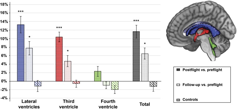
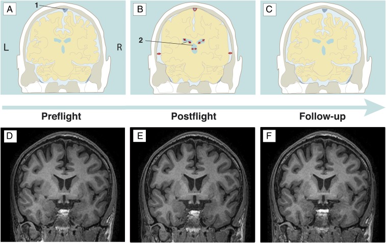
Significance: Long-duration spaceflight induces detrimental changes in human physiology due to microgravity. One example is a cephalic fluid shift. Here, we prospectively investigated the quantitative changes in cerebrospinal fluid (CSF) volume of the brain ventricular regions in space crew by means of a region of interest, observer-independent analysis on structural brain MRI scans. MRI scans were collected before the mission, shortly after and 7 mo after return to Earth. We found a significant increase in lateral and third ventricles at postflight and a trend to normalization at follow-up, but still significantly increased ventricular volumes. The observed spatiotemporal pattern of CSF compartment enlargement and recovery points to a reduced CSF resorption in microgravity as the underlying cause.
Overview
The claims and statistics of this paper haven’t been checked yet.
Free: it reads the open-access text and checks the first 5 claims that cite a source we can check. Create a free account to check every claim.
Integrity signals
Registry
Cites
Statistics
Figures
Verification
Claims the paper makes
The claims and statistics of this paper haven’t been checked yet.
Free: it reads the open-access text and checks the first 5 claims that cite a source we can check. Create a free account to check every claim.
Claims other papers make about this one
Each is checked against this paper’s own text: does it say what the citing paper says it does?
The source backs part of the claim, not all of it — or backs it for a different population, condition or comparison.
In the citing paper
“Most notably, a 10.7–14.6% ventricular enlargement (i.e., approximately a 2–3 ml increase) has been detected in astronauts and cosmonauts by multiple investigators56–59.”
From Red risks for a journey to the red planet: The highest priority human health risks for a mission to Mars (2020) · cites this paper as [57]
Experimental — accuracy not yet measured
Self-check
Percentages, sums and reported differences re-computed from the paper’s own numbers.
The self-check needs the paper’s full text.
Citations
Cites
Voxel-Based Morphometry—The Methods
Ashburner, J., & Friston, K. J. (2000) · NeuroImage · doi:10.1006/nimg.2000.0582 · Cited by 8,676
MRIQC: Advancing the automatic prediction of image quality in MRI from unseen sites
Esteban, O., Birman, D., Schaer, M., Koyejo, O. O., Poldrack, R. A., & Gorgolewski, K. J. (2017) · PLOS ONE · doi:10.1371/journal.pone.0184661 · Cited by 920
Anatomy and physiology of cerebrospinal fluid
Sakka, L., Coll, G., & Chazal, J. (2011) · European Annals of Otorhinolaryngology, Head and Neck Diseases · doi:10.1016/j.anorl.2011.03.002 · Cited by 815 · cited 1×
“This upward shift of the brain could lead to a compression of the superior sagittal sinus and Pachioni’s granulations, two structures responsible for the majority of CSF resorption (11).”
BackgroundMader, T. H., Gibson, C. R., Pass, A. F., Kramer, L. A., Lee, A. G., Fogarty, J., Tarver, W. J., Dervay, J. P., Hamilton, D. R., Sargsyan, A., et al. (2011) · Ophthalmology · doi:10.1016/j.ophtha.2011.06.021 · Cited by 748 · cited 3×
“2 and 3 for an overview), with a potential risk of permanent visual acuity changes (5).”
BackgroundEffects of Spaceflight on Astronaut Brain Structure as Indicated on MRI
Roberts, D. R., Albrecht, M. H., Collins, H. R., Asemani, D., Chatterjee, A. R., Spampinato, M. V., Zhu, X., Chimowitz, M. I., & Antonucci, M. U. (2017) · New England Journal of Medicine · doi:10.1056/nejmoa1705129 · Cited by 318 · cited 6×
“Another MRI study in ISS astronauts also observed a ventricular enlargement after spaceflight in combination with a narrowing of the central sulcus (6).”
BackgroundMethodInfluentialFundamentals of Space Medicine
Clément, G. (2011) · doi:10.1007/978-1-4419-9905-4 · Cited by 237
Microgravity-Induced Fluid Shift and Ophthalmic Changes
Nelson, E., Mulugeta, L., & Myers, J. (2014) · Life · doi:10.3390/life4040621 · Cited by 167 · cited 1×
“, arterial and venous structures) (2).”
Space flight-associated neuro-ocular syndrome (SANS)
Lee, A. G., Mader, T. H., Gibson, C. R., Brunstetter, T. J., & Tarver, W. J. (2018) · Eye · doi:10.1038/s41433-018-0070-y · Cited by 164 · cited 11×
“In addition, risk factors, such as age, body mass index, head size, gender, CSF turnover rate, and craniospinal compliance (12), should be assessed to determine a specific individual’s likelihood to develop VIIP/SANS.”
BackgroundInfluentialBrain Tissue–Volume Changes in Cosmonauts
Ombergen, A. V., Jillings, S., Jeurissen, B., Tomilovskaya, E., Rühl, R. M., Rumshiskaya, A. D., Nosikova, I., Litvinova, L., Annen, J., Pechenkova, E., et al. (2018) · New England Journal of Medicine · doi:10.1056/nejmc1809011 · Cited by 135 · cited 5×
“Previously, we reported alterations in total gray matter, white matter, and CSF tissue volumes after long-duration spaceflight (7).”
BackgroundMethodInfluentialSelf-citationSpaceflight-induced changes in white matter hyperintensity burden in astronauts
Alperin, N., Bagci, A. M., & Lee, S. H. (2017) · Neurology · doi:10.1212/wnl.0000000000004475 · Cited by 73 · cited 3×
“Lastly, the periventricular white matter changes previously reported in astronauts (16) might be a direct consequence of transependymal water diffusion from the enlarged ventricles into periventricular structures, due to pressure peaks in a backed up CSF circulation.”
BackgroundInfluential
Research works printed but not linked: 1
- Tarver W, Otto C (2012) NASA’s spaceflight visual impairment intracranial pressure (VIIP) risk: Clinical correlations and pathophysiology. Aerospace Medicine Grand Rounds (NASA, Washington, DC).
Cited by
Patel, Z. S., Brunstetter, T. J., Tarver, W. J., Whitmire, A. M., Zwart, S. R., Smith, S. M., & Huff, J. L. (2020) · npj Microgravity · doi:10.1038/s41526-020-00124-6 · Cited by 373
Challenges to the central nervous system during human spaceflight missions to Mars
Clement, G. R., Boyle, R. D., George, K. A., Nelson, G. A., Reschke, M. F., Williams, T. J., & Paloski, W. H. (2020) · Journal of Neurophysiology · doi:10.1152/jn.00476.2019 · Cited by 165
Imaging perivascular space structure and function using brain MRI
Barisano, G., Lynch, K. M., Sibilia, F., Lan, H., Shih, N., Sepehrband, F., & Choupan, J. (2022) · NeuroImage · doi:10.1016/j.neuroimage.2022.119329 · Cited by 158
Roy-O’Reilly, M. A., Mulavara, A. P., & Williams, T. J. (2021) · npj Microgravity · doi:10.1038/s41526-021-00133-z · Cited by 141
“Given the association between increased ventricular volumes and the smooth periventricular WMH and the absent deep WMH after spaceflight, it is possible that these alterations may represent trans-ependymal CSF diapedesis that are at least partially reversible after spaceflight.(14,21) Alperin et al.”
Intracranial Effects of Microgravity: A Prospective Longitudinal MRI Study
Kramer, L. A., Hasan, K. M., Stenger, M. B., Sargsyan, A., Laurie, S. S., Otto, C., Ploutz-Snyder, R. J., Marshall-Goebel, K., Riascos, R. F., & Macias, B. R. (2020) · Radiology · doi:10.1148/radiol.2020191413 · Cited by 123
Metadata
- Authors
- Van Ombergen, A., Jillings, S., Jeurissen, B., Tomilovskaya, E., Rumshiskaya, A., Litvinova, L., Nosikova, I., Pechenkova, E., Rukavishnikov, I., Manko, O., Danylichev, S., Rühl, R. M., Kozlovskaya, I. B., Sunaert, S., Parizel, P. M., Sinitsyn, V., Laureys, S., Sijbers, J., zu Eulenburg, P., & Wuyts, F. L.
- Affiliations
- Lab for Equilibrium Investigations and Aerospace, University of Antwerp, 2610 Antwerp, Belgium;
- University of Antwerp (BE)
- Russian Academy of Sciences (RU)
- Radiology Department, Federal Center of Treatment and Rehabilitation, 125367 Moscow, Russia;
- National Research University Higher School of Economics (RU)
- Gagarin Research and Test Cosmonaut Training Center (RU)
- Ludwig-Maximilians-Universität München (DE)
- KU Leuven (BE)
- Lomonosov Moscow State University (RU)
- University of Liège (BE)
- Department of Neurology, Ludwig-Maximilians-University Munich, 81377 Munich, Germany;
- ORCID
- Venue
- Proceedings of the National Academy of Sciences
- Publisher
- National Academy of Sciences
- Published
- 2019-05-06
- Volume
- 116
- Issue
- 21
- Pages
- 10531-10536
- ISSN
- 0027-8424 (ISSN-L)
- 1091-6490
- PMC
- PMC6535034
- OpenAlex
- W2944370899
- Cited by
- 148
- References
- 16
- Citations by year
- 2026: 14 · 2025: 24 · 2024: 18 · 2023: 19 · 2022: 22 · 2021: 27 · 2020: 19 · 2019: 5
- FWCI
- 12.51
- Type
- journal article
- Language
- en
- Open access
- hybrid
- doi.org
- Licence
- Locations
- Proceedings of the National Academy of Sciences (publishedVersion, cc-by-nc-nd)
- Europe PMC (PubMed Central) (publishedVersion, cc-by-nc-nd)
- PubMed Central (publishedVersion, cc-by-nc-nd)
- PubMed (publishedVersion)
- Lirias (KU Leuven) (submittedVersion, cc-by-nc-nd)
- Open Repository and Bibliography (University of Liège) (submittedVersion, cc-by-nc-nd)
- Funders
- Bundesministerium für Bildung und Forschung (EO 0901, 01EO0901)
- Fonds Wetenschappelijk Onderzoek
- Russian Science Foundation
- Vlaamse regering
- Topics
- Health Sciences › Medicine › Physiology (medical) › Spaceflight effects on biology
- Health Sciences › Medicine › Clinical Neurology › Cerebrospinal fluid and hydrocephalus
- Health Sciences › Medicine › Cardiology and Cardiovascular Medicine › Cardiovascular Syncope and Autonomic Disorders
- Keywords
- long-duration spaceflight, microgravity, cerebrospinal fluid, lateral ventricle, third ventricle, post-flight recovery, space crews, structural MRI
- MeSH terms
- Adult, Cerebral Ventricles, Humans, Magnetic Resonance Imaging, Middle Aged, Prospective Studies, Space Flight, Case-Control Studies
About these checks
Registry, retraction, Cites and statistics results are computed, not judged, with one exception: a printed reference that no index lists is read once by a model before it counts as a citation error. Claims are extracted by a model and kept only when their quote appears verbatim in the text; each cited source is then checked, with a model’s help, for whether it says what the claim relies on. Those verdicts can be wrong — read the claim’s quote and the cited source before relying on one.
Statistics and arithmetic are recomputed, not judged.
Confidence bands are shown only for fields calibrated on our annotated test set. None is calibrated yet, so no band is shown.
What counts as a claim ›How we measure confidence ›How we check this ›
Are you an author, or do you see a problem with this page? Request a review
How to cite
Cite CoChat for the checks on this page, and the paper for its own findings.
- This page
- CoChat. (2026). Citation check: Brain ventricular volume changes induced by long-duration spaceflight. https://app.cochat.ai/papers/10.1073/pnas.1820354116
- The paper
- Van Ombergen, A., Jillings, S., Jeurissen, B., Tomilovskaya, E., Rumshiskaya, A., Litvinova, L., Nosikova, I., Pechenkova, E., Rukavishnikov, I., Manko, O., Danylichev, S., Rühl, R. M., Kozlovskaya, I. B., Sunaert, S., Parizel, P. M., Sinitsyn, V., Laureys, S., Sijbers, J., zu Eulenburg, P., & Wuyts, F. L. (2019). Brain ventricular volume changes induced by long-duration spaceflight. Proceedings of the National Academy of Sciences. https://doi.org/10.1073/pnas.1820354116
Data sources
Bibliographic data: Semantic Scholar, OpenAlex, Crossref.
Contains information from the Semantic Scholar Academic Graph Datasets, made available under the ODC Attribution License (ODC-BY 1.0).
Semantic Scholar’s data is described in: Kinney, R., et al. (2023). The Semantic Scholar Open Data Platform. arXiv:2301.10140.
