

The DOI matches its Crossref record; 2 of the 2 references it cites were found in a registry or an index.
High-resolution structures of Myosin-IC reveal a unique actin-binding orientation, ADP release pathway, and power stroke trajectory
Open accessPublished version of a preprint
Abstract
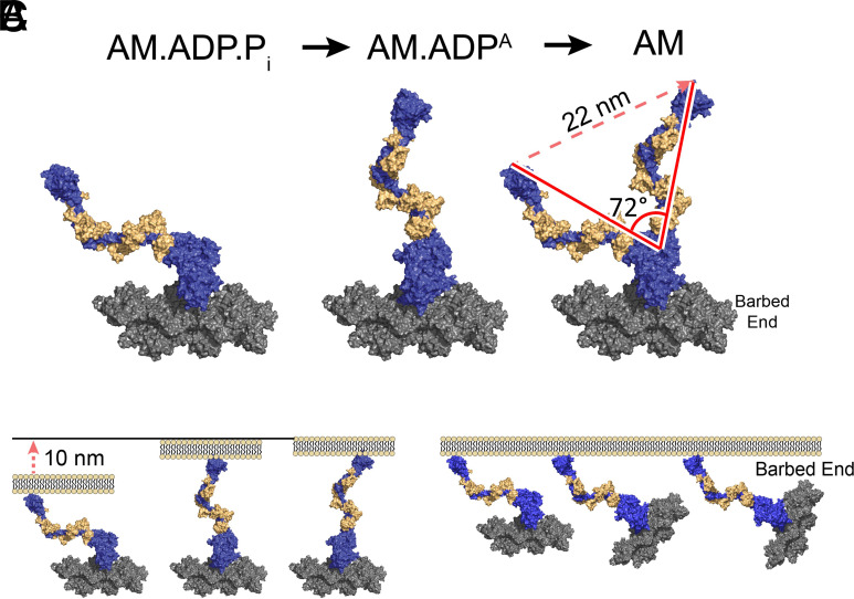
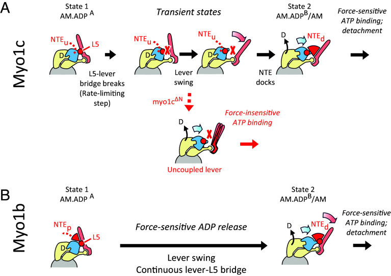
Myosin-IC (myo1c) is a class-I myosin that supports transport and remodeling of the plasma membrane and membrane-bound vesicles. Like other members of the myosin family, its biochemical kinetics are altered in response to changes in mechanical loads that resist the power stroke. However, myo1c is unique in that the primary force-sensitive kinetic transition is the isomerization that follows ATP binding, not ADP release as in other slow myosins. Myo1c also powers actin gliding along curved paths, propelling actin filaments in leftward circles. To understand the origins of this unique force-sensing and motile behavior, we solved actin-bound myo1c cryo-EM structures in the presence and absence of ADP. Our structures reveal that in contrast with other myosins, the myo1c lever arm swing is skewed, partly due to a different actin interface that reorients the motor domain on actin. The structures also reveal unique nucleotide-dependent behavior of both the nucleotide pocket as well as an element called the N-terminal extension (NTE). We incorporate these observations into a model that explains why force primarily regulates ATP binding in myo1c, rather than ADP release as in other myosins. Integrating our cryo-EM data with available crystallography structures allows the modeling of full-length myo1c during force generation, supplying insights into its role in membrane remodeling. These results highlight how relatively minor sequence differences in members of the myosin superfamily can significantly alter power stroke geometry and force-sensing properties, with important implications for biological function.
Overview
The claims and statistics of this paper haven’t been checked yet.
Run the check — free, no account needed. It reads the open-access text and checks the sources it cites.
Integrity signals
Registry
Cites
Statistics
Figures
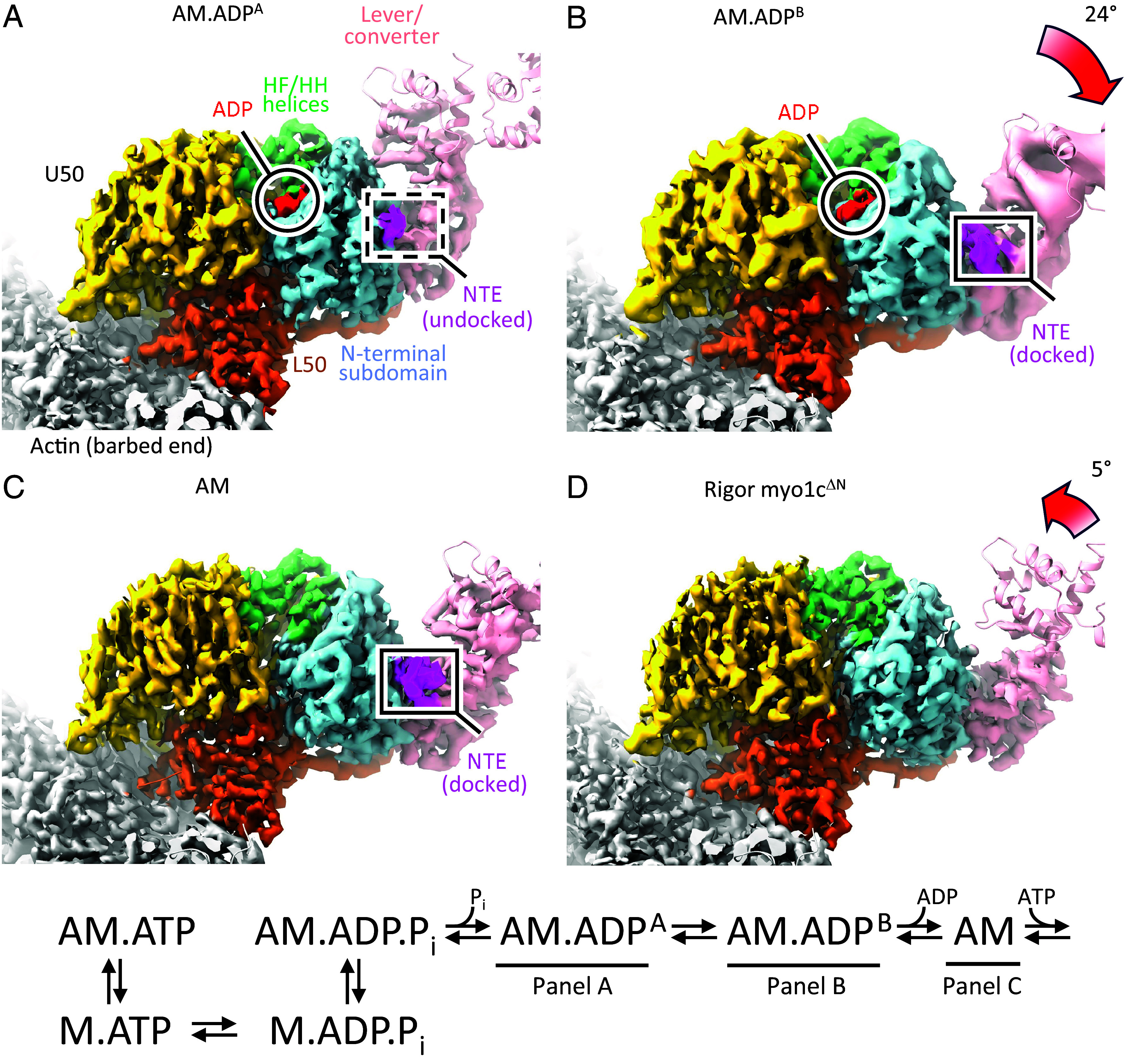
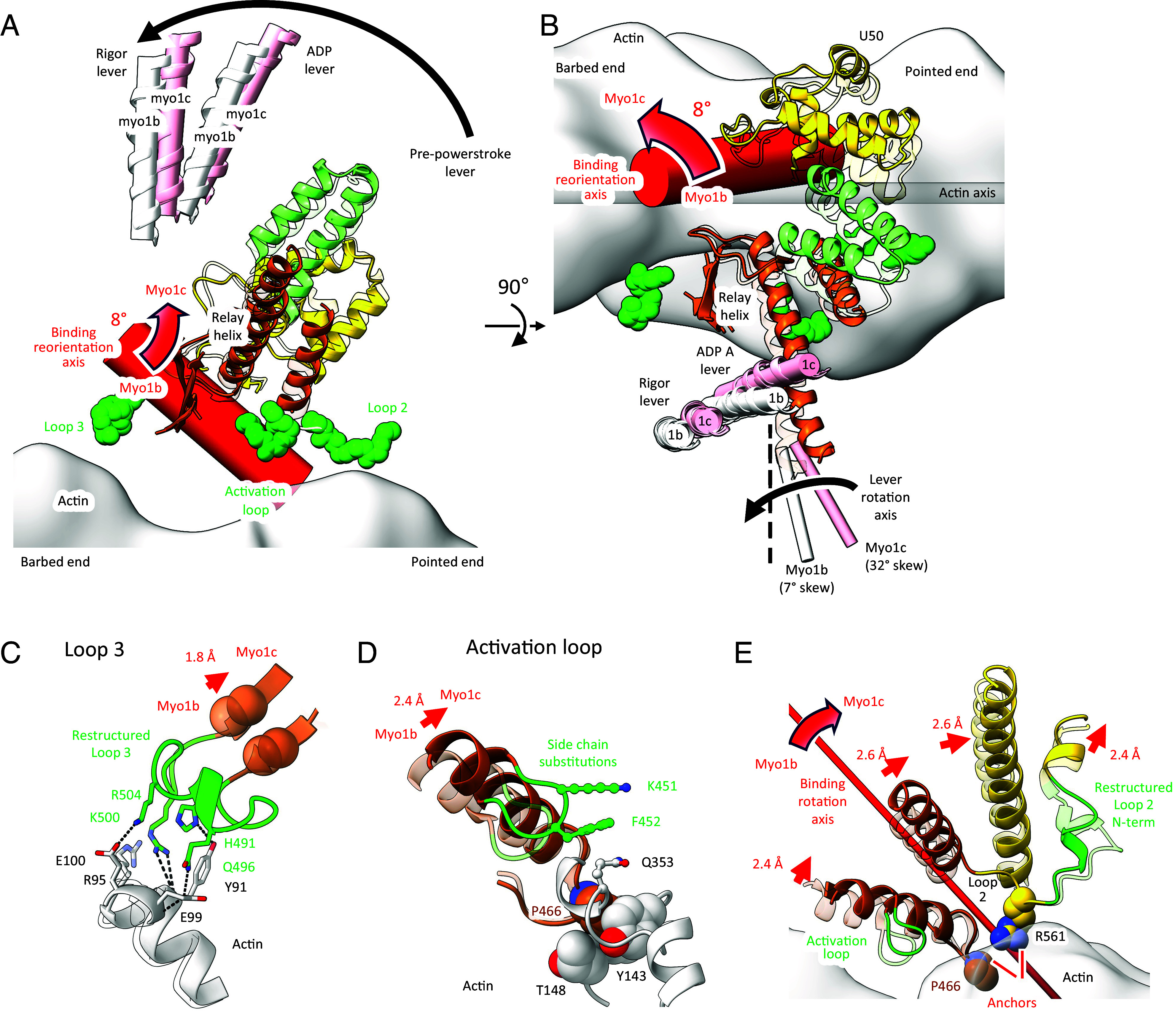
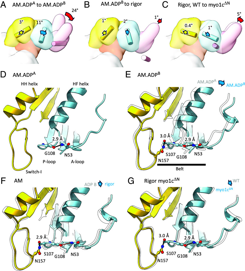
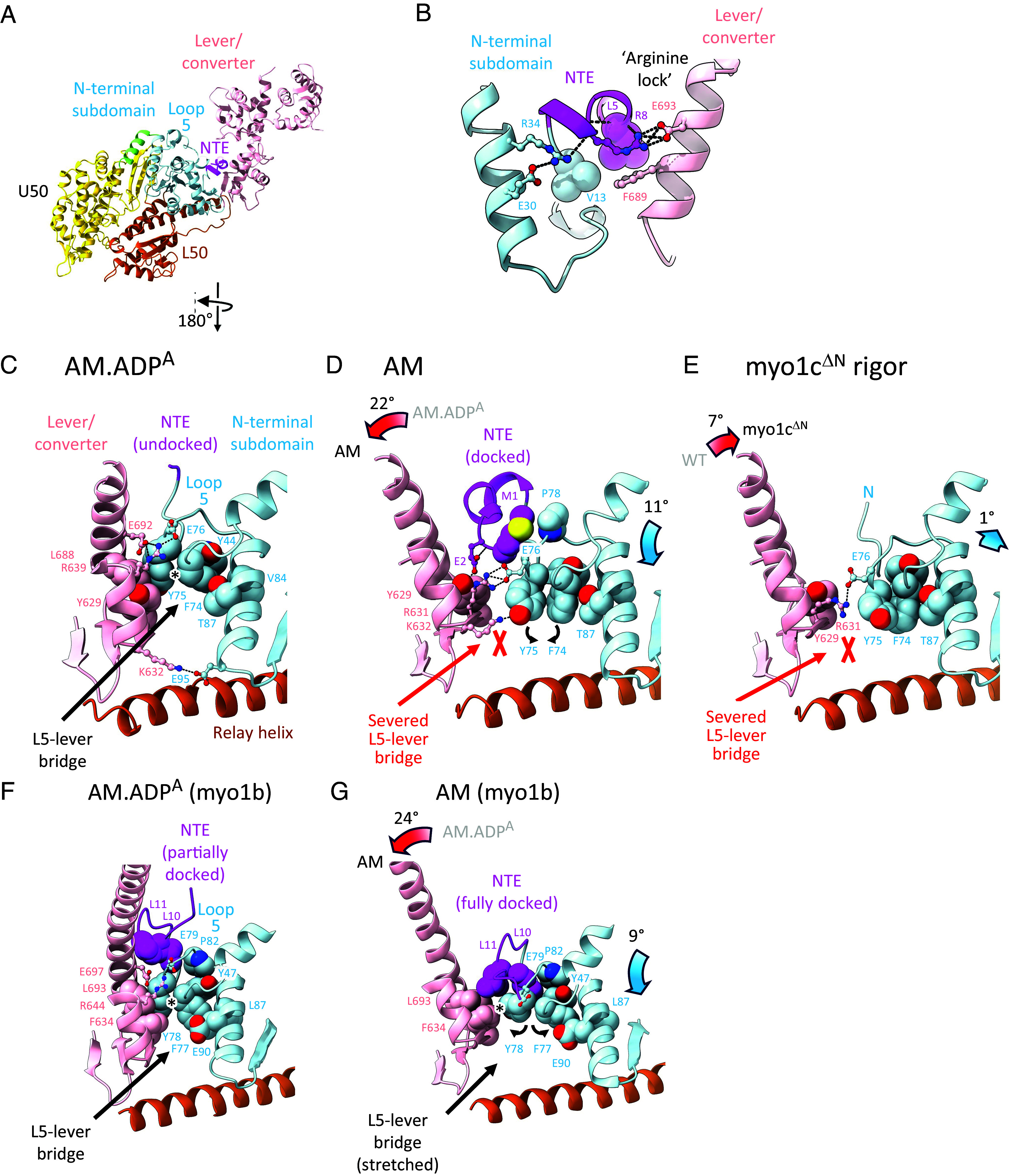
Verification
Claims the paper makes
The claims and statistics of this paper haven’t been checked yet.
Run the check — free, no account needed. It reads the open-access text and checks the sources it cites.
Self-check
Percentages, sums and reported differences re-computed from the paper’s own numbers.
The self-check needs the paper’s full text.
Citations
Cites
Highly accurate protein structure prediction with AlphaFold
Jumper, J., Evans, R., Pritzel, A., Green, T., Figurnov, M., Ronneberger, O., Tunyasuvunakool, K., Bates, R., Žídek, A., Potapenko, A., et al. (2021) · Nature · doi:10.1038/s41586-021-03819-2 · Cited by 43,764
Automated model building and protein identification in cryo-EM maps
Jamali, K., Käll, L., Zhang, R., Brown, A., Kimanius, D., & Scheres, S. H. W. (2023) · doi:10.1101/2023.05.16.541002 · Cited by 686
Research works printed but not linked: 63
- 10.1016/j.bpj.2016.05.021 · DOI
- 10.1111/tra.12388 · DOI
- 10.1016/j.ceb.2003.11.011 · DOI
- 10.1242/jcs.186403 · DOI
- 10.1016/j.tcb.2012.10.008 · DOI
- 10.1021/bi0611917 · DOI
- 10.1074/jbc.274.31.21575 · DOI
- 10.1073/pnas.1207811109 · DOI
- 10.1038/sj.emboj.7600169 · DOI
- 10.1126/science.1159419 · DOI
- 10.1073/pnas.0911426107 · DOI
- 10.1016/j.cub.2012.06.069 · DOI
- 10.1126/science.aat8642 · DOI
- 10.1073/pnas.1718316115 · DOI
- 10.1073/pnas.1321022111 · DOI
- 10.1073/pnas.1506633112 · DOI
- 10.1038/s41467-019-11120-0 · DOI
- 10.1074/jbc.M605171200 · DOI
- 10.1038/nature01927 · DOI
- 10.1038/ncb1060 · DOI
- 10.1016/j.bpj.2014.11.005 · DOI
- 10.7554/eLife.49266 · DOI
- 10.1038/nmeth.2152 · DOI
- 10.1038/ncb1287 · DOI
- 10.1038/s41467-024-46179-x · DOI
- 10.1002/pro.4792 · DOI
- 10.1083/jcb.141.1.155 · DOI
- 10.1038/s41467-021-22093-4 · DOI
- 10.1021/acs.chemrev.9b00264 · DOI
- 10.1016/j.jmb.2014.03.004 · DOI
- 10.1073/pnas.93.6.2285 · DOI
- 10.1038/nsmb.2216 · DOI
- 10.1016/j.devcel.2015.03.025 · DOI
- 10.1126/sciadv.abl4733 · DOI · Paper page
- 10.1016/j.jbc.2024.107539 · DOI
- 10.1111/tra.12342 · DOI
- 10.1038/19104 · DOI
- 10.7554/eLife.73724 · DOI
- 10.1038/nsmb.2923 · DOI
- J. Howard, Mechanics of Motor Proteins and the Cytoskeleton (Sinauer Associates Inc, Sunderland, 2001), p. 367.
- 10.1146/annurev.biochem.68.1.687 · DOI
- 10.1038/sj.emboj.7600458 · DOI
- 10.1126/science.8316857 · DOI
- 10.1073/pnas.0505685103 · DOI
- 10.1091/mbc.e06-05-0449 · DOI
- 10.1126/sciadv.ado5788 · DOI
- 10.1016/j.jbc.2023.104961 · DOI
- 10.1021/bi1016777 · DOI
- 10.1016/S0021-9258(18)62016-2 · DOI
- 10.1083/jcb.200612090 · DOI · Paper page
- 10.1038/nmeth.4169 · DOI · Paper page
- 10.1107/S2059798318002425 · DOI
- 10.1107/S0907444910007493 · DOI · Paper page
- 10.1107/S0907444909052925 · DOI · Paper page
- 10.1002/pro.3943 · DOI
- S. S. Chavali C. V. Sindelar E. M. Ostap Cryo-EM structure of myosin-1c bound to F-actin in the ADP-A state. Worldwide Protein Data Bank. https://doi.org/10.2210/pdb9cfu/pdb. Deposited 28 June 2024. · DOI
- S. S. Chavali C. V. Sindelar E. M. Ostap Cryo-EM structure of myosin-1c bound to F-actin in the ADP-A state. Electron Microscopy Data Bank. https://www.ebi.ac.uk/emdb/EMD-45563. Deposited 28 June 2024. · DOI
- S. S. Chavali C. V. Sindelar E. M. Ostap Cryo-EM structure of myosin-1c bound to F-actin in the ADP-B state. Worldwide Protein Data Bank. https://doi.org/10.2210/pdb9cfw/pdb. Deposited 28 June 2024. · DOI
- S. S. Chavali C. V. Sindelar E. M. Ostap Cryo-EM structure of myosin-1c bound to F-actin in the ADP-B state. Electron Microscopy Data Bank. https://www.ebi.ac.uk/emdb/EMD-45565. Deposited 28 June 2024. · DOI
- S. S. Chavali C. V. Sindelar E. M. Ostap Cryo-EM structure of myosin-1c bound to F-actin in the Rigor state. Worldwide Protein Data Bank. https://doi.org/10.2210/pdb9cfx/pdb. Deposited 28 June 2024. · DOI
- S. S. Chavali C. V. Sindelar E. M. Ostap Cryo-EM structure of myosin-1c bound to F-actin in the Rigor state. Electron Microscopy Data Bank. https://www.ebi.ac.uk/emdb/EMD-45566. Deposited 28 June 2024. · DOI
- S. S. Chavali C. V. Sindelar E. M. Ostap Cryo-EM structure of delta-NTR myosin-1c bound to F-actin. Worldwide Protein Data Bank. https://doi.org/10.2210/pdb9cfv/pdb. Deposited 28 June 2024. · DOI
- S. S. Chavali C. V. Sindelar E. M. Ostap Cryo-EM structure of delta-NTR myosin-1c bound to F-actin. Electron Microscopy Data Bank. https://www.ebi.ac.uk/emdb/EMD-45564. Deposited 28 June 2024. · DOI
Metadata
- Authors
- Chavali, S. S., Carman, P. J., Shuman, H., Ostap, E. M., & Sindelar, C. V.
- Affiliations
- Yale University (US)
- University of Pennsylvania (US)
- Corresponding author
- Ostap, E. Michael
- Sindelar, Charles V.
- Venue
- Proceedings of the National Academy of Sciences
- Publisher
- National Academy of Sciences
- Published
- 2025-02-27
- Volume
- 122
- Issue
- 9
- Pages
- e2415457122-e2415457122
- ISSN
- 0027-8424 (ISSN-L)
- 1091-6490
- PMC
- PMC11892617
- OpenAlex
- W4408019650
- Cited by
- 12
- References
- 2
- Citations by year
- 2026: 9 · 2025: 3
- FWCI
- 8.32
- Type
- journal article
- Language
- en
- Open access
- hybrid
- doi.org
- Licence
- Locations
- Proceedings of the National Academy of Sciences (publishedVersion, cc-by-nc-nd)
- bioRxiv (Cold Spring Harbor Laboratory) (acceptedVersion, cc-by-nc-nd)
- Europe PMC (PubMed Central) (publishedVersion, cc-by-nc-nd)
- PubMed Central (publishedVersion, cc-by-nc-nd)
- PubMed (publishedVersion)
- PubMed Central (submittedVersion, cc-by-nc-nd)
- Funders
- HHS | NIH | National Institute of General Medical Sciences (R37 GM057247, R01 GM110530)
- NSF (CMMI Grant 15-48571)
- Topics
- Health Sciences › Medicine › Cardiology and Cardiovascular Medicine › Cardiomyopathy and Myosin Studies
- Life Sciences › Biochemistry, Genetics and Molecular Biology › Molecular Biology › Protein Structure and Dynamics
- Life Sciences › Biochemistry, Genetics and Molecular Biology › Cell Biology › Microtubule and mitosis dynamics
- Keywords
- Myo1c, class I myosin, force sensing, ATP binding, N-terminal extension, membrane remodeling, cryo-EM structure
About these checks
Registry, retraction, Cites and statistics results are computed, not judged, with one exception: a printed reference that no index lists is read once by a model before it counts as a citation error. Claims are extracted by a model and kept only when their quote appears verbatim in the text; each cited source is then checked, with a model’s help, for whether it says what the claim relies on. Those verdicts can be wrong — read the claim’s quote and the cited source before relying on one.
Statistics and arithmetic are recomputed, not judged.
Confidence bands are shown only for fields calibrated on our annotated test set. None is calibrated yet, so no band is shown.
What counts as a claim ›How we measure confidence ›How we check this ›
Are you an author, or do you see a problem with this page? Request a review
