

The PEA3 Ets Transcription Factor Comprises Multiple Domains That Regulate Transactivation and DNA Binding
Open accessThe DOI matches its Crossref record; 41 of the 41 references it cites were found in a registry or an index.
Abstract
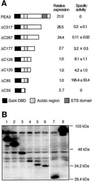
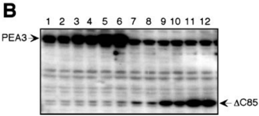
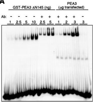
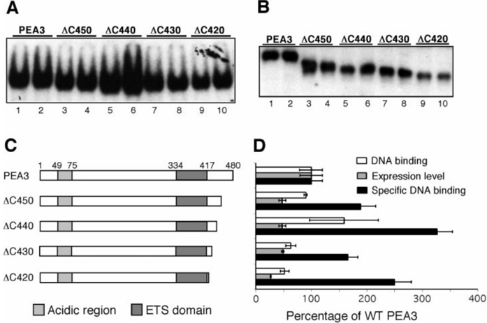
PEA3, a member of the Ets family of transcription factors, is a nuclear phosphoprotein capable of activating transcription. Mouse PEA3 comprises 480 amino acids and bears an approximately 85-amino acid ETS domain near its carboxyl terminus. Whereas analyses of bacterially expressed PEA3 revealed that the ETS domain is required for sequence-specific DNA binding, little is known of the functional domains in the protein required for its activity in mammalian cells. To this end, we defined the location of the PEA3 functional domains in COS cells. PEA3 bears a strong activation domain near its amino terminus, which is flanked by two regions that independently negatively regulate its activity. PEA3 expressed in COS cells was incapable of binding to DNA in vitro. However, DNA binding activity could be unmasked by incubation with a PEA3-specific antibody. Analyses of the DNA binding activity of PEA3 deletion mutants revealed that two regions flanking the ETS domain independently inhibited DNA binding; deletion of both regions was required to detect DNA binding in the absence of a PEA3-specific antibody. Under these conditions, the ETS domain was sufficient for sequence-specific DNA binding. These findings suggest that the activity of PEA3 is exquisitely controlled at multiple functional levels.
Overview
The claims and statistics of this paper haven’t been checked yet.
Free: it reads the open-access text and checks the first 5 claims that cite a source we can check. Create a free account to check every claim.
Integrity signals
Registry
Cites
Statistics
Figures







Verification
Claims the paper makes
The claims and statistics of this paper haven’t been checked yet.
Free: it reads the open-access text and checks the first 5 claims that cite a source we can check. Create a free account to check every claim.
Claims other papers make about this one
Each is checked against this paper’s own text: does it say what the citing paper says it does?
The cited source says what the claim attributes to it.
In the citing paper
“Members of the PEA3 subfamily, including ETV4 (12, 84) and ETV5 (85), contain autoinhibitory sequences appended to both sides of their ETS domains.”
From Genomic and biochemical insights into the specificity of ETS transcription factors (2011) · cites this paper as [12]
In the citing paper
“Although examined in only a few cases by biophysical methods (12–15), the remaining regions of the ETS factors, including those functioning as transactivation domains, likely display minimal stable secondary or tertiary structure.”
From Genomic and biochemical insights into the specificity of ETS transcription factors (2011) · cites this paper as [12]
Self-check
Percentages, sums and reported differences re-computed from the paper’s own numbers.
The self-check needs the paper’s full text.
Citations
Cites
Schreiber, E., Matthias, P., Müller, M. M., & Schaffner, W. (1989) · Nucleic Acids Research · doi:10.1093/nar/17.15.6419 · Cited by 4,179
Prediction of Protein Secondary Structure at Better than 70% Accuracy
Rost, B., & Sander, C. (1993) · Journal of Molecular Biology · doi:10.1006/jmbi.1993.1413 · Cited by 2,910
Gene fusion with an ETS DNA-binding domain caused by chromosome translocation in human tumours
Delattre, O., Zucman, J., Plougastel, B., Desmaze, C., Melot, T., Peter, M., Kovar, H., Joubert, I., de Jong, P., Rouleau, G., et al. (1992) · Nature · doi:10.1038/359162a0 · Cited by 1,907
Structural features in eukaryotic mRNAs that modulate the initiation of translation.
Kozak, M. (1991) · Journal of Biological Chemistry · doi:10.1016/s0021-9258(18)54860-2 · Cited by 1,836
Combining evolutionary information and neural networks to predict protein secondary structure
Rost, B., & Sander, C. (1994) · Proteins: Structure, Function, and Bioinformatics · doi:10.1002/prot.340190108 · Cited by 1,452
At least six nucleotides preceding the AUG initiator codon enhance translation in mammalian cells
Kozak, M. (1987) · Journal of Molecular Biology · doi:10.1016/0022-2836(87)90418-9 · Cited by 1,182
Transcription activation by the adenovirus E1a protein
Lillie, J. W., & Green, M. R. (1989) · Nature · doi:10.1038/338039a0 · Cited by 632
May, W. A., Gishizky, M. L., Lessnick, S. L., Lunsford, L. B., Lewis, B. C., Delattre, O., Zucman, J., Thomas, G., & Denny, C. T. (1993) · Proceedings of the National Academy of Sciences · doi:10.1073/pnas.90.12.5752 · Cited by 574
A vector for expressing GAL4(1–147) fusions tn mammalian cells
Sadowski, I., & Ptashne, M. (1989) · Nucleic Acids Research · doi:10.1093/nar/17.18.7539 · Cited by 542
The ETS-domain: a new DNA-binding motif that recognizes a purine-rich core DNA sequence.
Karim, F. D., Urness, L. D., Thummel, C. S., Klemsz, M. J., McKercher, S. R., Celada, A., Van Beveren, C., Maki, R. A., Gunther, C. V., & Nye, J. A. (1990) · Genes & Development · doi:10.1101/gad.4.9.1451 · Cited by 499
May, W. A., Lessnick, S. L., Braun, B. S., Klemsz, M., Lewis, B. C., Lunsford, L. B., Hromas, R., & Denny, C. T. (1993) · Molecular and Cellular Biology · doi:10.1128/mcb.13.12.7393 · Cited by 497
A variant Ewing's sarcoma translocation (7;22) fuses the EWS gene to the ETS gene ETV1.
Jeon, I. S., Davis, J. N., Braun, B. S., Sublett, J. E., Roussel, M. F., Denny, C. T., & Shapiro, D. N. (1995) · PubMed · Cited by 471
Specificity within the ets Family of Transcription Factors
(1998) · Advances in Cancer Research · doi:10.1016/s0065-230x(08)60738-1 · Cited by 344
Xin, J. H., Cowie, A., Lachance, P., & Hassell, J. A. (1992) · Genes & Development · doi:10.1101/gad.6.3.481 · Cited by 330
Self-citationA new pattern for helix–turn–helix recognition revealed by the PU.l ETS–domain–DNA complex
Kodandapani, R., Pio, F., Ni, C.-Z., Piccialli, G., Klemsz, M., McKercher, S., Maki, R. A., & Ely, K. R. (1996) · Nature · doi:10.1038/380456a0 · Cited by 315
Bailly, R.-A., Bosselut, R., Zucman, J., Cormier, F., Delattre, O., Roussel, M., Thomas, G., & Ghysdael, J. (1994) · Molecular and Cellular Biology · doi:10.1128/mcb.14.5.3230-3241.1994 · Cited by 297
Brown, T. A., & McKnight, S. L. (1992) · Genes & Development · doi:10.1101/gad.6.12b.2502 · Cited by 265
Brass, A. L., Kehrli, E., Eisenbeis, C. F., Storb, U., & Singh, H. (1996) · Genes & Development · doi:10.1101/gad.10.18.2335 · Cited by 242
Kaneko, Y., Yoshida, K., Handa, M., Toyoda, Y., Nishihira, H., Tanaka, Y., Sasaki, Y., Ishida, S., Higashino, F., & Fujinaga, K. (1996) · Genes, Chromosomes and Cancer · doi:10.1002/(sici)1098-2264(199602)15:2<115::aid-gcc6>3.0.co;2-6 · Cited by 239
Multiple domains mediate transformation by the Ewing's sarcoma EWS/FLI-1 fusion gene.
Lessnick, S. L., Braun, B. S., Denny, C. T., & May, W. A. (1995) · PubMed · Cited by 181
Urano, F., Umezawa, A., Hong, W., Kikuchi, H., & Hata, J.-I. (1996) · Biochemical and Biophysical Research Communications · doi:10.1006/bbrc.1996.0281 · Cited by 175
Solution structure of the ETS domain from murine Ets-1: a winged helix-turn-helix DNA binding motif.
Donaldson, L. W., Petersen, J. M., Graves, B. J., & McIntosh, L. P. (1996) · The EMBO Journal · doi:10.1002/j.1460-2075.1996.tb00340.x · Cited by 161
Benz, C. C., O’Hagan, R. C., Richter, B. D., Scott, G. K., Chang, C.-H., Xiong, X., Chew, K., Ljung, B., Edgerton, S. M., Thor, A. D., et al. (1997) · Oncogene · doi:10.1038/sj.onc.1201331 · Cited by 155
Self-citationChotteau‐Lelièvre, A., Desbiens, X., Pelczar, H., Defossez, P., & de Launoit, Y. (1997) · Oncogene · doi:10.1038/sj.onc.1201261 · Cited by 153
Molecular phylogeny of the ETS gene family
Laudet, V., Hänni, C., Stéhelin, D., & Duterque-Coquillaud, M. (1999) · Oncogene · doi:10.1038/sj.onc.1202444 · Cited by 139
Analysis of the ERK-Stimulated ETS Transcription Factor ER81
Janknecht, R. (1996) · Molecular and Cellular Biology · doi:10.1128/mcb.16.4.1550 · Cited by 124
Monté, D., Baert, J., Defossez, P., de Launoit, Y., & Stéhelin, D. (1994) · PubMed · Cited by 106
Functional dissection of the transcription factor Elk-1.
Janknecht, R., Zinck, R., Ernst, W. H., & Nordheim, A. (1994) · PubMed · Cited by 92
Structure–Function Relationships of the PEA3 Group of Ets-Related Transcription Factors
de Launoit, Y., Baert, J.-L., Chotteau, A., Monte, D., Defossez, P.-A., Coutte, L., Pelczar, H., & Leenders, F. (1997) · Biochemical and Molecular Medicine · doi:10.1006/bmme.1997.2605 · Cited by 90
Higashino, F., Yoshida, K., Fujinaga, Y., Kamio, K., & Fujinaga, K. (1993) · Nucleic Acids Research · doi:10.1093/nar/21.3.547 · Cited by 90
Donaldson, L. W., Petersen, J. M., Graves, B. J., & McIntosh, L. P. (1994) · Biochemistry · doi:10.1021/bi00250a001 · Cited by 80
PEA3 is overexpressed in mouse metastatic mammary adenocarcinomas.
Trimble, M. S., Xin, J.-H., Guy, C. T., Muller, W., & Hassell, J. A. (1993) · PubMed · Cited by 80
Self-citationMolecular characterization of the ets-related human transcription factor ER81.
Monté, D., Coutte, L., Baert, J., Angeli, I., Stéhelin, D., & de Launoit, Y. (1995) · PubMed · Cited by 78
Structural coupling of the inhibitory regions flanking the ETS domain of murine Ets‐1
Donaldson, L. W., Mcintosh, L. P., Skalicky, J. J., Petersen, J. M., & Graves, B. J. (1996) · Protein Science · doi:10.1002/pro.5560050214 · Cited by 65
Molecular characterization of the zebrafish PEA3 ETS-domain transcription factor
Brown, L. A., Amores, A., Schilling, T. F., Jowett, T., Baert, J., de Launoit, Y., & Sharrocks, A. D. (1998) · Oncogene · doi:10.1038/sj.onc.1201911 · Cited by 61
Liang, H., Olejniczak, E. T., Mao, X., Nettesheim, D. G., Yu, L., Thompson, C. B., & Fesik, S. W. (1994) · Proceedings of the National Academy of Sciences · doi:10.1073/pnas.91.24.11655 · Cited by 49
Two functionally distinct domains responsible for transactivation by the Ets family member ERM.
Laget, M.-P., Defossez, P., Albagli, O., Baert, J., Dewitte, F., Stéhelin, D., & de Launoit, Y. (1996) · PubMed · Cited by 48
The ETS family member ERM contains an alpha-helical acidic activation domain that contacts TAFII60
Defossez, P. (1997) · Nucleic Acids Research · doi:10.1093/nar/25.22.4455 · Cited by 40
Autoinhibition as a Transcriptional Regulatory Mechanism
GRAVES, B. J., COWLEY, D. O., GOETZ, T. L., PETERSEN, J. M., JONSEN, M. D., & GILLESPIE, M. E. (1998) · Cold Spring Harbor Symposia on Quantitative Biology · doi:10.1101/sqb.1998.63.621 · Cited by 36
Genomic organization of the human ERM (ETV5) gene, a PEA3 group member of ETS transcription factors
Monté, D., Coutte, L., Dewitte, F., Defossez, P.-A., Le Coniat, M., Stéhelin, D., Berger, R., & de Launoit, Y. (1996) · Genomics · doi:10.1006/geno.1996.0345 · Cited by 29
Phylogenetically interrelated ETS genes, ETV1, ERM and E1A-F locate on different chromosomes
Jeon, I., & Shapiro, D. N. (1998) · Journal of Korean Medical Science · doi:10.3346/jkms.1998.13.4.355 · Cited by 6
Research works printed but not linked: 9
- Jeon, I. S., Davis, J. N., Braun, B. S., Sublett, J. E., Roussel, M. F., Denny, C. T., and Shapiro, D. N. (1995) Oncogene 10, 1229 -1234
- Monte, D., Coutte, L., Baert, J. L., Angeli, I., Stehelin, D., and de Launoit, Y. (1995) Oncogene 11, 771-779
- Monte, D., Baert, J. L., Defossez, P. A., de Launoit, Y., and Stehelin, D. (1994) Oncogene 9, 1397-1406
- Trimble, M. S., Xin, J. H., Guy, C. T., Muller, W. J., and Hassell, J. A. (1993) Oncogene 8, 3037-3042
- Bailly, R. A., Bosselut, R., Zucman, J., Cormier, F., Delattre, O., Roussel, M., Thomas, G., and Ghysdael, J. (1994) Mol. Cell. Biol. 14, 3230 -3241
- May, W. A., Lessnick, S. L., Braun, B. S., Klemsz, M., Lewis, B. C., Lunsford, L. B., Hromas, R., and Denny, C. T. (1993) Mol. Cell. Biol. 13, 7393-7398
- Lessnick, S. L., Braun, B. S., Denny, C. T., and May, W. A. (1995) Oncogene 10, 423-431
- Laget, M. P., Defossez, P. A., Albagli, O., Baert, J. L., Dewitte, F., Stehelin, D., and de Launoit, Y. (1996) Oncogene 12, 1325-1336
- Janknecht, R., Zinck, R., Ernst, W. H., and Nordheim, A. (1994) Oncogene 9, 1273-1278
Cited by
A Developmental Switch in the Response of DRG Neurons to ETS Transcription Factor Signaling
Hippenmeyer, S., Vrieseling, E., Sigrist, M., Portmann, T., Laengle, C., Ladle, D. R., & Arber, S. (2005) · PLoS Biology · doi:10.1371/journal.pbio.0030159 · Cited by 1,088
“(J) Transcriptional transactivation of luciferase expression from a minimal reporter construct containing five consensus ETS DNA-binding sites (GCCGGAAGC; [18,19]) and a minimal TK promoter upon transient transfection of Er81 (n 7; 3.”
BackgroundMethodGenomic and biochemical insights into the specificity of ETS transcription factors
Hollenhorst, P. C., McIntosh, L. P., & Graves, B. J. (2011) · Annual Review of Biochemistry · doi:10.1146/annurev.biochem.79.081507.103945 · Cited by 490
“Chromosomal rearrangements that result in the overexpression of members of the PEA3 subfamily (ETV1, ETV4, or ETV5) are found less frequently (36, 170, 171).”
InfluentialETV1, 4 and 5: An oncogenic subfamily of ETS transcription factors
Oh, S., Shin, S., & Janknecht, R. (2012) · Biochimica et Biophysica Acta (BBA) - Reviews on Cancer · doi:10.1016/j.bbcan.2012.02.002 · Cited by 249
“The N-terminal activation domain appears to be more potent than the C-terminal one and sequences flanking both sides of the N-terminal acidic domain of ETV4 inhibit its activity [12].”
BackgroundInfluentialAutoinhibitory Domains: Modular Effectors of Cellular Regulation
Pufall, M. A., & Graves, B. J. (2002) · Annual Review of Cell and Developmental Biology · doi:10.1146/annurev.cellbio.18.031502.133614 · Cited by 236
“The PEA3 Ets transcription factor comprises multiple domains that regulate transactivation and DNA binding.”
BackgroundBrent, A. E., & Tabin, C. J. (2004) · Development · doi:10.1242/dev.01275 · Cited by 202
“Two out of the four FGF receptors are expressed in the somite at the time of Scx induction: Fgfr1, which is expressed broadly throughout the somite, slightly reduced in the myotome and slightly increased at the site of Scxexpression (Fig.”
BackgroundInfluential
Metadata
- Authors
- Bojović, B. B., & Hassell, J. A.
- Affiliations
- McMaster University (CA)
- Corresponding author
- Hassell, John A.
- Venue
- Journal of Biological Chemistry
- Publisher
- Elsevier BV
- Published
- 2001-02
- Volume
- 276
- Issue
- 6
- Pages
- 4509-4521
- ISSN
- 0021-9258 (ISSN-L)
- OpenAlex
- W2076111240
- Cited by
- 68
- References
- 41
- Citations by year
- 2023: 1 · 2022: 1 · 2020: 2 · 2018: 2 · 2017: 3 · 2016: 1 · 2015: 2 · 2014: 4 · 2013: 2 · 2012: 4
- FWCI
- 2.54
- Type
- journal article
- Language
- en
- Open access
- hybrid
- doi.org
- Licence
- cc-by-nc-nd
- www.elsevier.com/tdm/userlicense/1.0/ (tdm)
- creativecommons.org/licenses/by/4.0/ (vor, 7193 d)
- Locations
- Journal of Biological Chemistry (publishedVersion, cc-by-nc-nd)
- PubMed (publishedVersion)
- Radboud Repository (Radboud University) (submittedVersion)
- TSpace (University of Toronto) (submittedVersion)
- Radboud Repository (Radboud University) (submittedVersion, other-oa)
- Funders
- Canadian Institutes of Health Research
- National Cancer Institute
- Topics
- Life Sciences › Biochemistry, Genetics and Molecular Biology › Molecular Biology › Genomics and Chromatin Dynamics
- Life Sciences › Biochemistry, Genetics and Molecular Biology › Endocrinology › Estrogen and related hormone effects
- Life Sciences › Biochemistry, Genetics and Molecular Biology › Molecular Biology › Protein Structure and Dynamics
- Keywords
- PEA3, ETS transcription factors, ETS domain, functional domains, transcriptional activation, DNA binding, transactivation domain, negative regulation, deletion mutants, transcriptional regulation
- MeSH terms
- Animals, Base Sequence, DNA, DNA-Binding Proteins, Fungal Proteins, Protein Binding, Recombinant Fusion Proteins, Transcription Factors, Transcriptional Activation, Mutagenesis, Sequence Deletion, DNA Primers, COS Cells, Saccharomyces cerevisiae Proteins, Mice
About these checks
Registry, retraction, Cites and statistics results are computed, not judged, with one exception: a printed reference that no index lists is read once by a model before it counts as a citation error. Claims are extracted by a model and kept only when their quote appears verbatim in the text; each cited source is then checked, with a model’s help, for whether it says what the claim relies on. Those verdicts can be wrong — read the claim’s quote and the cited source before relying on one.
Statistics and arithmetic are recomputed, not judged.
Confidence bands are shown only for fields calibrated on our annotated test set. None is calibrated yet, so no band is shown.
What counts as a claim ›How we measure confidence ›How we check this ›
Are you an author, or do you see a problem with this page? Request a review
Data sources
Bibliographic data: Semantic Scholar, OpenAlex, Crossref.
Contains information from the Semantic Scholar Academic Graph Datasets, made available under the ODC Attribution License (ODC-BY 1.0).
Semantic Scholar’s data is described in: Kinney, R., et al. (2023). The Semantic Scholar Open Data Platform. arXiv:2301.10140.
