

The DOI matches its Crossref record; 68 of the 68 references it cites were found in a registry or an index.
Voltage-dependent Anion Channel 1-based Peptides Interact with Bcl-2 to Prevent Antiapoptotic Activity
Open accessAbstract
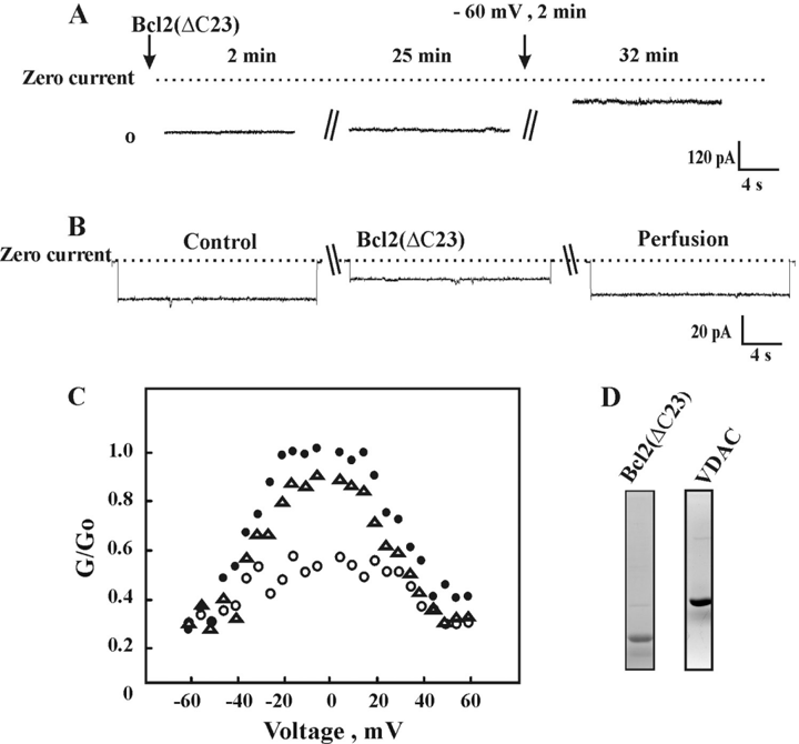
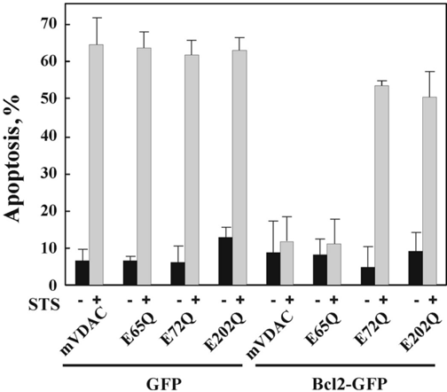
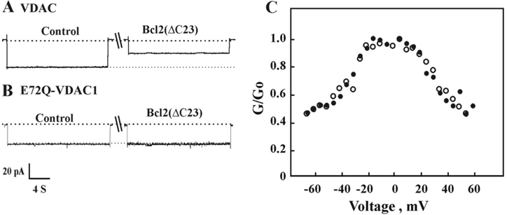
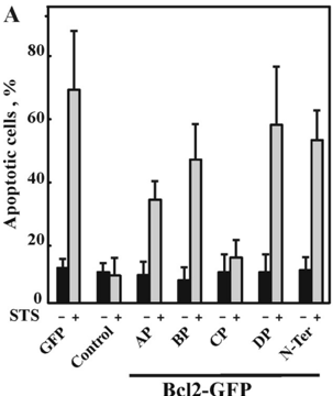
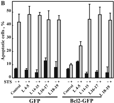
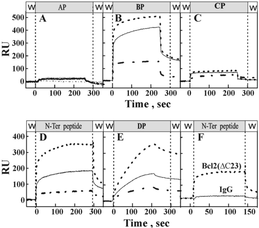
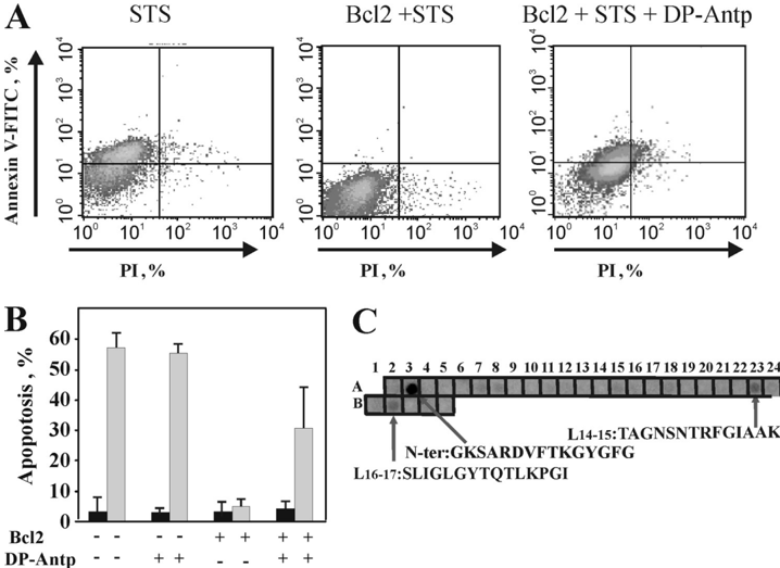
The antiapoptotic proteins of the Bcl-2 family are expressed at high levels in many types of cancer. However, the mechanism by which Bcl-2 family proteins regulate apoptosis is not fully understood. Here, we demonstrate the interaction of Bcl-2 with the outer mitochondrial membrane protein, voltage-dependent anion channel 1 (VDAC1). A direct interaction of Bcl-2 with bilayer-reconstituted purified VDAC was demonstrated, with Bcl-2 decreasing channel conductance. Expression of Bcl-2-GFP prevented apoptosis in cells expressing native but not certain VDAC1 mutants. VDAC1 sequences and amino acid residues important for interaction with Bcl-2 were defined through site-directed mutagenesis. Synthetic peptides corresponding to the VDAC1 N-terminal region and selected sequences bound specifically, in a concentration- and time-dependent manner, to immobilized Bcl-2, as revealed by the real-time surface plasmon resonance. Moreover, expression of the VDAC1-based peptides in cells over-expressing Bcl-2 prevented Bcl-2-mediated protection against staurosporine-induced apoptotic cell death. Similarly, a cell-permeable VDAC1-based synthetic peptide was also found to prevent Bcl-2-GFP-mediated protection against apoptosis. These results point to Bcl-2 as promoting tumor cell survival through binding to VDAC1, thereby inhibiting cytochrome c release and apoptotic cell death. Moreover, these findings suggest that interfering with the binding of Bcl-2 to mitochondria by VDAC1-based peptides may serve to potentiate the efficacy of conventional chemotherapeutic agents.
Overview
The claims and statistics of this paper haven’t been checked yet.
Run the check — free, no account needed. It reads the open-access text and checks the sources it cites.
Integrity signals
Registry
Cites
Statistics
Figures

Verification
Claims the paper makes
The claims and statistics of this paper haven’t been checked yet.
Run the check — free, no account needed. It reads the open-access text and checks the sources it cites.
Claims other papers make about this one
Each is checked against this paper’s own text: does it say what the citing paper says it does?
The source backs part of the claim, not all of it — or backs it for a different population, condition or comparison.
In the citing paper
“The N-terminal domain of VDAC1 interacts with several Bcl-2 family proteins that regulate apoptosis and is involved in gating the channel [96,97,98,99,100,101].”
From Mitochondrial Oxidative and Nitrosative Stress and Alzheimer Disease (2020) · cites this paper as [100]
Experimental — accuracy not yet measured
Self-check
Percentages, sums and reported differences re-computed from the paper’s own numbers.
The self-check needs the paper’s full text.
Citations
Cites
Cleavage of Structural Proteins during the Assembly of the Head of Bacteriophage T4
LAEMMLI, U. K. (1970) · Nature · doi:10.1038/227680a0 · Cited by 249,961
Green, D. R., & Reed, J. C. (1998) · Science · doi:10.1126/science.281.5381.1309 · Cited by 9,051
The BCL-2 protein family: opposing activities that mediate cell death
Youle, R. J., & Strasser, A. (2007) · Nature Reviews Molecular Cell Biology · doi:10.1038/nrm2308 · Cited by 4,806
Danial, N. N., & Korsmeyer, S. J. (2004) · Cell · doi:10.1016/s0092-8674(04)00046-7 · Cited by 4,651
The Bcl2 family: regulators of the cellular life-or-death switch
Cory, S., & Adams, J. M. (2002) · Nature Reviews Cancer · doi:10.1038/nrc883 · Cited by 4,032
BCL-2 family members and the mitochondria in apoptosisPreprint
Gross, A., McDonnell, J. M., & Korsmeyer, S. J. (1999) · Genes & Development · doi:10.1101/gad.13.15.1899 · Cited by 3,642
Mitochondrial membrane permeabilization in cell death
Kroemer, G., Galluzzi, L., & Brenner, C. (2007) · Physiological Reviews · doi:10.1152/physrev.00013.2006 · Cited by 3,598
The mitochondrial permeability transition pore and its role in cell death
CROMPTON, M. (1999) · Biochemical Journal · doi:10.1042/bj3410233 · Cited by 2,432
The Bcl-2 apoptotic switch in cancer development and therapy
Adams, J. M., & Cory, S. (2007) · Oncogene · doi:10.1038/sj.onc.1210220 · Cited by 2,256
Shimizu, S., Narita, M., Tsujimoto, Y., & Tsujimoto, Y. (1999) · Nature · doi:10.1038/20959 · Cited by 2,178
Research works printed but not linked: 4
- Krajewski, 1993, Cancer Res., 53, 4701
- De Pinto, 2003, Ital. J. Biochem., 52, 17
- Dias, 2002, Mol. Cancer Ther., 1, 347
- Leone, 2003, Cancer Res., 63, 8118
Cited by
Sorted by citations
VDAC, a multi-functional mitochondrial protein regulating cell life and death
Shoshan‐Barmatz, V., Pinto, V. D., Zweckstetter, M., Raviv, Z., Keinan, N., & Arbel, N. (2010) · Molecular Aspects of Medicine · doi:10.1016/j.mam.2010.03.002 · Cited by 824
The endoplasmic reticulum–mitochondria connection: One touch, multiple functions
Marchi, S., Patergnani, S., & Pinton, P. (2014) · Biochimica et Biophysica Acta (BBA) - Bioenergetics · doi:10.1016/j.bbabio.2013.10.015 · Cited by 475
“The antiapoptotic protein Bcl-2 interacts with VDAC1 [118], decreasing channel conductance and likely limiting mitochondrial Ca2+ entry and ROS production.”
BackgroundInfluentialMitochondrial Channels: Ion Fluxes and More
Szabó, I., & Zoratti, M. (2014) · Physiological Reviews · doi:10.1152/physrev.00021.2013 · Cited by 330
Misfolded Mutant SOD1 Directly Inhibits VDAC1 Conductance in a Mouse Model of Inherited ALS
Israelson, A., Arbel, N., Cruz, S. D., Ilieva, H. S., Yamanaka, K., Shoshan‐Barmatz, V., & Cleveland, D. W. (2010) · Neuron · doi:10.1016/j.neuron.2010.07.019 · Cited by 329
“Taken together, our results establish a direct link between misfolded mutant SOD1 and mitochondrial dysfunction in this form of inherited ALS.”
BackgroundVDAC1, mitochondrial dysfunction, and Alzheimer’s disease
Shoshan-Barmatz, V., Nahon-Crystal, E., Shteinfer-Kuzmine, A., & Gupta, R. (2018) · Pharmacological Research · doi:10.1016/j.phrs.2018.03.010 · Cited by 286
Self-citationRovere, R. M. L. L., Roest, G., Bultynck, G., & Parys, J. B. (2016) · Cell Calcium · doi:10.1016/j.ceca.2016.04.005 · Cited by 266
VDAC, a multi-functional mitochondrial protein as a pharmacological target
Shoshan‐Barmatz, V., & Ben-Hail, D. (2011) · Mitochondrion · doi:10.1016/j.mito.2011.04.001 · Cited by 257
“, 2009) and when expressed in cells expressing Bcl-2, the same peptide prevented Bcl-2 antiapoptotic activity (Arbel and Shoshan-Barmatz, 2009).”
BackgroundInfluentialThe mitochondrial voltage-dependent anion channel 1 in tumor cells
Shoshan‐Barmatz, V., Ben-Hail, D., Admoni, L., Krelin, Y., & Tripathi, S. S. (2014) · Biochimica et Biophysica Acta (BBA) - Biomembranes · doi:10.1016/j.bbamem.2014.10.040 · Cited by 255
“and endostatin-induced apoptosis [188]; (f) VDAC1was shown to be involved in Cyto c release from proteoliposomes in vitro [162,192,193]; (g) VDAC1-deficient mitochondria from mutant yeast did not exhibit Bax/Bak-induced Cyto c release or Δψ loss [192,194]; (h) the BH4 domain of Bcl-xL was found sufficient for inhibiti…”
InfluentialVDAC1 at the Intersection of Cell Metabolism, Apoptosis, and Diseases
Shoshan‐Barmatz, V., Shteinfer‐Kuzmine, A., & Verma, A. (2020) · Biomolecules · doi:10.3390/biom10111485 · Cited by 246
“Importantly, we have been able to develop VDAC1-based peptides, which can interfere with these interactions, leading to impaired cell metabolism and apoptosis [27,34,39,199,200].”
BackgroundInfluentialApoptosis Regulators Bcl-2 and Caspase-3
Hussar, P. (2022) · Encyclopedia · doi:10.3390/encyclopedia2040111 · Cited by 228
“In animal cells, Bcl-2 proteins have been found on the outer mitochondrial membrane, which is supposed to form a complex with the voltage-gated anion channel porin (VDAC) [64].”
BackgroundVDAC1: from structure to cancer therapy
Shoshan‐Barmatz, V., & Mizrachi, D. (2012) · Frontiers in Oncology · doi:10.3389/fonc.2012.00164 · Cited by 200
“Purified C-terminally truncated Bcl2 and Bcl-xL interact with VDAC1 and reduced the channel conductance of wild type but not of certain mutated forms of VDAC1 (Arbel and Shoshan-Barmatz, 2010; Arbel et al., 2012) or N-terminally truncated VDAC (Abu-Hamad et al.”
BackgroundInfluentialMitochondrial quality control as a key determinant of cell survival
Sedlackova, L., & Korolchuk, V. I. (2018) · Biochimica et Biophysica Acta (BBA) - Molecular Cell Research · doi:10.1016/j.bbamcr.2018.12.012 · Cited by 187
VDAC1 as Pharmacological Target in Cancer and Neurodegeneration: Focus on Its Role in Apoptosis
Magrì, A., Reina, S., & Pinto, V. D. (2018) · Frontiers in Chemistry · doi:10.3389/fchem.2018.00108 · Cited by 186
“Interestingly, modified VDAC1-based peptides were proved to reduce the anti-apoptotic effects of Bcl2 or Bcl-xL (Arbel and Shoshan-Barmatz, 2010; Arbel et al., 2012) and selectively kill Chronic Lymphocytic Leukemia cells (Prezma et al.”
BackgroundMediation of the antiapoptotic activity of Bcl-xL protein upon interaction with VDAC1 protein
Arbel, N., Ben-Hail, D., & Shoshan-Barmatz, V. (2012) · Journal of Biological Chemistry · doi:10.1074/jbc.m112.345918 · Cited by 179
“These include theVDAC1mutants, E72Q and E202Q, which prevented the antiapoptotic activity of both proteins, and mutant E65Q, which affects the activity of Bcl2 but not of Bcl-xL (11) (Fig.”
BackgroundInfluentialVDAC1 at the crossroads of cell metabolism, apoptosis and cell stress
Shoshan-Barmatz, V., N. Maldonado, E., & Krelin, Y. (2017) · Cell Stress · doi:10.15698/cst2017.10.104 · Cited by 170
Bcl-2 proteins and calcium signaling: complexity beneath the surface
Vervliet, T., Parys, J. B., & Bultynck, G. (2016) · Oncogene · doi:10.1038/onc.2016.31 · Cited by 173
Huang, H., Shah, K., Bradbury, N. A., Li, C., & White, C. (2014) · Cell Death and Disease · doi:10.1038/cddis.2014.419 · Cited by 174
Mazure, N. M. (2017) · Biochimica et Biophysica Acta (BBA) - Bioenergetics · doi:10.1016/j.bbabio.2017.03.002 · Cited by 166
Kerkhofs, M., Bittremieux, M., Morciano, G., Giorgi, C., Pinton, P., Parys, J. B., & Bultynck, G. (2018) · Cell Death and Disease · doi:10.1038/s41419-017-0179-0 · Cited by 165
VDAC1 functions in Ca2+ homeostasis and cell life and death in health and disease
Shoshan‐Barmatz, V., Krelin, Y., & Shteinfer‐Kuzmine, A. (2017) · Cell Calcium · doi:10.1016/j.ceca.2017.06.007 · Cited by 165
Brain-specific knockdown of miR-29 results in neuronal cell death and ataxia in mice
Roshan, R., Shridhar, S., Sarangdhar, M. A., Banik, A., Chawla, M., Garg, M., Singh, V. P., & Pillai, B. (2014) · RNA · doi:10.1261/rna.044008.113 · Cited by 149
“It is believed that VDAC1 interacts with Bcl-2 family members (Arbel and Shoshan-Barmatz 2010) and is the major channel through which cytochrome c release into the cytoplasm is mediated in apoptotic cells (Shimizu et al. 1999).”
BackgroundHuang, H., Hu, X., Eno, C. O., Zhao, G., Li, C., & White, C. (2013) · Journal of Biological Chemistry · doi:10.1074/jbc.m112.448290 · Cited by 161
“Based on an analysis of the binding determinants, peptides representing sections of the VDAC1 sequence were shown previously to be highly effective in disrupting the interaction between either Bcl-2 or Bcl-xL and VDAC1 (20, 35).”
BackgroundMethodShoshan‐Barmatz, V., Keinan, N., Abu‐Hamad, S., Tyomkin, D., & Aram, L. (2010) · Biochimica et Biophysica Acta (BBA) - Bioenergetics · doi:10.1016/j.bbabio.2010.03.003 · Cited by 155
Smilansky, A., Dangoor, L., Nakdimon, I., Ben-Hail, D., Mizrachi, D., & Shoshan-Barmatz, V. (2015) · Journal of Biological Chemistry · doi:10.1074/jbc.m115.691493 · Cited by 143
“VDAC1 Reconstitution into a Planar Lipid Bilayer and Analysis of Channel Activity—VDAC1 purified from rat liver mitochondria was used for channel reconstitution into a planar lipid bilayer (PLB) prepared from soybean asolectin dissolved in n-decane (30 mg/ml) (18).”
MethodSelf-citationMitochondrial VDAC1: Function in Cell Life and Death and a Target for Cancer Therapy
Shoshan‐Barmatz, V., & Golan, M. (2012) · Current Medicinal Chemistry · doi:10.2174/092986712798992110 · Cited by 142
Voltage-Dependent Anion Channel 1 As an Emerging Drug Target for Novel Anti-Cancer Therapeutics
Shoshan‐Barmatz, V., Krelin, Y., Shteinfer‐Kuzmine, A., & Arif, T. (2017) · Frontiers in Oncology · doi:10.3389/fonc.2017.00154 · Cited by 135
“N-Ter-Antp peptide interacted with HK, Bcl-2, and Bcl-xL and inhibited their anti-apoptotic effects (16–20, 133).”
BackgroundInfluentialMonaco, G., Decrock, E., Arbel, N., van Vliet, A. R., Rovere, R. M. L. L., SMEDT, H. D., Parys, J. B., Agostinis, P., Leybaert, L., Shoshan‐Barmatz, V., et al. (2015) · Journal of Biological Chemistry · doi:10.1074/jbc.m114.622514 · Cited by 144
“Next, we examined whether the isolated BH4 domains of Bcl-2 and Bcl-XL differ in their VDAC1-binding properties.”
BackgroundMethodInfluentialControl of mitochondrial physiology and cell death by the Bcl-2 family proteins Bax and Bok
D’Orsi, B., Mateyka, J., & Prehn, J. H. M. (2017) · Neurochemistry International · doi:10.1016/j.neuint.2017.03.010 · Cited by 134
“…the rate of InsP(3)-mediated Ca2þ release (Eckenrode et al., 2010) T-REx-293 cells Intensifies mitochondrial Ca2þ uptake through VDAC1 interaction (Arbel and Shoshan-Barmatz, 2010) Mouse heart Reduces the extrusion of Ca2þ from the mitochondrial matrix (Zhu et al., 2001) Mouse Pancreatic…”
Role of Mitochondria-Associated ER Membranes in Calcium Regulation in Cancer-Specific Settings
Morciano, G., Marchi, S., Morganti, C., Sbano, L., Bittremieux, M., Kerkhofs, M., Corricelli, M., Danese, A., Karkucińska‐Więckowska, A., Wieckowski, M. R., et al. (2018) · Neoplasia · doi:10.1016/j.neo.2018.03.005 · Cited by 136
The Mitochondrial Voltage-Dependent Anion Channel 1, Ca2+ Transport, Apoptosis, and Their Regulation
Shoshan‐Barmatz, V., De, S., & Meir, A. (2017) · Frontiers in Oncology · doi:10.3389/fonc.2017.00060 · Cited by 127
VDAC2-specific cellular functions and the underlying structure
Naghdi, S., & Hajnóczky, G. (2016) · Biochimica et Biophysica Acta (BBA) - Molecular Cell Research · doi:10.1016/j.bbamcr.2016.04.020 · Cited by 122
“Bcl-2 has also been shown to interact with VDAC1 through its N- terminal and inhibit cytochrome c release and apoptosis [108, 109].”
Tadić, V., Prell, T., Lautenschläger, J., & Großkreutz, J. (2014) · Frontiers in Cellular Neuroscience · doi:10.3389/fncel.2014.00147 · Cited by 115
“VDAC is regulated by Bcl-2 and both can form toxic complexes with mutated SOD1 (Pasinelli et al., 2004; Arbel and Shoshan-Barmatz, 2010; Israelson et al., 2010; Pedrini et al., 2010).”
BackgroundAkl, H., & Bultynck, G. (2012) · Biochimica et Biophysica Acta (BBA) - Reviews on Cancer · doi:10.1016/j.bbcan.2012.12.001 · Cited by 110
“channel [122], the VDAC isoform particularly involved in pro-apoptotic Ca signaling [123].”
BackgroundThe MAMs Structure and Its Role in Cell Death
Wang, N., Wang, C., Zhao, H., He, Y., Lan, B., Sun, L., & Gao, Y. (2021) · Cells · doi:10.3390/cells10030657 · Cited by 110
“Bcl2 and Bcl-xl can directly bind to the central regulatory domain of IP3Rs or indirectly affect the phosphorylation level of IP3Rs to inhibit the function of IP3Rs; they can also interact with VDAC1 to reduce the uptake of Ca2+ by the mitochondria [13,95,96].”
BackgroundCa2+-mediated regulation of VDAC1 expression levels is associated with cell death induction
Algor, S. W., Keinan, N., Ben-Hail, D., Arif, T., & Shoshan‐Barmatz, V. (2014) · Biochimica et Biophysica Acta (BBA) - Molecular Cell Research · doi:10.1016/j.bbamcr.2014.03.021 · Cited by 108
Geula, S., Ben-Hail, D., & Shoshan-Barmatz, V. (2012) · Biochemical Journal · doi:10.1042/bj20112079 · Cited by 105
Mitochondria-Associated Membranes As Networking Platforms and Regulators of Cancer Cell Fate
Sassano, M. L., van Vliet, A. R., & Agostinis, P. (2017) · Frontiers in Oncology · doi:10.3389/fonc.2017.00174 · Cited by 100
Rocha, S., Freitas, A., Guimarães, S., Vitorino, R. M. P., Aroso, M., & Gómez-Lázaro, M. (2017) · Antioxidants · doi:10.3390/antiox7010001 · Cited by 93
“Furthermore, these channels interact with Bcl-2 proteins, therefore enabling cell survival [192,193].”
BackgroundMartinvalet, D. (2018) · Cell Death and Disease · doi:10.1038/s41419-017-0237-7 · Cited by 94
Oligomerization of the Mitochondrial Protein VDAC1
Shoshan‐Barmatz, V., Mizrachi, D., & Keinan, N. (2013) · Progress in molecular biology and translational science · doi:10.1016/b978-0-12-386931-9.00011-8 · Cited by 91
Mitochondrial Oxidative and Nitrosative Stress and Alzheimer Disease
Butterfield, D. A., & Boyd-Kimball, D. (2020) · Antioxidants · doi:10.3390/antiox9090818 · Cited by 85
Targeting Liver Cancer and Associated Pathologies in Mice with a Mitochondrial VDAC1-Based Peptide
Pittala, S., Krelin, Y., & Shoshan‐Barmatz, V. (2018) · Neoplasia · doi:10.1016/j.neo.2018.02.012 · Cited by 86
“VDAC1 directly interacts with Bcl2 and Bcl-xL, leading to protection against apoptosis [19].”
The mystery of mitochondria-ER contact sites in physiology and pathology: A cancer perspective
Simões, I. C. M., Morciano, G., Lebiedzińska, M., Aguiari, G., Pinton, P., Potes, Y., & Wieckowski, M. R. (2020) · Biochimica et Biophysica Acta (BBA) - Molecular Basis of Disease · doi:10.1016/j.bbadis.2020.165834 · Cited by 84
“In addition, the N-terminal portion of VDAC1 is responsible for the release of cyt c from mitochondria; in mitochondria, the BCL-2–VDAC1 interaction counteracts the action of cyt c, protecting cells from apoptosis [101].”
BackgroundSander, P., Gudermann, T. W., & Schredelseker, J. (2021) · International Journal of Molecular Sciences · doi:10.3390/ijms22020946 · Cited by 84
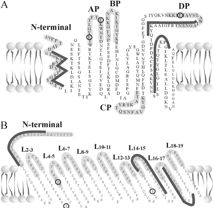
“In particular, members of the Bcl-2 family were described to influence mitochondrial Ca2+ uptake by binding to VDAC, presumably at a structure formed by the N-terminal helix and the loop connecting beta sheets 14 and 15 [36,69].”
BackgroundBcl-2-Protein Family as Modulators of IP 3 Receptors and Other Organellar Ca 2+ ChannelsPreprint
Ivanova, H., Vervliet, T., Monaco, G., Terry, L. E., Rosa, N., Baker, M. R., Parys, J. B., Serysheva, I. I., Yule, D. I., & Bultynck, G. (2019) · Cold Spring Harbor Perspectives in Biology · doi:10.1101/cshperspect.a035089 · Cited by 83
“Bcl-2, Bcl-Xl, and Mcl-1 target the amino terminus of VDAC1, as shown by using VDAC1-amino-terminal derived (N-Ter) peptides, which also interfered with the antiapoptotic function of all three proteins when intracellularly delivered in multiple cell models (Abu-Hamad et al. 2009; Arbel and Shoshan-Barmatz 2010; Arbel…”
BackgroundInfluentialTargeting mitochondrial ion channels for cancer therapy
Szabó, I., Zoratti, M., & Biasutto, L. (2020) · Redox Biology · doi:10.1016/j.redox.2020.101846 · Cited by 78
“VDAC1 also binds pro-apoptotic proteins: Bax and Bak, Bim, Bid whose interactions can also be antagonized by suitable peptides [263, 268].”
BackgroundPrezma, T., Shteinfer‐Kuzmine, A., Admoni, L., Raviv, Z., Sela, I., Levi, I., & Shoshan‐Barmatz, V. (2013) · Cell Death and Disease · doi:10.1038/cddis.2013.316 · Cited by 77
“However, when the peptide interferes with their association with VDAC1, apoptosis ensues.(14,15) Indeed, cells treated with the peptide show characteristic features of”
BackgroundMitochondria: Insights into Crucial Features to Overcome Cancer Chemoresistance
Genovese, I., Carinci, M., Modesti, L., Aguiari, G., Pinton, P., & Giorgi, C. (2021) · International Journal of Molecular Sciences · doi:10.3390/ijms22094770 · Cited by 77
Tomasello, M. F., Guarino, F., Reina, S., Messina, A. A., & Pinto, V. D. (2013) · PLoS ONE · doi:10.1371/journal.pone.0081522 · Cited by 72
“Circled in red the residues found involved with the HK-VDAC1 binding [34]; the blue circles mark HKI binding sites as defined by SPR and peptide competition assay [35]; the yellow triangles show the position of peptides anti-Bax [36]; the purple squares mark the loop involved in the binding to Bcl 2 [37].”
BackgroundVDAC2 and the BCL-2 family of proteins
Yuan, Z., Dewson, G., Czabotar, P. E., & Birkinshaw, R. W. (2021) · Biochemical Society Transactions · doi:10.1042/bst20210753 · Cited by 82
Alves, C. J., Dariolli, R., Jorge, F. M. H., Monteiro, M. R., Maximino, J. R., Martins, R. S., Strauss, B. E., Krieger, J. E., Callegaro, D., & Chadi, G. (2015) · Frontiers in Cellular Neuroscience · doi:10.3389/fncel.2015.00289 · Cited by 71
Bcl-2 binds to and inhibits ryanodine receptors
Vervliet, T., Decrock, E., Molgó, J., Sorrentino, V., Missiaen, L., Leybaert, L., SMEDT, H. D., Kasri, N. N., Parys, J. B., & Bultynck, G. (2014) · Journal of Cell Science · doi:10.1242/jcs.150011 · Cited by 69
“At the mitochondrial outer membranes, Bcl-2 proteins target the voltage-dependent anion channels (VDACs) (Arbel and Shoshan- Barmatz, 2010; Arbel et al., 2012; Plötz et al., 2012).”
InfluentialIdentification of new channels by systematic analysis of the mitochondrial outer membrane
Krüger, V., Becker, T., Becker, L., Montilla‐Martinez, M., Ellenrieder, L., Vögtle, F. ‐., Meyer, H. E., Ryan, M. T., Wiedemann, N., Warscheid, B., et al. (2017) · The Journal of Cell Biology · doi:10.1083/jcb.201706043 · Cited by 70
Apoptotic proteins with non-apoptotic activity: expression and function in cancer
Shoshan‐Barmatz, V., Arif, T., & Shteinfer‐Kuzmine, A. (2023) · APOPTOSIS · doi:10.1007/s10495-023-01835-3 · Cited by 73
IP3, a small molecule with a powerful message
Decrock, E., Bock, M. D., Wang, N., Gadicherla, A. K., Bol, M., Delvaeye, T., Vandenabeele, P., Vinken, M., Bultynck, G., Krysko, D. V., et al. (2013) · Biochimica et Biophysica Acta (BBA) - Molecular Cell Research · doi:10.1016/j.bbamcr.2012.12.016 · Cited by 64
Shteinfer‐Kuzmine, A., Amsalem, Z., Arif, T., Zooravlov, A., & Shoshan‐Barmatz, V. (2018) · Molecular Oncology · doi:10.1002/1878-0261.12313 · Cited by 64
“In our previous studies, key sequences and amino acid residues of VDAC1 required for Bcl-2, Bcl-xL, and HK binding and protection against cell death were identified (Abu-Hamad et al., 2009; Arbel and Shoshan-Barmatz, 2010; Arzoine et al., 2009; Shi et al., 2003; Shoshan-Barmatz et al., 2009).”
BackgroundInfluentialS-Nitrosylation in Alzheimer's disease
Zhao, Q., Yu, J., & Tan, L. (2014) · Molecular Neurobiology · doi:10.1007/s12035-014-8672-2 · Cited by 63
“Pathological stimuli, such as oxidative and nitrosative stress, could affect the VDAC-mediated mitochondrial membrane permeability, partially regulated by Bcl-2 family gene products via binding to VDAC thus regulate the open state of VDAC channel [68].”
Tan, W., Naniche, N., Bogush, A. I., Pedrini, S., Trotti, D., & Pasinelli, P. (2013) · Journal of Neuroscience · doi:10.1523/jneurosci.5385-12.2013 · Cited by 61
“In light of the evidence that Bcl-2 binds VDAC1 and regulates its gating properties (Arbel and Shoshan-Barmatz, 2010) and based on our findings that in mouse and human spinal cord mitochondria, mutSOD1 aberrantly binds Bcl-2 and changes its conformation, we hypothesized that rather than directly the mutSOD1 affects VD…”
BackgroundInfluentialZhang, G., Jiang, G., Wang, C., Zhong, K., Zhang, J., Xue, Q., Li, X., Jin, H. D., & Li, B. (2016) · Oncotarget · doi:10.18632/oncotarget.9943 · Cited by 60
“VDAC1 also regulates apoptosis as an anchor point for mitochondria-interacting proteins, such as hexokinase (HK), Bcl2 and Bcl-xL, some of which are highly expressed in many cancers [10–12, 13].”
BackgroundRelevance of the endoplasmic reticulum-mitochondria axis in cancer diagnosis and therapy
An, G., Park, J., Song, J., Hong, T., Song, G., & Lim, W. (2024) · Experimental & Molecular Medicine · doi:10.1038/s12276-023-01137-3 · Cited by 57
“Furthermore, the interaction between BCL2 and VDAC1 interferes with the export of cytochrome c from mitochondria and thus hinders apoptosis 98,102 .”
BackgroundA New Fungal Diterpene Induces VDAC1-dependent Apoptosis in Bax/Bak-deficient Cells
Huang, L., Han, J., Ben-Hail, D., He, L., Li, B., Chen, Z., Wang, Y., Yang, Y., Liu, L., Zhu, Y., et al. (2015) · Journal of Biological Chemistry · doi:10.1074/jbc.m115.648774 · Cited by 57
“Bcl-2 Protects against Cyathin-R-induced Apoptosis—Bcl-2 and Bcl-xL are important antagonists of MOMP and apoptosis and were shown to directly interact with VDAC1 (25, 48, 50, 51).”
BackgroundNiedźwiecka, K., Tisi, R., Penna, S., Lichocka, M., Płochocka, D., & Kucharczyk, R. (2017) · Biochimica et Biophysica Acta (BBA) - Molecular Cell Research · doi:10.1016/j.bbamcr.2017.10.003 · Cited by 58
Mitochondrial VDAC, the Na+/Ca2+ Exchanger, and the Ca2+ Uniporter in Ca2+ Dynamics and Signaling
Shoshan‐Barmatz, V., & De, S. (2017) · Advances in experimental medicine and biology · doi:10.1007/978-3-319-55858-5_13 · Cited by 56
Zhang, Z., He, H., Yang, Q., Huang, J., Wu, Z., Xu, P., Yin, D., & He, M. L. (2018) · Oxidative Medicine and Cellular Longevity · doi:10.1155/2018/3583921 · Cited by 54
“The Bcl-2 family of proteins plays an important role in mitochondria-mediated apoptosis [13].”
BackgroundRegulation of Cell Death by Mitochondrial Transport Systems of Calcium and Bcl-2 Proteins
Naumova, N., & Šachl, R. (2020) · Membranes · doi:10.3390/membranes10100299 · Cited by 53
Targeting Acute Myeloid Leukemia Stem Cells through Perturbation of Mitochondrial Calcium
Sheth, A. I., Engel, K. L., Tolison, H., Althoff, M. J., Amaya, M. L., Krug, A. E., Young, T. N., Pei, S., Patel, S. B., Minhajuddin, M., et al. (2024) · Cancer Discovery · doi:10.1158/2159-8290.cd-23-1145 · Cited by 56
Vervliet, T., Clerix, E., Seitaj, B., Ivanova, H., Monaco, G., & Bultynck, G. (2017) · Frontiers in Oncology · doi:10.3389/fonc.2017.00075 · Cited by 55
“Both Bcl-2 and Bcl-XL have been reported to inhibit VDAC1-mediated Ca2+ uptake into the mitochondria, thereby protecting cells from Ca2+-induced MOMP (45, 54, 55, 71).”
BackgroundInfluentialPharmacological modulation of mitochondrial ion channels
Leanza, L., Checchetto, V., Biasutto, L., Rossa, A., Costa, R., Bachmann, M., Zoratti, M., & Szabó, I. (2018) · British Journal of Pharmacology · doi:10.1111/bph.14544 · Cited by 50
“Bcl2 and Bcl-xL) and with hexokinase (HK) (Shimizu et al., 2000; Arbel and Shoshan-Barmatz, 2010).”
BackgroundGielecińska, A., Kciuk, M., & Kontek, R. (2024) · International Journal of Molecular Sciences · doi:10.3390/ijms252413727 · Cited by 53
“Membrane-localized voltage-dependent anion channels (VDACs), RyRs, and plasma membrane calcium ATPases (PMCAs) also interact with BCL-2, constituting a complex network of regulation of cellular calcium homeostasis [77–80].”
Balancing life and death: BCL‐2 family members at diverse ER–mitochondrial contact sites
Means, R. E., & Katz, S. G. (2021) · FEBS Journal · doi:10.1111/febs.16241 · Cited by 50
Liu, X., He, B., Xu, T., Pan, Y., Hu, X., Chen, X., & Wang, S. (2018) · Journal of Cancer · doi:10.7150/jca.23662 · Cited by 48
“VDAC1 could serve as a hub protein and provide structural and functional anchoring sites for anti-apoptosis proteins, including HK, Bcl2 and Bcl-xL [14, 36, 37], inhibiting the release of apoptosis-inducing proteins from mitochondria to the cytosol, such as Cyto c, AIF and HTRA2/OMI [38-40].”
BackgroundLee, M. W., Bassiouni, R., Sparrow, N. A., Iketani, A., Boohaker, R. J., Moskowitz, C., Vishnubhotla, P., Khaled, A. S., Oyer, J. L., Copik, A. J., et al. (2014) · Cell Death and Disease · doi:10.1038/cddis.2014.225 · Cited by 46
Bcl-2 inhibitors as anti-cancer therapeutics: The impact of and on calcium signaling
Vervloessem, T., Kerkhofs, M., Rovere, R. M. L. L., Sneyers, F., Parys, J. B., & Bultynck, G. (2017) · Cell Calcium · doi:10.1016/j.ceca.2017.05.014 · Cited by 46
Xu, L., Xie, Q., Qi, L., Wang, C., Xu, N., Liu, W., Yu, Y., Li, S., & Xu, Y. (2017) · Oncology Reports · doi:10.3892/or.2017.6164 · Cited by 45
“Anti-cleaved caspase-3 (ab2302; rabbit Ab; 1:200 dilution), anti-cleaved caspase-9 (ab2324; rabbit Ab; 1:400 dilution), anti-PDI (ab2792; murine Ab; 1:1,000 dilution), anti-VDAC1 (ab14734; murine Ab; 1:1,000 dilution), anti-CHOP (ab11419; murine Ab; 1:1,000 dilution), anti-IP3R (ab5804; rabbit Ab; 1:1,000 dilution) an…”
BackgroundInfluentialZhang, D., Yip, Y. M., & Li, L. (2016) · Mitochondrion · doi:10.1016/j.mito.2016.08.009 · Cited by 44
Tong, Z., Xie, Y., He, M. L., Ma, W., Zhou, Y., Lai, S., Meng, Y., & Liao, Z. (2017) · Biomedicine & Pharmacotherapy · doi:10.1016/j.biopha.2017.08.046 · Cited by 43
“Purified Bcl-2 was found to reduce the channel conductance of native VDAC1 [23].”
BackgroundShteinfer‐Kuzmine, A., Argueti-Ostrovsky, S., Gupta, R., Shvil, N., Abu‐Hamad, S., Gropper, Y., Hoeber, J., Magrì, A., Messina, A. A., Kozlova, E. N., et al. (2019) · Frontiers in Cellular Neuroscience · doi:10.3389/fncel.2019.00346 · Cited by 42
Zhang, F., Angelova, A., Garamus, V. M., Angelov, B., Tu, S., Kong, L., Zhang, X., Li, N., & Zou, A. (2021) · ACS Applied Materials & Interfaces · doi:10.1021/acsami.1c04385 · Cited by 42
Shoshan‐Barmatz, V., Anand, U., Crystal, E. N., Carlo, M. D., & Shteinfer‐Kuzmine, A. (2021) · Frontiers in Physiology · doi:10.3389/fphys.2021.730048 · Cited by 42
Cyclin D1 Inhibits Mitochondrial Activity in B Cells
Tchakarska, G., Roussel, M., Troussard, X., & Sola, B. (2011) · Cancer Research · doi:10.1158/0008-5472.can-10-2564 · Cited by 42
“The VDAC seems to serve as an anchoring point for proteins of the BCL2 family and forms part of the cytochrome c release channel (41).”
Calcium signaling: A therapeutic target to overcome resistance to therapies in cancer
Romito, O., Guéguinou, M., Raoul, W., Champion, O., Robert, A., Trebak, M., Goupille, C., & Potier‐Cartereau, M. (2022) · Cell Calcium · doi:10.1016/j.ceca.2022.102673 · Cited by 43
Vervliet, T., Lemmens, I. H., Vandermarliere, E., Decrock, E., Ivanova, H., Monaco, G., Sorrentino, V., Kasri, N. N., Missiaen, L., Martens, L., et al. (2015) · Scientific Reports · doi:10.1038/srep09641 · Cited by 41
The selective BH4-domain biology of Bcl-2-family members: IP3Rs and beyond
Monaco, G., Vervliet, T., Akl, H., & Bultynck, G. (2012) · Cellular and Molecular Life Sciences · doi:10.1007/s00018-012-1118-y · Cited by 40
“In addition, anti-apoptotic Bcl-2 proteins can regulate VDAC1-channel activity by directly binding, among other domains, its N-terminal tail, a region important for VDAC-1’s pro-apoptotic activity [57].”
BackgroundContribution of Mitochondrial Ion Channels to Chemo-Resistance in Cancer Cells
Peruzzo, R., & Szabó, I. (2019) · Cancers · doi:10.3390/cancers11060761 · Cited by 40
“VDAC1 was also demonstrated to serve as anchoring site for BCL-2 and BCL-XL anti-apoptotic proteins with BCL-2 able to decrease VDAC1 channel conductance [74].”
BackgroundThe functions of voltage-dependent anion channels in plants
Takahashi, Y., & Tateda, C. (2013) · APOPTOSIS · doi:10.1007/s10495-013-0845-3 · Cited by 40
Mitochondria‐Associated Endoplasmic Reticulum Membranes in Human Health and Diseases
Liu, Y., Mao, Z., Huang, J., Wang, H., Zhang, X., Zhou, X., Xu, Y., Pan, S., Liu, D., Liu, Z., et al. (2025) · MedComm · doi:10.1002/mco2.70259 · Cited by 42
Miura, N., Takemori, N., Kikugawa, T., Tanji, N., Higashiyama, S., & Yokoyama, M. (2012) · Molecular Oncology · doi:10.1016/j.molonc.2011.12.002 · Cited by 39
p53 and Ca2+ signaling from the endoplasmic reticulum: partners in anti-cancer therapies
Bittremieux, M., & Bultynck, G. (2015) · Oncoscience · doi:10.18632/oncoscience.139 · Cited by 38
“There is now a growing list of oncogenes and tumor suppressors that appear to be present at the ER and/or MAMs, thereby directly targeting and controlling IP3Rs, VDAC1 and the sarco/endoplasmic reticulum Ca 2+ ATPase (SERCA), responsible for pumping Ca2+ into the ER [9, 22].”
BackgroundInfluentialMolecular determinants as therapeutic targets in cancer chemotherapy: An update
Haider, T., Tiwari, R., Vyas, S. P., & Soni, V. (2019) · Pharmacology & Therapeutics · doi:10.1016/j.pharmthera.2019.04.011 · Cited by 38
Misfolded SOD1 and ALS: Zeroing in on mitochondria
Pickles, S. R., & Velde, C. V. (2012) · Amyotrophic Lateral Sclerosis · doi:10.3109/17482968.2012.648645 · Cited by 38
Cheng, W. W. L., Budelier, M. M., Sugasawa, Y., Bergdoll, L. A., Queralt-Martín, M., Rosencrans, W. M., Rostovtseva, T. K., Chen, Z.-W., Abramson, J., Krishnan, K., et al. (2019) · Biochimica et Biophysica Acta (BBA) - Molecular and Cell Biology of Lipids · doi:10.1016/j.bbalip.2019.06.004 · Cited by 38
“The β-barrel outer mitochondrial membrane porin, VDAC1, is regulated by a diverse number of ligands and proteins [1,34,35], and its roles in mitochondrial metabolism and apoptosis make it a potential therapeutic target in cancer and neurodegenerative disorders [9,36].”
BackgroundArif, T., Krelin, Y., & Shoshan‐Barmatz, V. (2016) · Biochimica et Biophysica Acta (BBA) - Bioenergetics · doi:10.1016/j.bbabio.2016.04.005 · Cited by 36
Mitochondrial Ca2+ Remodeling is a Prime Factor in Oncogenic Behavior
Rimessi, A., Patergnani, S., Bonora, M., Wieckowski, M. R., & Pinton, P. (2015) · Frontiers in Oncology · doi:10.3389/fonc.2015.00143 · Cited by 35
“The mechanism by which Bcl-2 family members control apoptosis remains unclear; however, their activity involves controlling OMM permeability, which also requires interactions with OMM proteins such as VDACs (51).”
BackgroundInfluentialMitochondrial VDAC1-based peptides: Attacking oncogenic properties in glioblastoma
Shteinfer‐Kuzmine, A., Arif, T., Krelin, Y., Tripathi, S. S., Paul, A., & Shoshan‐Barmatz, V. (2017) · Oncotarget · doi:10.18632/oncotarget.15455 · Cited by 34
“We have identified VDAC1 domains and amino acid residues important for such interactions and have designed VDAC1-based peptides targeting these interactions, thereby inhibiting the anti-apoptotic effects of these proteins [17, 18, 20–22].”
Chien, C., Fan, S.-C., Lin, S.-C., Kuo, C., Yang, C., Yu, T., Lee, S., Cheng, D.-Y., & Li, P.-C. (2014) · Thrombosis and Haemostasis · doi:10.1160/th14-03-0258 · Cited by 31
“In contrast, interfering with the binding of Bcl-2 to mitochondria by VDAC-based peptides (37) or upregulation of Bcl-2 by infection with adenovirus containing the Bcl-2 gene (38) inhibits cytochrome C release and apoptotic cell death.”
BackgroundFunctional Implications of the spectrum of BCL2 mutations in Lymphoma
Singh, K., & Briggs, J. M. (2016) · Mutation Research/Reviews in Mutation Research · doi:10.1016/j.mrrev.2016.06.001 · Cited by 32
Apoptotic signals at the endoplasmic reticulum-mitochondria interface
Giamogante, F., Poggio, E., Barazzuol, L., Covallero, A., & Calí, T. (2021) · Advances in protein chemistry and structural biology · doi:10.1016/bs.apcsb.2021.02.007 · Cited by 30
Mitochondrial dysfunction in familial amyotrophic lateral sclerosis
Faes, L., & Callewaert, G. (2011) · Journal of Bioenergetics and Biomembranes · doi:10.1007/s10863-011-9393-0 · Cited by 31
“2008), an enzyme required for mitDNAencoded protein translation, members of the Bcl-2 family of proteins (Arbel and Shoshan-Barmatz 2010) and the voltage-dependent anion channel VDAC1 (Israelson et al.”
BackgroundPaschon, V., Morena, B. C., Correia, F. F., Beltrame, G. R., dos Santos, G. B., Cristante, A. F., & Kihara, A. H. (2019) · Scientific Reports · doi:10.1038/s41598-019-50506-4 · Cited by 30
Danese, A., Marchi, S., Vitto, V. A. M., Modesti, L., Leo, S., Wieckowski, M. R., Giorgi, C., & Pinton, P. (2020) · Reviews of physiology, biochemistry and pharmacology · doi:10.1007/112_2020_43 · Cited by 30
Metadata
- Authors
- Arbel, N., & Shoshan-Barmatz, V.
- Affiliations
- Ben-Gurion University of the Negev (IL)
- Venue
- Journal of Biological Chemistry
- Publisher
- Elsevier BV
- Published
- 2010-02
- Volume
- 285
- Issue
- 9
- Pages
- 6053-6062
- ISSN
- 0021-9258 (ISSN-L)
- PMC
- PMC2825399
- OpenAlex
- W1998225262
- Cited by
- 168
- References
- 68
- Citations by year
- 2026: 5 · 2025: 8 · 2024: 8 · 2023: 5 · 2022: 8 · 2021: 11 · 2020: 14 · 2019: 8 · 2018: 18 · 2017: 18 · 2016: 11 · 2015: 9 · 2014: 15 · 2013: 9 · 2012: 11
- FWCI
- 2.82
- Type
- journal article
- Language
- en
- Open access
- hybrid
- doi.org
- Licence
- Locations
- Journal of Biological Chemistry (publishedVersion, cc-by)
- Europe PMC (PubMed Central) (publishedVersion)
- PubMed Central (publishedVersion)
- PubMed (publishedVersion)
- Funders
- Israel Science Foundation
- Topics
- Life Sciences › Biochemistry, Genetics and Molecular Biology › Molecular Biology › Cell death mechanisms and regulation
- Life Sciences › Biochemistry, Genetics and Molecular Biology › Molecular Biology › Mitochondrial Function and Pathology
- Health Sciences › Medicine › Cardiology and Cardiovascular Medicine › Cardiac Ischemia and Reperfusion
- Keywords
- VDAC1, Bcl-2, Bcl-2 family proteins, apoptosis, anti-apoptotic effects, mitochondrial outer membrane, cytochrome c release, staurosporine-induced apoptosis, tumor cell survival, chemosensitization, channel conductance
- MeSH terms
- Animals, Cell Line, Electric Conductivity, Electrophysiology, Humans, Mitochondria, Peptide Fragments, Protein Binding, Sheep, Mutagenesis, Site-Directed, Apoptosis, Proto-Oncogene Proteins c-bcl-2, Surface Plasmon Resonance, Voltage-Dependent Anion Channel 1
About these checks
Registry, retraction, Cites and statistics results are computed, not judged, with one exception: a printed reference that no index lists is read once by a model before it counts as a citation error. Claims are extracted by a model and kept only when their quote appears verbatim in the text; each cited source is then checked, with a model’s help, for whether it says what the claim relies on. Those verdicts can be wrong — read the claim’s quote and the cited source before relying on one.
Statistics and arithmetic are recomputed, not judged.
Confidence bands are shown only for fields calibrated on our annotated test set. None is calibrated yet, so no band is shown.
What counts as a claim ›How we measure confidence ›How we check this ›
Are you an author, or do you see a problem with this page? Request a review
