

The DOI matches its Crossref record; 59 of the 61 references it cites were found in a registry or an index; 3 of the 80 sources its claims cite were checked for support.
A Unique Carboxyl-terminal Insert Domain in the Hematopoietic-specific, GTPase-deficient Rho GTPase RhoH Regulates Post-translational Processing
Open accessAbstract
RhoH is a hematopoietic-specific, GTPase-deficient member of the Rho GTPase family that was first identified as a hypermutable gene in human B lineage lymphomas. RhoH remains in a constitutively active state and thus its effects are regulated by expression levels or post-translational modifications. Similar to other small GTPases, intracellular localization of RhoH is dependent upon the conserved "CAAX" box and surrounding sequences within the carboxyl (C) terminus. However, RhoH also contains a unique C-terminal "insert" domain of yet undetermined function. RhoH serves as adaptor molecule in T cell receptor signaling and RhoH expression correlates with the unfavorable prognostic marker ZAP70 in human chronic lymphocytic leukemia. Disease progression is attenuated in a Rhoh(-/-) mouse model of chronic lymphocytic leukemia and treatment of primary human chronic lymphocytic leukemia cells with Lenalidomide results in reduced RhoH protein levels. Thus, RhoH is a potential therapeutic target in B cell malignancies. In the current studies, we demonstrate that deletion of the insert domain (LFSINE) results in significant cytoplasmic protein accumulation. Using inhibitors of degradation pathways, we show that LFSINE regulates lysosomal RhoH uptake and degradation via chaperone-mediated autophagy. Whereas the C-terminal prenylation site is critical for ZAP70 interaction, subcellular localization and rescue of the Rhoh(-/-) T cell defect in vivo, the insert domain appears dispensable for these functions. Taken together, our findings suggest that the insert domain regulates protein stability and activity without otherwise affecting RhoH function.
Overview
CoChat Summary
Written by AI from the paper’s full text.
TL;DR
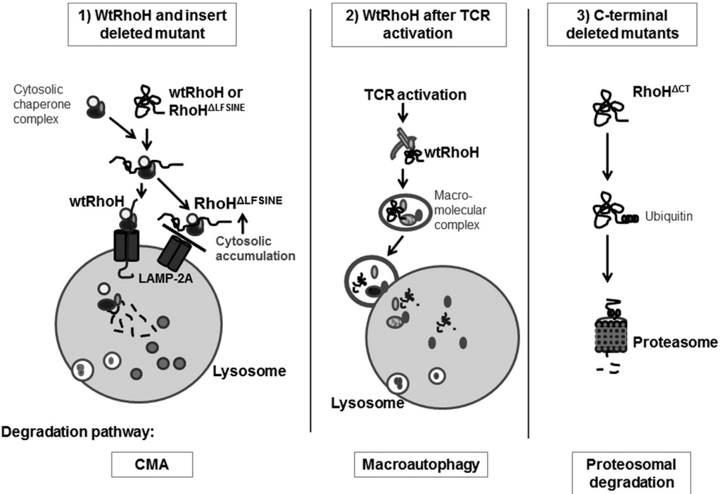
The RhoH C-terminal insert domain LFSINE regulates RhoH protein stability by promoting lysosomal degradation via chaperone-mediated autophagy, while being dispensable for ZAP70 interaction, membrane localization, and T cell reconstitution. Targeting this domain may allow alteration of RhoH protein levels without impairing vital TCR functions.
Key findings
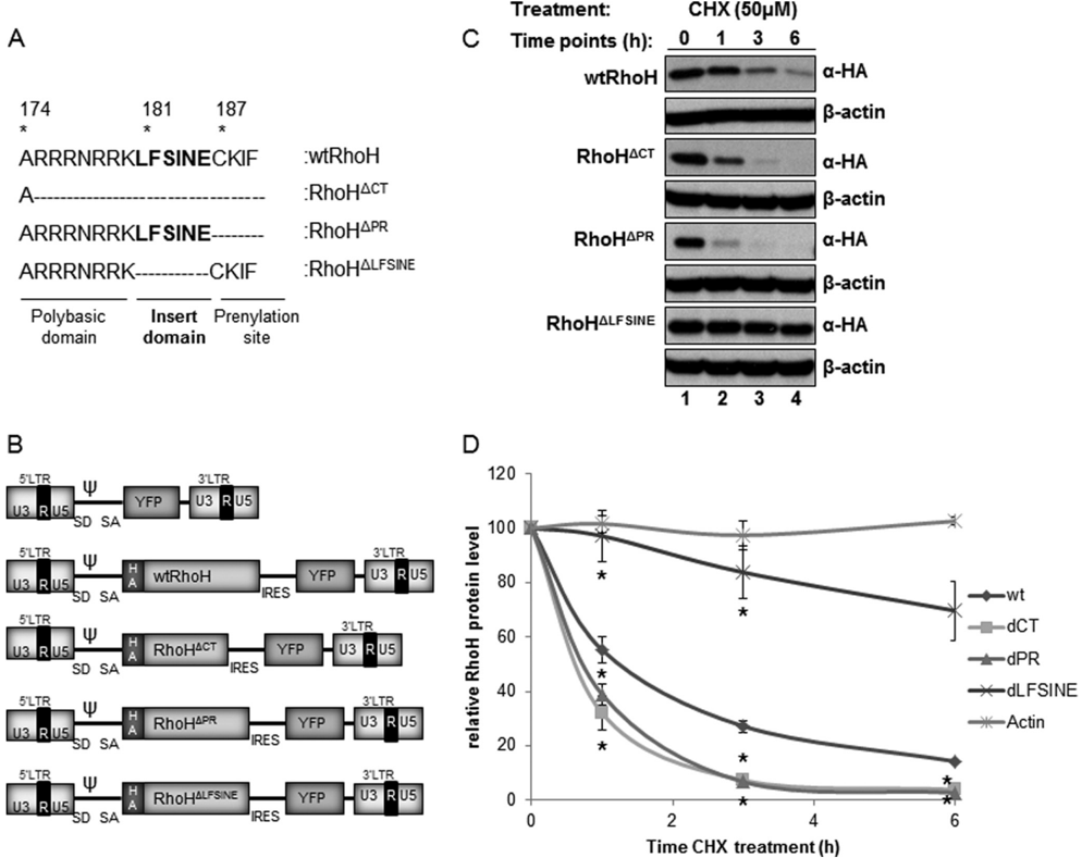
- wtRhoH has a protein half-life of less than 3 h; deletion of the insert domain (RhoH ΔLFSINE) substantially enhanced stability, with protein levels unaffected by cycloheximide for up to 6 h. Loss of the prenylation site or polybasic domain (RhoH ΔCT and RhoH ΔPR) reduced half-life by ~50%.
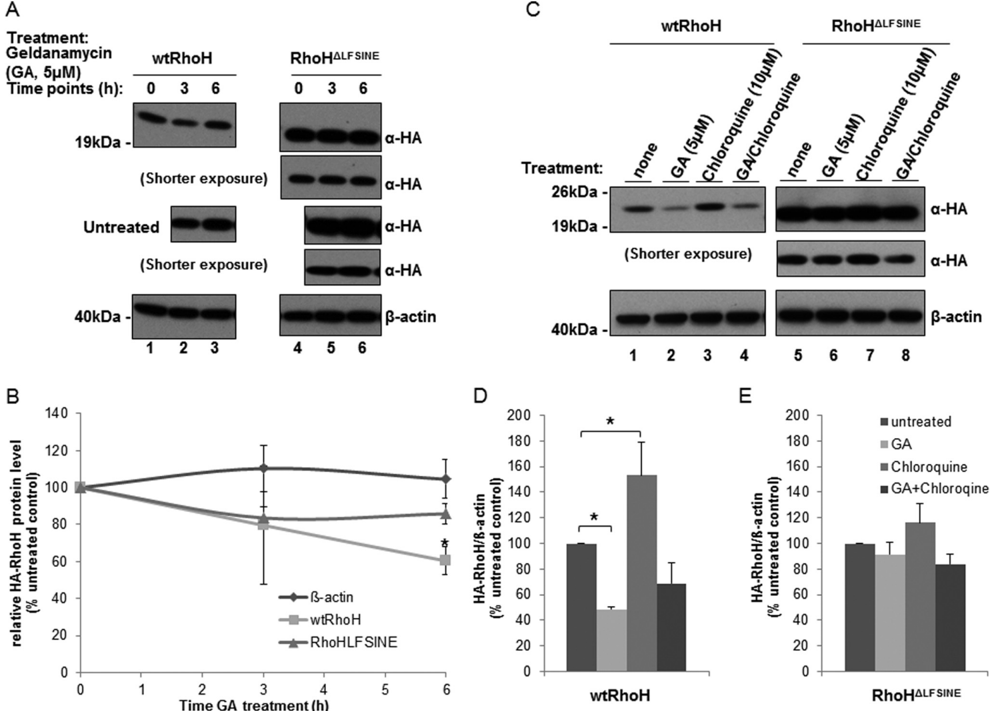
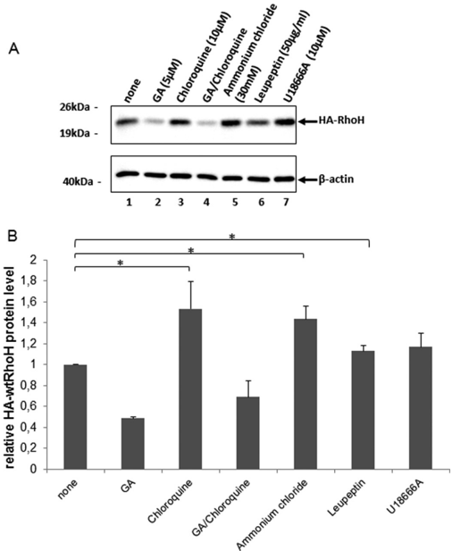
- wtRhoH is mainly degraded via chaperone-mediated autophagy/lysosomal pathways: chloroquine increased wtRhoH 1.4-fold (p = 0.044), ammonium chloride 1.4-fold (p = 0.012), and leupeptin 1.12-fold (p = 0.032); geldanamycin decreased wtRhoH at 6 h (p = 0.02) but had negligible effect on RhoH ΔLFSINE.
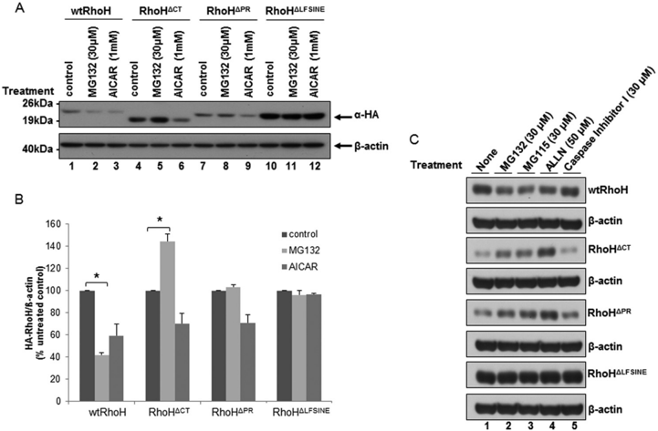
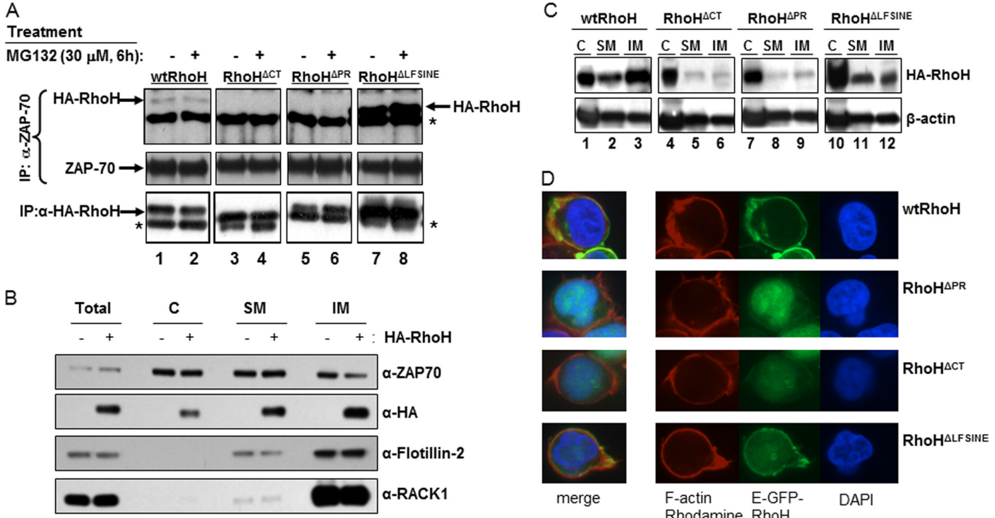
- C-terminal/prenylation-deleted mutants were mislocalized, lost ZAP70 binding, and were degraded mainly by proteasomes; MG132 increased RhoH ΔCT levels but had no effect on RhoH ΔLFSINE.
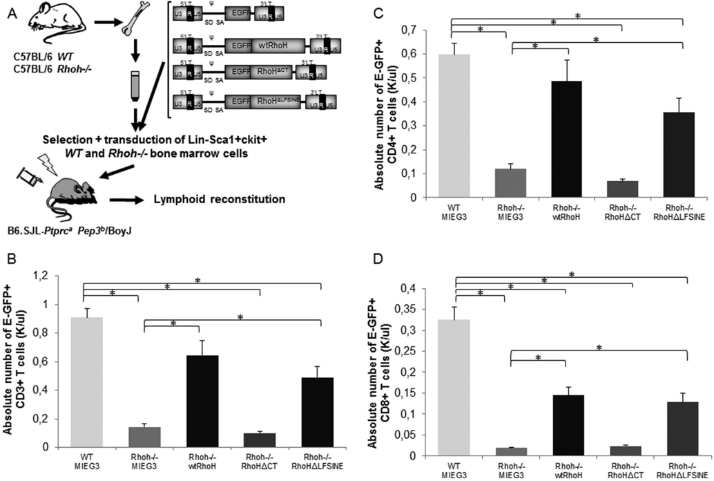
- In vivo, Rhoh−/− LSK cells reconstituted with empty vector or RhoH ΔCT remained T cell-deficient (0.1 ± 0.02 and 0.1 ± 0.01 EGFP+ T cells (K/l), respectively; n = 4–5), whereas wtRhoH rescued T lymphopenia (0.6 ± 0.1 versus WT BM 0.9 ± 0.1 EGFP+ T cells (K/l)) and RhoH ΔLFSINE reconstituted similarly to wtRhoH (0.5 ± 0.1 versus 0.6 ± 0.1 EGFP+ T cells (K/l)).
Methods
- HA-tagged wtRhoH and C-terminal mutants (RhoH ΔCT, RhoH ΔPR, RhoH ΔLFSINE) were expressed in Jurkat T cells via retroviral transduction; protein stability was assessed by cycloheximide chase and immunoblot, with inhibitors of proteasomal, lysosomal, autophagy, and chaperone pathways (MG132, MG115, ALLN, caspase inhibitor, AICAR, geldanamycin, ammonium chloride, chloroquine, leupeptin, U18666A, FTI-277).
- Interaction and localization were analyzed by anti-ZAP70 immunoprecipitation, subcellular fractionation into cytosolic/detergent-soluble/detergent-insoluble membrane fractions, and immunofluorescence confocal microscopy.
- In vivo rescue used sorted WT and Rhoh−/− Lin−Sca1+ckit+ (LSK) cells transduced with EGFP-fused wtRhoH, mutants, or empty vector and transplanted into lethally irradiated B6.SJL-Ptprc a Pep3 b/BoyJ recipients; peripheral blood T and B cells were analyzed by flow cytometry at 2 and 3 months, with Student’s t test or Mann Whitney U test and p < 0.05 considered significant.
Limitations
- The paper does not include a dedicated limitations section and states no explicit limitations.
- Evident from the text: mechanistic degradation studies rely largely on Jurkat T cells, and primary human CLL cells were not used to test the LFSINE/CMA pathway directly.
- Evident from the text: the in vivo rescue experiments used n = 4–5 recipients with 2–3 month follow-up, and the model proposes that LFSINE may contain a ubiquitination site or lysosomal recognition sequence without directly mapping it.
Model-written summary. Check it against the paper before relying on it.
Integrity signals
Registry
Cites
Statistics
Figures
Verification
Claims the paper makes
61 Own result · 35 Own interpretation · 15 Not checked yet · 11 Check pending · 5 No full text of the sources · 5 Own method · 2 Supported · 1 Partly supported · accuracy not yet measured
Every source the paper cites for this claim was checked and supports it.
In the paper
“Thus, RhoH-dependent cellular functions appear to be regulated by intracellular protein levels (51).”
§ A Unique Carboxyl-terminal Insert Domain in the Hematopoietic-specific, GTPase-deficient Rho GTPase RhoH Regulates Post-translational Processing * > DISCUSSION
What the sources say
- Supportedcites [51] Gu, 2005, RhoH GTPase. A key regulator of hematopoietic cell proliferation and apoptosis?, Cell Cycle source
The Abstract states that RhoH activity is likely regulated by its level of expression rather than by the usual GTP-binding/GTP-hydrolysis cycle. It also reports that overexpression and knockdown have opposite effects on hematopoietic progenitor-cell functions, supporting the hedged claim. (LLM, conf=0.93)
hedgedstates a possible cause- Asserted: passes — The authors put it forward as true.
- Checkable: passes — It can be checked against evidence.
- Load-bearing: passes — The paper’s conclusions depend on it.
- Based: passes — The paper gives a reason why it holds.
- Supported
A result the paper reports from its own data — it cites no other source for it, so there is none to check. The quote shows it is in the paper.
In the paper
“MG132 treatment enhanced protein levels of C terminus-deleted RhoH mutants in Jurkat T cells at 6 h (Fig. 2, A, lane 5, and B)”
§ A Unique Carboxyl-terminal Insert Domain in the Hematopoietic-specific, GTPase-deficient Rho GTPase RhoH Regulates Post-translational Processing * > Deletion of the Insert Domain Significantly Increases RhoH Protein Stability Contributing to Its Cytoplasmic Accumulation-
plainstates a cause- Asserted: passes — The authors put it forward as true.
- Checkable: passes — It can be checked against evidence.
- Load-bearing: passes — The paper’s conclusions depend on it.
- Based: passes — The paper gives a reason why it holds.
A result the paper reports from its own data — it cites no other source for it, so there is none to check. The quote shows it is in the paper.
In the paper
“Surprisingly, MG132 treatment was associated with a significant decrease in wtRhoH protein (Fig. 2, A, lane 2, and B)”
§ A Unique Carboxyl-terminal Insert Domain in the Hematopoietic-specific, GTPase-deficient Rho GTPase RhoH Regulates Post-translational Processing * > Deletion of the Insert Domain Significantly Increases RhoH Protein Stability Contributing to Its Cytoplasmic Accumulation-
plainstates an association- Asserted: passes — The authors put it forward as true.
- Checkable: passes — It can be checked against evidence.
- Load-bearing: passes — The paper’s conclusions depend on it.
- Based: passes — The paper gives a reason why it holds.
A result the paper reports from its own data — it cites no other source for it, so there is none to check. The quote shows it is in the paper.
In the paper
“whereas this treatment had little or modest effects on prenylation site-deleted RhoH ⌬PR (Fig. 2A, lane 8, and B)”
§ A Unique Carboxyl-terminal Insert Domain in the Hematopoietic-specific, GTPase-deficient Rho GTPase RhoH Regulates Post-translational Processing * > Deletion of the Insert Domain Significantly Increases RhoH Protein Stability Contributing to Its Cytoplasmic Accumulation-
plain- Asserted: passes — The authors put it forward as true.
- Checkable: passes — It can be checked against evidence.
- Load-bearing: passes — The paper’s conclusions depend on it.
- Based: passes — The paper gives a reason why it holds.
A result the paper reports from its own data — it cites no other source for it, so there is none to check. The quote shows it is in the paper.
In the paper
“in in vivo biotinylation experiments we have identified members of the heat shock protein family including Hsp90 and Hsp70 as novel interaction partners of RhoH”
§ A Unique Carboxyl-terminal Insert Domain in the Hematopoietic-specific, GTPase-deficient Rho GTPase RhoH Regulates Post-translational Processing * > Deletion of the Insert Domain Significantly Increases RhoH Protein Stability Contributing to Its Cytoplasmic Accumulation-
boostedstates an association- Asserted: passes — The authors put it forward as true.
- Checkable: passes — It can be checked against evidence.
- Load-bearing: passes — The paper’s conclusions depend on it.
- Based: passes — The paper gives a reason why it holds.
- Probably not a substantial claim. 22
- It may state two separate claims as one. 27
Create a free account to see these claims.
Create a free account to see these assumptions.
89 more claims
Create a free account to see all 135 claims and to verify the 39 references still open.
Claims other papers make about this one
Each is checked against this paper’s own text: does it say what the citing paper says it does?
In the citing paper
“Alternatively, its activation can be regulated by other types of regulation, including lysosomal degradation, somatic mutation and transcriptional repressor, which also results in an altered protein expression.”
From The Role of RhoH in TCR Signalling and Its Involvement in Diseases (2021) · cites this paper as [26]
In the citing paper
“Additionally, RhoH activity can be regulated by lysosomal degradation through its unique C-terminal region, L182FSINE187 domain, located in between its polybasic region and CAAX box. This insert domain has been shown to regulate RhoH stability via chaperone-mediated autophagy (CMA) [26].”
From The Role of RhoH in TCR Signalling and Its Involvement in Diseases (2021) · cites this paper as [26]
In the citing paper
“while not affecting its function on both T cells and B cells [26].”
From The Role of RhoH in TCR Signalling and Its Involvement in Diseases (2021) · cites this paper as [26]
In the citing paper
“the presence of the inserted domain, LFSINE, at the C-terminal was shown to act as a recognition signal for lysosomal uptake and CMA-mediated degradation”
From The Role of RhoH in TCR Signalling and Its Involvement in Diseases (2021) · cites this paper as [26]
In the citing paper
“This approach can be applied to RhoH as the presence of the inserted domain, LFSINE, at the C-terminal was shown to act as a recognition signal for lysosomal uptake and CMA-mediated degradation, while not affecting its function on both T cells and B cells [26].”
From The Role of RhoH in TCR Signalling and Its Involvement in Diseases (2021) · cites this paper as [26]
Citations
Cites
Etienne-Manneville, S., & Hall, A. (2002) · Nature · doi:10.1038/nature01148 · Cited by 4,201
Rho GTPases and their effector proteins
BISHOP, A. L., & HALL, A. (2000) · Biochemical Journal · doi:10.1042/bj3480241 · Cited by 2,575
Whitesell, L., Mimnaugh, E. G., De Costa, B., Myers, C. E., & Neckers, L. M. (1994) · Proceedings of the National Academy of Sciences · doi:10.1073/pnas.91.18.8324 · Cited by 1,444
Hypermutation of multiple proto-oncogenes in B-cell diffuse large-cell lymphomas
Pasqualucci, L., Neumeister, P., Goossens, T., Nanjangud, G., Chaganti, R. S. K., Küppers, R., & Dalla-Favera, R. (2001) · Nature · doi:10.1038/35085588 · Cited by 1,032
Hsp90 inhibitors as novel cancer chemotherapeutic agents
Neckers, L. (2002) · Trends in Molecular Medicine · doi:10.1016/s1471-4914(02)02316-x · Cited by 677
Selective autophagy: ubiquitin-mediated recognition and beyond
Kraft, C., Peter, M., & Hofmann, K. (2010) · Nature Cell Biology · doi:10.1038/ncb0910-836 · Cited by 639
Human chronic lymphocytic leukemia modeled in mouse by targeted TCL1 expression
Bichi, R., Shinton, S. A., Martin, E. S., Koval, A., Calin, G. A., Cesari, R., Russo, G., Hardy, R. R., & Croce, C. M. (2002) · Proceedings of the National Academy of Sciences · doi:10.1073/pnas.102181599 · Cited by 582
Dice, J. F. (2007) · Autophagy · doi:10.4161/auto.4144 · Cited by 569
The family of ubiquitin‐conjugating enzymes (E2s): deciding between life and death of proteins
van Wijk, S. J. L., & Timmers, H. T. M. (2009) · The FASEB Journal · doi:10.1096/fj.09-136259 · Cited by 454
Regulation of Rho GTPase crosstalk, degradation and activity by RhoGDI1
Boulter, E., Garcia-Mata, R., Guilluy, C., Dubash, A., Rossi, G., Brennwald, P. J., & Burridge, K. (2010) · Nature Cell Biology · doi:10.1038/ncb2049 · Cited by 346
Cited by
The RHO Family GTPases: Mechanisms of Regulation and Signaling
Mosaddeghzadeh, N., & Ahmadian, M. R. (2021) · Cells · doi:10.3390/cells10071831 · Cited by 388
Identifying and analyzing different cancer subtypes using RNA-seq data of blood platelets
Zhang, Y.-H., Huang, T., Chen, L., Xu, Y., Hu, Y., Hu, L.-D., Cai, Y., & Kong, X. (2017) · Oncotarget · doi:10.18632/oncotarget.20903 · Cited by 54
The Role of RhoH in TCR Signalling and Its Involvement in Diseases
Ahmad Mokhtar, A. M., Hashim, I. F., Mohd Zaini Makhtar, M., Salikin, N. H., & Amin-Nordin, S. (2021) · Cells · doi:10.3390/cells10040950 · Cited by 25
Multiple regulatory mechanisms, functions and therapeutic potential of chaperone-mediated autophagy
Wang, Y., Liu, J., Wang, H., Jiang, P., Cao, L., Lu, S., Zhang, S., Yang, R., Feng, H., Cao, L., et al. (2025) · Theranostics · doi:10.7150/thno.107761 · Cited by 22
Overexpression of RhoH Permits to Bypass the Pre-TCR Checkpoint
Tamehiro, N., Oda, H., Shirai, M., & Suzuki, H. (2015) · PLOS ONE · doi:10.1371/journal.pone.0131047 · Cited by 14
Molecular basis and current insights of atypical Rho small GTPase in cancer
Huang, H., Wang, S., Guan, Y., Ren, J., & Liu, X. (2024) · Molecular Biology Reports · doi:10.1007/s11033-023-09140-7 · Cited by 13
Targeting Chaperone-Mediated Autophagy for Disease Therapy
Li, W., Dou, J., Yang, J., Xu, H., & She, H. (2018) · Current Pharmacology Reports · doi:10.1007/s40495-018-0138-1 · Cited by 10
Thyroid hormones alter the transcriptome of in vitro-produced bovine blastocysts
Ashkar, F. A., Révay, T., Rho, N.-Y., Madan, P., Dufort, I., Robert, C., Favetta, L. A., Schmidt, C., & King, W. A. (2015) · Zygote · doi:10.1017/s0967199415000167 · Cited by 6
Koch, J., Ruggia, A., Beha, C., Wipf, I., Zhakparov, D., Westermann, P., Schmelzer, S., Heider, A., Fröhlich, K., & Baerenfaller, K. (2025) · BMC Biology · doi:10.1186/s12915-025-02345-1 · Cited by 4
Classical Rho Proteins: Biochemistry of Molecular Switch Function and Regulation
Zhang, S.-C., Nouri, K., Amin, E., Taha, M. S., Nakhaeizadeh, H., Nakhaei‐Rad, S., Dvorský, R., & Ahmadian, M. R. (2014) · Springer eBooks · doi:10.1007/978-3-7091-1806-1_14 · Cited by 4
Horiguchi, H., Xu, H., Duvert, B., Ciuculescu, F., Yao, Q., Sinha, A., McGuinness, M., Harris, C., Brendel, C., Troeger, A., et al. (2022) · Small GTPases · doi:10.1080/21541248.2021.2019503 · Cited by 3
Self-citationd’Água, B. B., Infante, E., Riou, P., Tajadura, V., & Ridley, A. J. (2014) · Springer eBooks · doi:10.1007/978-3-7091-1806-1_15 · Cited by 2
Full-length RNA-Seq of the RHOH gene in human B cells reveals new exons and splicing patterns
Leprêtre, F., Meneboo, J., Villenet, C., Delestré, L., Quesnel, B., Shelley, C. S., Figeac, M., & Galiègue‐Zouitina, S. (2024) · Scientific Reports · doi:10.1038/s41598-024-79307-0 · Cited by 1
Cai, X.-X., & Yang, Y. (2025) · New cell · doi:10.61958/ncas2202
Non-canonical and constitutive activation of small GTPases: more than an exception to the rule?
Kiers, J., & Hordijk, P. L. (2026) · Biochemical Journal · doi:10.1042/bcj20250137
Metadata
- Authors
- Troeger, A., Chae, H.-D., Senturk, M., Wood, J., & Williams, D. A.
- Affiliations
- Boston Children's Hospital (US)
- Stanford Medicine (US)
- Corresponding author
- Williams, David A.
- Venue
- Journal of Biological Chemistry
- Publisher
- Elsevier BV
- Published
- 2013-12
- Volume
- 288
- Issue
- 51
- Pages
- 36451-36462
- ISSN
- 0021-9258 (ISSN-L)
- PMC
- PMC3868758
- OpenAlex
- W2088689128
- Semantic Scholar
- ab5a010bbc5c48bae8f4b3045a9e3b87b69f8b85
- Cited by
- 15
- References
- 61
- Citations by year
- 2026: 1 · 2025: 3 · 2024: 2 · 2022: 1 · 2021: 2 · 2018: 1 · 2017: 1 · 2015: 2 · 2014: 2
- FWCI
- 0.40
- Type
- journal article
- Language
- en
- Open access
- hybrid
- doi.org
- Licence
- Locations
- Journal of Biological Chemistry (publishedVersion, cc-by)
- PubMed (publishedVersion)
- PubMed Central (publishedVersion)
- Funders
- National Institutes of Health (U54TR001005)
- National Cancer Institute (R01CA113969)
- National Center for Advancing Translational Sciences
- Topics
- Life Sciences › Biochemistry, Genetics and Molecular Biology › Molecular Biology › Protein Kinase Regulation and GTPase Signaling
- Health Sciences › Medicine › Hematology › Chronic Lymphocytic Leukemia Research
- Life Sciences › Biochemistry, Genetics and Molecular Biology › Cell Biology › Cellular transport and secretion
- Keywords
- RhoH, Rho GTPases, CAAX box, protein stability, chaperone-mediated autophagy, lysosomal degradation, ZAP-70, T cell receptor signaling, chronic lymphocytic leukemia, B-cell malignancies, lenalidomide, prenylation, subcellular localization
- MeSH terms
- Animals, Bone Marrow Cells, Cell Membrane, Cytoplasm, Humans, Lysosomes, Mice, Inbred C57BL, Mutation, Protein Processing, Post-Translational, Transcription Factors, Protein Structure, Tertiary, Jurkat Cells, rho GTP-Binding Proteins, Amino Acid Motifs, Protein Transport, Mice, Prenylation, Protein Stability, Proteolysis
About these checks
Registry, retraction, Cites and statistics results are computed, not judged, with one exception: a printed reference that no index lists is read once by a model before it counts as a citation error. Claims are extracted by a model and kept only when their quote appears verbatim in the text; each cited source is then checked, with a model’s help, for whether it says what the claim relies on. Those verdicts can be wrong — read the claim’s quote and the cited source before relying on one.
A language model drew the claims from the paper on 2026-11-12, under version 3 of the claim definition (model deepseek-v4.1-flash, prompt extract-v3).
A second model labelled each claim on 2026-11-12 (model jev-1.13-20260917, definition revision gold-guideline-v0.2).
Cited sources are checked with a language model’s help (gpt-6-sol).
Statistics and arithmetic are recomputed, not judged.
Confidence bands are shown only for fields calibrated on our annotated test set. None is calibrated yet, so no band is shown.
What counts as a claim ›How we measure confidence ›How we check this ›
Are you an author, or do you see a problem with this page? Request a review
