Student or educator? Sign up with your school email and get 6 months of Pro free.
CoChat

The DOI matches its Crossref record; 36 of the 36 references it cites were found in a registry or an index.

Activated pp60c-Src Leads to Elevated Hypoxia-inducible Factor (HIF)-1α Expression under Normoxia

Open access
Karni, Rotem, Dor, Yuval, Keshet, Eli, Meyuhas, Oded, Levitzki, Alexander · 2002 · Journal of Biological Chemistry

Abstract

Hypoxia-inducible factor (HIF)-1 is a master transcription factor, which up-regulates glycolysis, erythropoiesis, and angiogenesis under hypoxia. HIF-1alpha accumulates in normoxic tumor cells, leading to glycolysis under aerobic conditions. This phenomenon, known as the "Warburg effect," is caused by a yet unknown mechanism. Here we show that transformed cells that express constitutively active pp60(c-Src) (Src) express HIF-1alpha protein under normoxia, which results in the expression of multiple HIF-1alpha target genes. We show that this occurrence is due to an enhanced rate of HIF-1alpha protein synthesis and not due to reduced HIF-1alpha degradation. Furthermore, we show that the Src-induced increase in protein synthesis is due to the global increase in the rate of cap-dependent translation and does not involve inhibition of HIF-1alpha degradation.

Overview

The claims and statistics of this paper haven’t been checked yet.

Free: it reads the open-access text and checks the first 5 claims that cite a source we can check. Create a free account to check every claim.

A few free checks a day · papers already verified open instantly · results are public

Integrity signals

Registry

Crossref · 10.1074/jbc.m206141200

Cites

36 of 36 references verified0 citation errors · 0 not checkable

Statistics

Figures

FIG. 1 Src kinase activity regulates the mRNA levels of hypoxia-regulated genes and the transcriptional activity of HIF1 𝛂 . A , the levels of VEGF and PGK1 mRNA transcripts in NIH3T3 and CSH12 cells (NIH3T3 overexpressing EGFR-HER-2 chimeric receptor and possessing constitutive Src activity). B , the levels of mRNA transcripts of HIF-1 𝛂 , Glut-1, PGK-1, LDH-A, or VEGF in CSH12 cells and HT29 cells. HT29 and CSH12 cells were seeded and treated with PP1 at the indicated concentrations (see 'Material and Methods'). C and E , 3 × 10 5 (HT29) or 8 × 10 4 (CSH12) cells were seeded on six-well plates (Nunc) and 24 h later were co-transfected with the HRE reporter plasmid (p-tk p -luc), and the CMV-lac-Z plasmid as an internal control. 24 h after transfection the medium was replaced with medium containing PP1 at the concentrations indicated for an additional 24 h. Cells were then lysed and luciferase activity was measured using a luciferase kit (Promega). Luciferase activity was normalized to 𝛃 -galactosidase activity. The vehicle ( Veh ) in which PP1 was dissolved was 0.02% Me2SO, 0.4% ethanol. Cobalt chloride (CoCl 2 ) is known to stabilize HIF-1 𝛂 and to induce HRE. D and F , cells were co-transfected with p-tk p -luc and the CMV-lac-Z plasmids together with Src constructs (empty plasmid ( pUSE ( -) , wild type c-Src ( pUSE-WT Src ), kinasedead Src (K297R) ( pUSE -KD Src ), or active Src (Y529F) ( pUSE-Active Src )) as indicated in the figure and luciferase and 𝛃 -galactosidase activity was measured 48 h after transfection.
FIG. 1 Src kinase activity regulates the mRNA levels of hypoxia-regulated genes and the transcriptional activity of HIF1 𝛂 . A , the levels of VEGF and PGK1 mRNA transcripts in NIH3T3 and CSH12 cells (NIH3T3 overexpressing EGFR-HER-2 chimeric receptor and possessing constitutive Src activity). B , the levels of mRNA transcripts of HIF-1 𝛂 , Glut-1, PGK-1, LDH-A, or VEGF in CSH12 cells and HT29 cells. HT29 and CSH12 cells were seeded and treated with PP1 at the indicated concentrations (see 'Material and Methods'). C and E , 3 × 10 5 (HT29) or 8 × 10 4 (CSH12) cells were seeded on six-well plates (Nunc) and 24 h later were co-transfected with the HRE reporter plasmid (p-tk p -luc), and the CMV-lac-Z plasmid as an internal control. 24 h after transfection the medium was replaced with medium containing PP1 at the concentrations indicated for an additional 24 h. Cells were then lysed and luciferase activity was measured using a luciferase kit (Promega). Luciferase activity was normalized to 𝛃 -galactosidase activity. The vehicle ( Veh ) in which PP1 was dissolved was 0.02% Me2SO, 0.4% ethanol. Cobalt chloride (CoCl 2 ) is known to stabilize HIF-1 𝛂 and to induce HRE. D and F , cells were co-transfected with p-tk p -luc and the CMV-lac-Z plasmids together with Src constructs (empty plasmid ( pUSE ( -) , wild type c-Src ( pUSE-WT Src ), kinasedead Src (K297R) ( pUSE -KD Src ), or active Src (Y529F) ( pUSE-Active Src )) as indicated in the figure and luciferase and 𝛃 -galactosidase activity was measured 48 h after transfection.CC-BY · From the PDF
FIG. 2 Expression of active c-Src elevates the protein levels of HIF-1 𝛂 with no effect on its mRNA levels or protein stability. A , Saos-2 clones stably expressing empty plasmid ( pUSE (-)), wild type c-Src ( pUSE-WT Src ), kinase-dead Src (K297R) ( pUSE -KD Src ), or active Src (Y529F) ( pUSE-Active Src ) were seeded on six-well plates (2 × 10 5 cells) or 10-cm dishes (2 × 10 6 cells). Cells were lysed with sample buffer, and after Western blotting the blot was probed with antibodies against HIF-1 𝛂 (1:250 Transduction Laboratories), c-Src (mAb 327), and actin (1:2000 Santa Cruz). B , total RNA from the Saos-2 clones (10 𝛍 g) was subjected to Northern blotting. The blot was hybridized with a HIF-1 𝛂 probe. To verify equal loading and transfer, the levels of ribosomal RNA were detected by methylene blue. C , Saos-2 clones were incubated in the presence of 100 𝛍 M cycloheximide ( CHX ) for the indicated times. Cells were then lysed with sample buffer, and after Western blotting the blot was probed with antibodies to HIF-1 𝛂 and actin as in A . D , HT29 cells were grown on 60-mm dishes (10 6 ), and 24 h later 100 𝛍 M cycloheximide was added for the indicated times in the presence or absence of 20 𝛍 M PP1. Cells were then lysed with sample buffer and subjected to SDS-PAGE and Western blotting. The blots were probed as in A . Calculation of t 1/2 was performed after quantification of HIF-1 𝛂 levels normalized to actin levels by the NIHIMAGE program and was done by the 'Regression' program (Blackwell Scientific Software, Osney Mead, Oxford, UK).
FIG. 2 Expression of active c-Src elevates the protein levels of HIF-1 𝛂 with no effect on its mRNA levels or protein stability. A , Saos-2 clones stably expressing empty plasmid ( pUSE (-)), wild type c-Src ( pUSE-WT Src ), kinase-dead Src (K297R) ( pUSE -KD Src ), or active Src (Y529F) ( pUSE-Active Src ) were seeded on six-well plates (2 × 10 5 cells) or 10-cm dishes (2 × 10 6 cells). Cells were lysed with sample buffer, and after Western blotting the blot was probed with antibodies against HIF-1 𝛂 (1:250 Transduction Laboratories), c-Src (mAb 327), and actin (1:2000 Santa Cruz). B , total RNA from the Saos-2 clones (10 𝛍 g) was subjected to Northern blotting. The blot was hybridized with a HIF-1 𝛂 probe. To verify equal loading and transfer, the levels of ribosomal RNA were detected by methylene blue. C , Saos-2 clones were incubated in the presence of 100 𝛍 M cycloheximide ( CHX ) for the indicated times. Cells were then lysed with sample buffer, and after Western blotting the blot was probed with antibodies to HIF-1 𝛂 and actin as in A . D , HT29 cells were grown on 60-mm dishes (10 6 ), and 24 h later 100 𝛍 M cycloheximide was added for the indicated times in the presence or absence of 20 𝛍 M PP1. Cells were then lysed with sample buffer and subjected to SDS-PAGE and Western blotting. The blots were probed as in A . Calculation of t 1/2 was performed after quantification of HIF-1 𝛂 levels normalized to actin levels by the NIHIMAGE program and was done by the 'Regression' program (Blackwell Scientific Software, Osney Mead, Oxford, UK).CC-BY · From the PDF
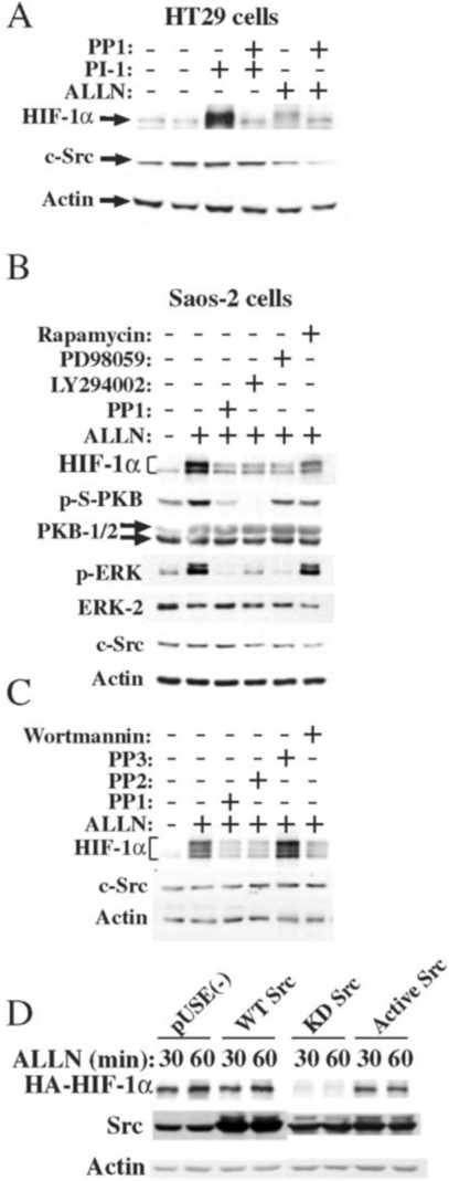
FIG. 3 Src, MEK1, mTOR, and PI3-kinase inhibitors block the accumulation of HIF-1 𝛂 protein. A , HT29 cells were seeded on six-well plates (Nunc) (4 × 10 5 cells/well). After 24 h, the medium was replaced with medium containing the inhibitor (20 𝛍 M PP1) for 30 min and then with medium containing the inhibitor and proteasome inhibitors (100 𝛍 M PI-1 (Proteasome Inhibitor-1) or 200 𝛍 M ALLN (Calbiochem)) for an additional 2 h. Cells were then lysed with sample buffer and after Western blotting were probed as in Fig. 2. B , Saos-2 cells were seeded on six-well plates (Nunc) (2 × 10 5 cells/well). After 24 h, the medium was replaced with medium containing the inhibitor (20 𝛍 M PP1, 20 𝛍 M LY294002, 50 𝛍 M PD98059, or 50 nM rapamycin) for 30 min and then with medium containing the inhibitors and the proteasome inhibitor ALLN (200 𝛍 M) (Calbiochem) for an additional 2 h. Cells were then lysed with sample buffer, and after Western blotting the blot was probed with antibodies against HIF-1 𝛂 (1:250 Transduction Laboratories), c-Src (mAb 327) and actin (1:2000 Santa Cruz), phosphorylated ERK (1:10000 Sigma), phospho-serine-Protein Kinase B (1:1000 NEB), ERK-2 (1:5000 Santa Cruz) and Akt-1, 2 ( PKB ) (1:1000 Santa Cruz). C ,
FIG. 3 Src, MEK1, mTOR, and PI3-kinase inhibitors block the accumulation of HIF-1 𝛂 protein. A , HT29 cells were seeded on six-well plates (Nunc) (4 × 10 5 cells/well). After 24 h, the medium was replaced with medium containing the inhibitor (20 𝛍 M PP1) for 30 min and then with medium containing the inhibitor and proteasome inhibitors (100 𝛍 M PI-1 (Proteasome Inhibitor-1) or 200 𝛍 M ALLN (Calbiochem)) for an additional 2 h. Cells were then lysed with sample buffer and after Western blotting were probed as in Fig. 2. B , Saos-2 cells were seeded on six-well plates (Nunc) (2 × 10 5 cells/well). After 24 h, the medium was replaced with medium containing the inhibitor (20 𝛍 M PP1, 20 𝛍 M LY294002, 50 𝛍 M PD98059, or 50 nM rapamycin) for 30 min and then with medium containing the inhibitors and the proteasome inhibitor ALLN (200 𝛍 M) (Calbiochem) for an additional 2 h. Cells were then lysed with sample buffer, and after Western blotting the blot was probed with antibodies against HIF-1 𝛂 (1:250 Transduction Laboratories), c-Src (mAb 327) and actin (1:2000 Santa Cruz), phosphorylated ERK (1:10000 Sigma), phospho-serine-Protein Kinase B (1:1000 NEB), ERK-2 (1:5000 Santa Cruz) and Akt-1, 2 ( PKB ) (1:1000 Santa Cruz). C ,CC-BY · From the PDF
FIG. 4 Src kinase activity regulates the synthesis of HIF-1 𝛂 by a cap-dependent mechanism in CSH12 cells. A , CSH12 cells were grown on 10-cm dishes and were co-transfected with HA-HIF-1 𝛂 expression plasmid (0.5 𝛍 g) and pUSE( -) plasmid (5 𝛍 g). After 24 h the medium was replaced with methionine- and cysteine-deficient medium for 1 h. 100 𝛍 Ci/ml of [ 35 S]Met/Cys Promix (Amersham Biosciences) was then added for 1 h in the presence of 200 𝛍 M ALLN, which prevents degradation of HA-HIF-1 𝛂 . HA-HIF-1 𝛂 was immunoprecipitated as described under 'Materials and Methods.' After Western blotting, the membrane was exposed for HA-HIF-1 𝛂 radioactivity and probed as in Fig. 3 for HA-HIF-1 𝛂 protein levels. B , CSH12 cells were seeded on 24-well plates (Nunc). 24 h later, cells were co-transfected with 0.5 𝛍 g of Src constructs as indicated and 0.1 𝛍 g of luc-IRES-SeAP plasmid (see 'Materials and Methods'), or luc and SeAP on separate plasmids. PP1 was added 24 h following transfection. 24 h after addition of PP1, cells were lysed with reporter lysis buffer (Promega), and luciferase and SeAP activities were measured. C , the HIF-1 𝛂 5 ′ -UTR-SeAP construct was co-transfected as described under 'Materials and Methods' with the indicated Src constructs.
FIG. 4 Src kinase activity regulates the synthesis of HIF-1 𝛂 by a cap-dependent mechanism in CSH12 cells. A , CSH12 cells were grown on 10-cm dishes and were co-transfected with HA-HIF-1 𝛂 expression plasmid (0.5 𝛍 g) and pUSE( -) plasmid (5 𝛍 g). After 24 h the medium was replaced with methionine- and cysteine-deficient medium for 1 h. 100 𝛍 Ci/ml of [ 35 S]Met/Cys Promix (Amersham Biosciences) was then added for 1 h in the presence of 200 𝛍 M ALLN, which prevents degradation of HA-HIF-1 𝛂 . HA-HIF-1 𝛂 was immunoprecipitated as described under 'Materials and Methods.' After Western blotting, the membrane was exposed for HA-HIF-1 𝛂 radioactivity and probed as in Fig. 3 for HA-HIF-1 𝛂 protein levels. B , CSH12 cells were seeded on 24-well plates (Nunc). 24 h later, cells were co-transfected with 0.5 𝛍 g of Src constructs as indicated and 0.1 𝛍 g of luc-IRES-SeAP plasmid (see 'Materials and Methods'), or luc and SeAP on separate plasmids. PP1 was added 24 h following transfection. 24 h after addition of PP1, cells were lysed with reporter lysis buffer (Promega), and luciferase and SeAP activities were measured. C , the HIF-1 𝛂 5 ′ -UTR-SeAP construct was co-transfected as described under 'Materials and Methods' with the indicated Src constructs.CC-BY · From the PDF
FIG. 5 Inhibition of Src kinase, PI3K/mTOR or Ras/MAPK pathways reduces protein synthesis rate. A , Saos-2 cells (3 × 10 5 cells/well) were metabolically labeled with [ 35 S]Met/Cys as described under 'Materials and Methods.' Equal amounts of protein from each sample were separated on 10% SDS-PAGE and exposed to film. An identical experiment was performed with HT29 cells (not shown). B , Saos-2 (3 × 10 5 cells/well) and HT29 (5 × 10 5 cells/well) were metabolically labeled with [ 35 S]Met/Cys as described under 'Materials and Methods.' Protein synthesis rate was calculated as described under 'Materials and Methods.' C , HeLa cells expressing empty plasmid (pCDNA3) or HA-eIF4E were seeded (2 × 10 5 ), and 24 h later were lysed with sample buffer. After Western blotting, the membrane was probed with antibodies against HIF-1 𝛂 and c-Src (as in Fig. 3), and anti-HA tag (1:1000, Roche Molecular Biochemicals).
FIG. 5 Inhibition of Src kinase, PI3K/mTOR or Ras/MAPK pathways reduces protein synthesis rate. A , Saos-2 cells (3 × 10 5 cells/well) were metabolically labeled with [ 35 S]Met/Cys as described under 'Materials and Methods.' Equal amounts of protein from each sample were separated on 10% SDS-PAGE and exposed to film. An identical experiment was performed with HT29 cells (not shown). B , Saos-2 (3 × 10 5 cells/well) and HT29 (5 × 10 5 cells/well) were metabolically labeled with [ 35 S]Met/Cys as described under 'Materials and Methods.' Protein synthesis rate was calculated as described under 'Materials and Methods.' C , HeLa cells expressing empty plasmid (pCDNA3) or HA-eIF4E were seeded (2 × 10 5 ), and 24 h later were lysed with sample buffer. After Western blotting, the membrane was probed with antibodies against HIF-1 𝛂 and c-Src (as in Fig. 3), and anti-HA tag (1:1000, Roche Molecular Biochemicals).CC-BY · From the PDF
FIG. 6 Inhibition of Src kinase, PI3K/mTOR, or Ras/MAPK pathways reduces the production of lactic acid. A , CSH12, Saos-2, and HT29 cells were grown on six-well plates, and the amounts of lactic acid in the medium were measured using a lactate detection kit (Sigma). Levels were measured at 6 and 19 h after medium was replaced by medium with dialyzed serum containing the kinase inhibitors as indicated. CSH12 and NIH3T3 cells were grown as in A , and the rate of lactate production was calculated from the slope of the accumulation curve over 2, 4, and 6 h. B , the steady state level of HIF-1 𝛂 protein is determined by the balance between the rates of degradation and synthesis. Any protein or oncogene that can enhance HIF-1 𝛂 translation or stimuli that inhibit its degradation will elevate its protein levels. The outcome will be elevated glycolysis and angiogenesis.
FIG. 6 Inhibition of Src kinase, PI3K/mTOR, or Ras/MAPK pathways reduces the production of lactic acid. A , CSH12, Saos-2, and HT29 cells were grown on six-well plates, and the amounts of lactic acid in the medium were measured using a lactate detection kit (Sigma). Levels were measured at 6 and 19 h after medium was replaced by medium with dialyzed serum containing the kinase inhibitors as indicated. CSH12 and NIH3T3 cells were grown as in A , and the rate of lactate production was calculated from the slope of the accumulation curve over 2, 4, and 6 h. B , the steady state level of HIF-1 𝛂 protein is determined by the balance between the rates of degradation and synthesis. Any protein or oncogene that can enhance HIF-1 𝛂 translation or stimuli that inhibit its degradation will elevate its protein levels. The outcome will be elevated glycolysis and angiogenesis.CC-BY · From the PDF

Verification

Claims the paper makes

The claims and statistics of this paper haven’t been checked yet.

Free: it reads the open-access text and checks the first 5 claims that cite a source we can check. Create a free account to check every claim.

A few free checks a day · papers already verified open instantly · results are public

Claims other papers make about this one

Each is checked against this paper’s own text: does it say what the citing paper says it does?

Self-check

Percentages, sums and reported differences re-computed from the paper’s own numbers.

The self-check needs the paper’s full text.

Citations

Cites

Show all 36 references

Research works printed but not linked: 5

  1. Jiang, 1997, Cancer Res., 57, 5328
  2. Krystal, 1998, Cancer Res., 58, 4660
  3. Li, 1996, Cell Biol. Int., 20, 723
  4. Feldser, 1999, Cancer Res., 59, 3915
  5. Zhong, 1999, Cancer Res., 59, 5830

Cited by

See all 117

Metadata

Authors
Karni, R., Dor, Y., Keshet, E., Meyuhas, O., & Levitzki, A.
Affiliations
Hebrew University of Jerusalem (IL)
Corresponding author
Levitzki, Alexander
Venue
Journal of Biological Chemistry
Publisher
Elsevier BV
Published
2002-11
Volume
277
Issue
45
Pages
42919-42925
ISSN
0021-9258 (ISSN-L)
OpenAlex
W2142210400
Cited by
116
References
36
Citations by year
2025: 1 · 2024: 1 · 2023: 4 · 2022: 3 · 2021: 3 · 2020: 2 · 2019: 3 · 2018: 1 · 2017: 2 · 2016: 3 · 2015: 3 · 2014: 3 · 2013: 3 · 2012: 11
FWCI
3.39
Type
journal article
Language
en
Open access
Licence
Locations
  • Journal of Biological Chemistry (publishedVersion, cc-by)
  • PubMed (publishedVersion)
Funders
James S. McDonnell Foundation
Topics
  • Life Sciences › Biochemistry, Genetics and Molecular Biology › Cancer Research › Cancer, Hypoxia, and Metabolism
  • Life Sciences › Biochemistry, Genetics and Molecular Biology › Molecular Biology › Metabolism, Diabetes, and Cancer
  • Life Sciences › Biochemistry, Genetics and Molecular Biology › Molecular Biology › PI3K/AKT/mTOR signaling in cancer
Keywords
Src kinase, HIF-1α, hypoxia-inducible factor, normoxia, Warburg effect, aerobic glycolysis, protein synthesis, cap-dependent translation, HIF-1α degradation, oncogenic transformation, HIF-1 target genes
MeSH terms
Animals, Base Sequence, Cell Line, Gene Expression Regulation, Half-Life, Humans, Kinetics, Recombinant Fusion Proteins, RNA, Messenger, Transcription Factors, Transfection, Protein Biosynthesis, Proto-Oncogene Proteins pp60(c-src), Genes, Reporter, DNA Primers, Hypoxia-Inducible Factor 1, alpha Subunit

About these checks

Registry, retraction, Cites and statistics results are computed, not judged, with one exception: a printed reference that no index lists is read once by a model before it counts as a citation error. Claims are extracted by a model and kept only when their quote appears verbatim in the text; each cited source is then checked, with a model’s help, for whether it says what the claim relies on. Those verdicts can be wrong — read the claim’s quote and the cited source before relying on one.

Statistics and arithmetic are recomputed, not judged.

Confidence bands are shown only for fields calibrated on our annotated test set. None is calibrated yet, so no band is shown.

What counts as a claim ›How we measure confidence ›How we check this ›

Are you an author, or do you see a problem with this page? Request a review

How to cite

Cite CoChat for the checks on this page, and the paper for its own findings.

This page
CoChat. (2026). Citation check: Activated pp60c-Src Leads to Elevated Hypoxia-inducible Factor (HIF)-1α Expression under Normoxia. https://app.cochat.ai/papers/10.1074/jbc.m206141200
The paper
Karni, R., Dor, Y., Keshet, E., Meyuhas, O., & Levitzki, A. (2002). Activated pp60c-Src Leads to Elevated Hypoxia-inducible Factor (HIF)-1α Expression under Normoxia. Journal of Biological Chemistry. https://doi.org/10.1074/jbc.m206141200

Data sources

Bibliographic data: Semantic Scholar, OpenAlex, Crossref.

Contains information from the Semantic Scholar Academic Graph Datasets, made available under the ODC Attribution License (ODC-BY 1.0).

Semantic Scholar’s data is described in: Kinney, R., et al. (2023). The Semantic Scholar Open Data Platform. arXiv:2301.10140.