

The DOI matches its Crossref record; 27 of the 27 references it cites were found in a registry or an index.
IRAK4 Kinase Activity Is Redundant for Interleukin-1 (IL-1) Receptor-associated Kinase Phosphorylation and IL-1 Responsiveness
Open accessAbstract
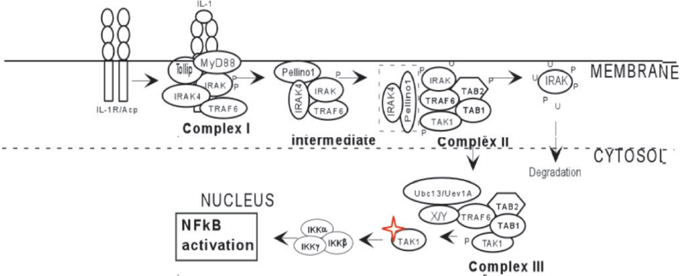
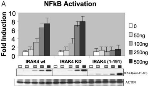
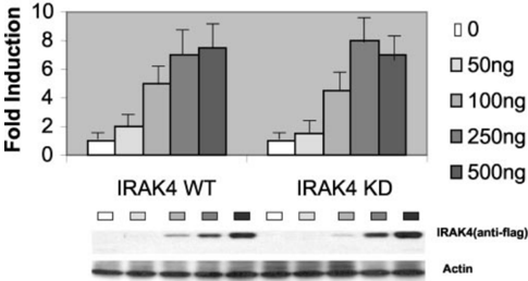
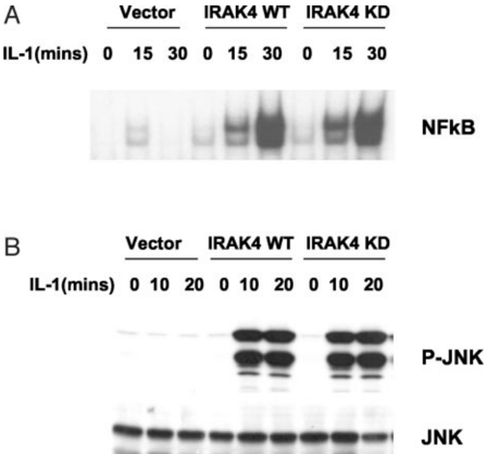
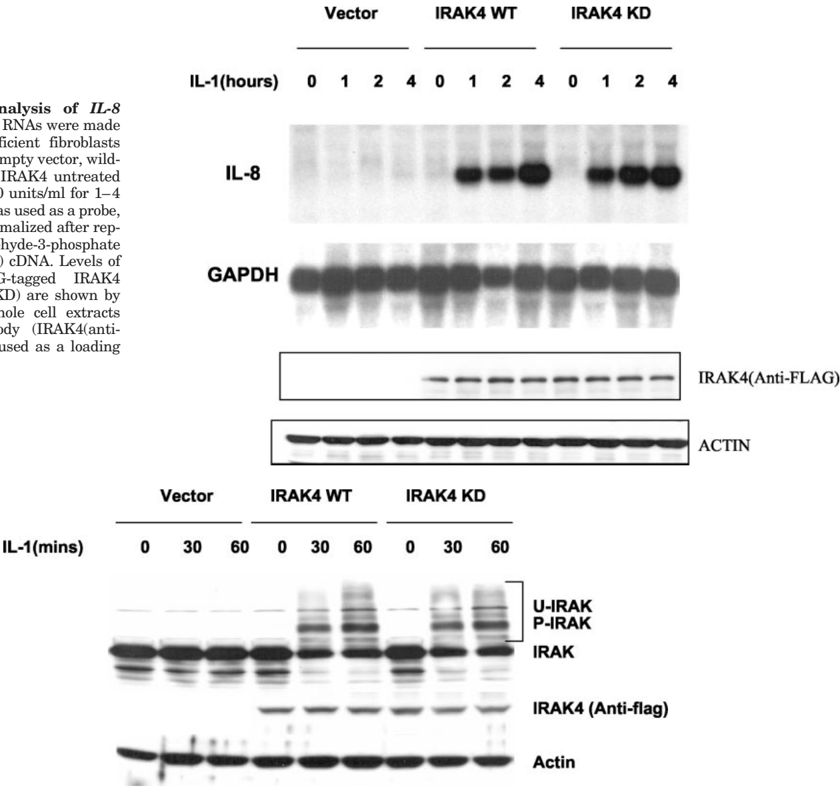
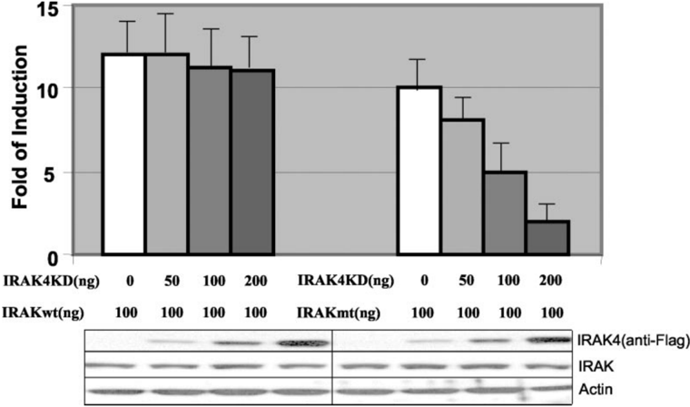
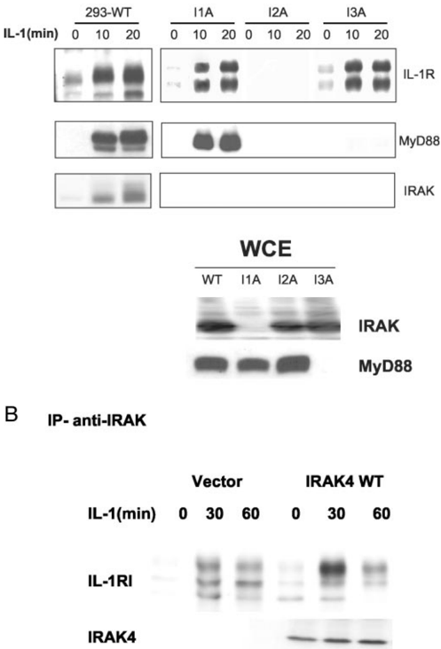
Interleukin-1 (IL-1) stimulation leads to the recruitment of interleukin-1 receptor-associated kinase (IRAK) to the IL-1 receptor, where IRAK is phosphorylated, ubiquitinated, and eventually degraded. Kinase-inactive mutant IRAK is still phosphorylated in response to IL-1 stimulation when it is transfected into IRAK-deficient cells, suggesting that there must be an IRAK kinase in the pathway. The fact that IRAK4, another IRAK family member necessary for the IL-1 pathway, is able to phosphorylate IRAK in vitro suggests that IRAK4 might be the IRAK kinase. However, we now found that the IRAK4 kinase-inactive mutant had the same ability as the wild-type IRAK4 in restoring IL-1-mediated signaling in human IRAK4-deficient cells, including NFkappaB-dependent reporter gene expression, the activation of NFkappaB and JNK, and endogenous IL-8 gene expression. These results strongly indicate that the kinase activity of human IRAK4 is not necessary for IL-1 signaling. Furthermore, we showed that the kinase activity of IRAK4 was not necessary for IL-1-induced IRAK phosphorylation, suggesting that IRAK phosphorylation can probably be achieved either by autophosphorylation or by trans-phosphorylation through IRAK4. In support of this, only the impairment of the kinase activity of both IRAK and IRAK4 efficiently abolished the IL-1 pathway, demonstrating that the kinase activity of IRAK and IRAK4 is redundant for IL-1-mediated signaling. Moreover, consistent with the fact that IRAK4 is a necessary component of the IL-1 pathway, we found that IRAK4 was required for the efficient recruitment of IRAK to the IL-1 receptor complex.
Overview
The claims and statistics of this paper haven’t been checked yet.
Run the check — free, no account needed. It reads the open-access text and checks the sources it cites.
Integrity signals
Registry
Cites
Statistics
Figures
Verification
Claims the paper makes
The claims and statistics of this paper haven’t been checked yet.
Run the check — free, no account needed. It reads the open-access text and checks the sources it cites.
Claims other papers make about this one
Each is checked against this paper’s own text: does it say what the citing paper says it does?
In the citing paper
“Particularly for IRAK4, its scaffolding role in Toll-like receptor (TLR) signalling around the myddosome is established165,172”
From PROTAC targeted protein degraders: the past is prologue (2022) · cites this paper as [172]
Self-check
Percentages, sums and reported differences re-computed from the paper’s own numbers.
The self-check needs the paper’s full text.
Citations
Cites
A Toll-like receptor recognizes bacterial DNA
Hemmi, H., Takeuchi, O., Kawai, T., Kaisho, T., Sato, S., Sanjo, H., Matsumoto, M., Hoshino, K., Wagner, H., Takeda, K., et al. (2000) · Nature · doi:10.1038/35047123 · Cited by 6,491
A human homologue of the Drosophila Toll protein signals activation of adaptive immunity
Medzhitov, R., Preston-Hurlburt, P., & Janeway, C. A. J. (1997) · Nature · doi:10.1038/41131 · Cited by 6,137
Nuclear Factor-κB — A Pivotal Transcription Factor in Chronic Inflammatory Diseases
Barnes, P. J., & Karin, M. (1997) · New England Journal of Medicine · doi:10.1056/nejm199704103361506 · Cited by 4,622
Biologic basis for interleukin-1 in disease.
Dinarello, C. (1996) · Blood · doi:10.1182/blood.v87.6.2095.bloodjournal8762095 · Cited by 4,344
IKK-1 and IKK-2: Cytokine-Activated IκB Kinases Essential for NF-κB Activation
Mercurio, F., Zhu, H., Murray, B. W., Shevchenko, A., Bennett, B. L., Li, J. W., Young, D. B., Barbosa, M. S., Mann, M., Manning, A. M., et al. (1997) · Science · doi:10.1126/science.278.5339.860 · Cited by 2,042
A family of human receptors structurally related to Drosophila Toll.
Rock, F., Hardiman, G., Timans, J., Kastelein, R., & Bazan, J. (1998) · Proceedings of the National Academy of Sciences of the United States of America · doi:10.1073/pnas.95.2.588 · Cited by 2,009
Zandi, E., Rothwarf, D. M., Delhase, M., Hayakawa, M., & Karin, M. (1997) · Cell · doi:10.1016/s0092-8674(00)80406-7 · Cited by 1,814
TRAF6 is a signal transducer for interleukin-1
Cao, Z., Xiong, J., Takeuchi, M., Kurama, T., & Goeddel, D. V. (1996) · Nature · doi:10.1038/383443a0 · Cited by 1,305
MyD88: an adapter that recruits IRAK to the IL-1 receptor complex.
Wesche, H., Henzel, W., Shillinglaw, W., Li, S., & Cao, Z.-D. (1997) · Immunity · doi:10.1016/s1074-7613(00)80402-1 · Cited by 1,247
Ninomiya‐Tsuji, J., Kishimoto, K., Hiyama, A., Inoue, J., Cao, Z., & Matsumoto, K. (1999) · Nature · doi:10.1038/18465 · Cited by 1,187
Research works printed but not linked: 2
- Chuang, 2000, Eur. Cytokine Netw., 11, 372
- Jiang, 2002, J. Biol. Chem.
Cited by
Sorted by citations
PROTAC targeted protein degraders: the past is prologue
Békés, M., Langley, D. R., & Crews, C. M. (2022) · Nature Reviews Drug Discovery · doi:10.1038/s41573-021-00371-6 · Cited by 3,395
Regulation and Function of IKK and IKK-Related Kinases
Häcker, H., & Karin, M. (2006) · Science s STKE · doi:10.1126/stke.3572006re13 · Cited by 1,298
Signal transduction by the lipopolysaccharide receptor, Toll-like receptor-4
Pålsson‐McDermott, E. M., & O'Neill, L. A. J. (2004) · Immunology · doi:10.1111/j.1365-2567.2004.01976.x · Cited by 1,254
Targeted protein degradation: mechanisms, strategies and application
Zhao, L., Zhao, J., Zhong, K., Tong, A., & Jia, D. (2022) · Signal Transduction and Targeted Therapy · doi:10.1038/s41392-022-00966-4 · Cited by 1,035
Structure and Function of Toll Receptors and Their Ligands
Gay, N. J., & Gangloff, M. (2007) · Annual Review of Biochemistry · doi:10.1146/annurev.biochem.76.060305.151318 · Cited by 690
Metadata
- Authors
- Qin, J., Jiang, Z., Qian, Y., Casanova, J.-L., & Li, X.
- Affiliations
- Cleveland State University (US)
- Cleveland Clinic (US)
- Inserm (FR)
- Corresponding author
- Li, Xiaoxia
- Venue
- Journal of Biological Chemistry
- Publisher
- Elsevier BV
- Published
- 2004-06
- Volume
- 279
- Issue
- 25
- Pages
- 26748-26753
- ISSN
- 0021-9258 (ISSN-L)
- OpenAlex
- W1964985092
- Cited by
- 110
- References
- 27
- Citations by year
- 2026: 3 · 2025: 2 · 2024: 1 · 2023: 3 · 2022: 5 · 2021: 3 · 2020: 5 · 2019: 2 · 2018: 8 · 2017: 2 · 2016: 4 · 2015: 1 · 2014: 6 · 2013: 2 · 2012: 1
- FWCI
- 4.88
- Type
- journal article
- Language
- en
- Open access
- hybrid
- doi.org
- Licence
- Locations
- Journal of Biological Chemistry (publishedVersion, cc-by)
- PubMed (publishedVersion)
- Funders
- National Institutes of Health
- National Institute of General Medical Sciences (GM 60020, R01GM060020)
- Topics
- Life Sciences › Biochemistry, Genetics and Molecular Biology › Cancer Research › NF-κB Signaling Pathways
- Life Sciences › Immunology and Microbiology › Immunology › Immune Response and Inflammation
- Life Sciences › Immunology and Microbiology › Immunology › Cytokine Signaling Pathways and Interactions
- Keywords
- IRAK4, IRAK, IL-1 signaling, IL-1 receptor, kinase redundancy, transphosphorylation, autophosphorylation, NF-κB activation, JNK activation, IL-8 expression
- MeSH terms
- Cell Line, Dose-Response Relationship, Drug, Enzyme Activation, Fibroblasts, Genetic Complementation Test, Humans, Interleukin-1, Luciferases, Models, Biological, Mutation, Phosphorylation, Plasmids, Precipitin Tests, Time Factors, Transfection, Blotting, Northern, Blotting, Western, Interleukin-8, NF-kappa B, Receptors, Interleukin-1, Phosphotransferases (Alcohol Group Acceptor), Genes, Reporter, E-Selectin, Mitogen-Activated Protein Kinase Kinases, Ubiquitin, JNK Mitogen-Activated Protein Kinases, MAP Kinase Kinase 4, Interleukin-1 Receptor-Associated Kinases
About these checks
Registry, retraction, Cites and statistics results are computed, not judged, with one exception: a printed reference that no index lists is read once by a model before it counts as a citation error. Claims are extracted by a model and kept only when their quote appears verbatim in the text; each cited source is then checked, with a model’s help, for whether it says what the claim relies on. Those verdicts can be wrong — read the claim’s quote and the cited source before relying on one.
Statistics and arithmetic are recomputed, not judged.
Confidence bands are shown only for fields calibrated on our annotated test set. None is calibrated yet, so no band is shown.
What counts as a claim ›How we measure confidence ›How we check this ›
Are you an author, or do you see a problem with this page? Request a review
