Cited by 55

The DOI matches its Crossref record; 1 of the 1 references it cites were found in a registry or an index.

The Role of the Ubiquitin Ligase E6-AP in Human Papillomavirus E6-mediated Degradation of PDZ Domain-containing Proteins

Kuballa, Petric, Matentzoglu, Konstantin, Scheffner, Martin·2007·Journal of Biological Chemistry

Overview

The claims and statistics of this paper haven’t been checked yet.

Run the check — free, no account needed. It reads the open-access text and checks the sources it cites.

A few free checks a day · papers already verified open instantly · results are public

Integrity signals

Registry

Crossref · 10.1074/jbc.m605117200

Cites

1 of 1 references verified0 citation errors · 0 not checkable

Statistics

Figures

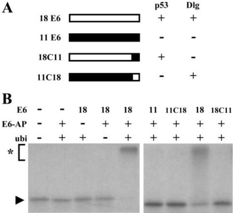
FIGURE 1 E6-AP facilitates E6-mediated ubiquitination of Dlg in vitro . A , schematic representation of the E6 proteins used and their ability to bind and degrade p53 and Dlg, respectively. 18C11, chimeric protein consisting of the 10 C-terminal amino acid residues of HPV11 E6 fused to amino acid residues 1-141 of HV18 E6 (consists of 158 residues); 11C18, chimeric protein consisting of the 17 C-terminal amino acid residues of HPV18 E6 fused to amino acid residues 1-140 of HV11 E6 (consists of 150 residues). + , binds to p53 and Dlg, respectively; -, does not bind to p53 and Dlg, respectively. B , bacterially expressed GST fusion proteins of the E6 proteins indicated were incubated with in vitro translated radiolabeled Dlg under standardubiquitinationconditionsfor2hinthepresenceorabsenceofbaculovirus-expressed E6-AP and ubiquitin ( ubi ), respectively, as indicated. The reaction products were analyzed by SDS-PAGE followed by fluorography. Running positions of the nonmodified form and the ubiquitinated forms of Dlg are indicated by an arrowhead and an asterisk , respectively.
FIGURE 1 E6-AP facilitates E6-mediated ubiquitination of Dlg in vitro . A , schematic representation of the E6 proteins used and their ability to bind and degrade p53 and Dlg, respectively. 18C11, chimeric protein consisting of the 10 C-terminal amino acid residues of HPV11 E6 fused to amino acid residues 1-141 of HV18 E6 (consists of 158 residues); 11C18, chimeric protein consisting of the 17 C-terminal amino acid residues of HPV18 E6 fused to amino acid residues 1-140 of HV11 E6 (consists of 150 residues). + , binds to p53 and Dlg, respectively; -, does not bind to p53 and Dlg, respectively. B , bacterially expressed GST fusion proteins of the E6 proteins indicated were incubated with in vitro translated radiolabeled Dlg under standardubiquitinationconditionsfor2hinthepresenceorabsenceofbaculovirus-expressed E6-AP and ubiquitin ( ubi ), respectively, as indicated. The reaction products were analyzed by SDS-PAGE followed by fluorography. Running positions of the nonmodified form and the ubiquitinated forms of Dlg are indicated by an arrowhead and an asterisk , respectively.CC-BY · From the PDF
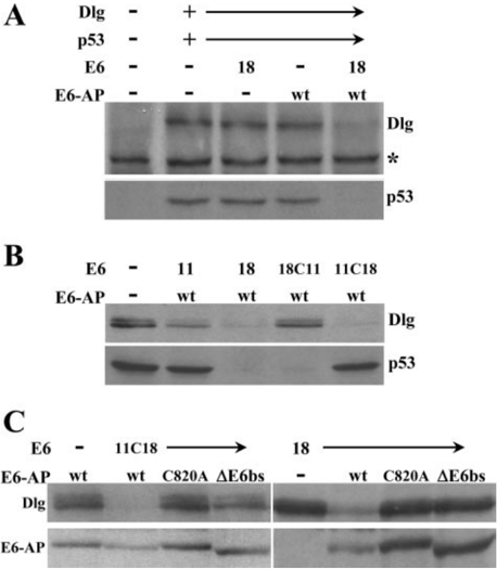
FIGURE 2 E6-AP is required for E6-induced degradation of Dlg in MEFs derived from E6-AP null mice. A-C , mouse embryo fibroblasts derived from E6-AP null mice (24) were cotransfected with expression constructs for p53 and HA-tagged Dlg in the absence and presence of expression constructs encoding various E6 proteins and different forms of E6-AP isoform 1 (28) as indicated. Protein extracts were prepared 24 h after transfection, and levels of p53, Dlg, and E6-AP were determined by Western blot analysis as indicated. Notethat the anti-p53 antibody used (DO1) recognizes ectopically expressed human p53 but not endogenous murine p53. wt , wild-type E6-AP isoform 1; C820A , catalytically inactive E6-AP with a substitution of the catalytic site cysteine residue at position 820 (numbering is according to E6-AP isoform 1) by alanine; 𝚫 E6bs , E6-AP isoform 1 with a deletion of the E6 binding region (amino acid residues 378-395). *, denotes a cross-reactive band serving as loading control.
FIGURE 2 E6-AP is required for E6-induced degradation of Dlg in MEFs derived from E6-AP null mice. A-C , mouse embryo fibroblasts derived from E6-AP null mice (24) were cotransfected with expression constructs for p53 and HA-tagged Dlg in the absence and presence of expression constructs encoding various E6 proteins and different forms of E6-AP isoform 1 (28) as indicated. Protein extracts were prepared 24 h after transfection, and levels of p53, Dlg, and E6-AP were determined by Western blot analysis as indicated. Notethat the anti-p53 antibody used (DO1) recognizes ectopically expressed human p53 but not endogenous murine p53. wt , wild-type E6-AP isoform 1; C820A , catalytically inactive E6-AP with a substitution of the catalytic site cysteine residue at position 820 (numbering is according to E6-AP isoform 1) by alanine; 𝚫 E6bs , E6-AP isoform 1 with a deletion of the E6 binding region (amino acid residues 378-395). *, denotes a cross-reactive band serving as loading control.CC-BY · From the PDF
Image 3
CC-BY · From the PDF
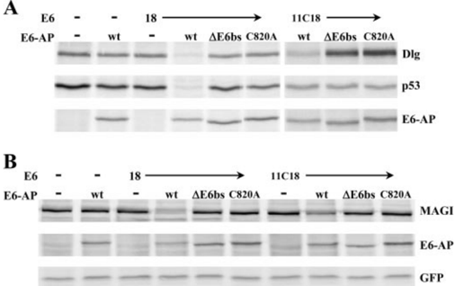
FIGURE 4 Reconstitution of E6-induced degradation of Dlg and MAGI-1 in E6-AP null H1299 cells. A , the E6-AP null H1299 clone K3 was cotransfected with expression constructs for p53 and HA-tagged Dlg in the absence and presence of expression constructs encoding HPV18 E6 and 11C18, respectively, and different forms of E6-AP isoform 1 (28) as indicated. Protein extracts were prepared 24 h after transfection, and levels of p53, Dlg, and E6-AP were determined by Western blot analysis as indicated. B , K3 cells were cotransfectedwithexpressionconstructsforFLAG-taggedMAGI-1andGFPin the absence and presence of expression constructs encoding HPV18 E6 and 11C18, respectively, and different forms of E6-AP isoform 1 (28) as indicated. Protein extracts were prepared 24 h after transfection, and levels of MAGI-1, GFP, and E6-AP were determined by Western blot analysis as indicated. wt , wild-type E6-AP isoform 1; C820A , catalytically inactive E6-AP with a substitution of the catalytic site cysteine residue at position 820 (numbering is according to E6-AP isoform 1) by alanine; 𝚫 E6bs , E6-AP isoform 1 with a deletion of the E6 binding region (amino acid residues 378-395).
FIGURE 4 Reconstitution of E6-induced degradation of Dlg and MAGI-1 in E6-AP null H1299 cells. A , the E6-AP null H1299 clone K3 was cotransfected with expression constructs for p53 and HA-tagged Dlg in the absence and presence of expression constructs encoding HPV18 E6 and 11C18, respectively, and different forms of E6-AP isoform 1 (28) as indicated. Protein extracts were prepared 24 h after transfection, and levels of p53, Dlg, and E6-AP were determined by Western blot analysis as indicated. B , K3 cells were cotransfectedwithexpressionconstructsforFLAG-taggedMAGI-1andGFPin the absence and presence of expression constructs encoding HPV18 E6 and 11C18, respectively, and different forms of E6-AP isoform 1 (28) as indicated. Protein extracts were prepared 24 h after transfection, and levels of MAGI-1, GFP, and E6-AP were determined by Western blot analysis as indicated. wt , wild-type E6-AP isoform 1; C820A , catalytically inactive E6-AP with a substitution of the catalytic site cysteine residue at position 820 (numbering is according to E6-AP isoform 1) by alanine; 𝚫 E6bs , E6-AP isoform 1 with a deletion of the E6 binding region (amino acid residues 378-395).CC-BY · From the PDF
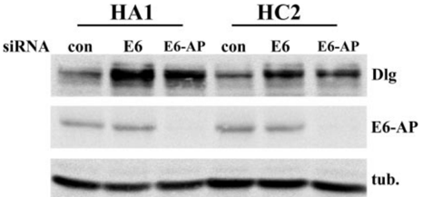
FIGURE 5 Down-regulation of E6-AP expression results in accumulation of Dlg in p53 null HeLa cells. The previously described p53 null HeLa clones HA1andHC2(9)weretransfected with chemically synthesized siRNAs specifically targeting HPV18 E6 ( E6 ) or E6-AP ( E6-AP ). As control, HA1 and HC2 cells were transfected with siRNA directed against Renilla luciferase ( con ). Protein extracts were prepared 72 h after transfection, and levels of Dlg, E6-AP, and tubulin ( tub ; loading control) were determined by Western blot analysis as indicated.
FIGURE 5 Down-regulation of E6-AP expression results in accumulation of Dlg in p53 null HeLa cells. The previously described p53 null HeLa clones HA1andHC2(9)weretransfected with chemically synthesized siRNAs specifically targeting HPV18 E6 ( E6 ) or E6-AP ( E6-AP ). As control, HA1 and HC2 cells were transfected with siRNA directed against Renilla luciferase ( con ). Protein extracts were prepared 72 h after transfection, and levels of Dlg, E6-AP, and tubulin ( tub ; loading control) were determined by Western blot analysis as indicated.CC-BY · From the PDF
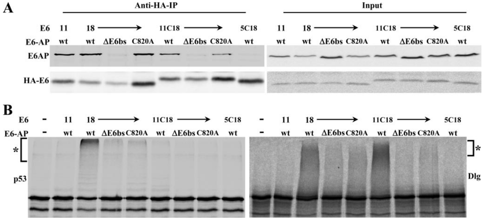
FIGURE6 Functional interaction of low risk HPV E6 proteins with E6-AP. HEK293cells were transfected with expression constructs encoding various forms of HA-tagged E6 and FLAG-tagged E6-AP, respectively, as indicated. Protein extracts were prepared 24 h after transfection. 5% of the respective extracts were directly used for determination of the expression levels of the various forms of E6 and E6-AP, respectively ( Input ). The remaining extracts were used for immunoprecipitation of E6 with an anti-HA antibody. A , 10% of the precipitates were analyzed by Western blot analysis to determine the levels of the respective immunoprecipitated E6 and of potentially coprecipitating E6-AP. B , the remaining precipitates were used for in vitro ubiquitination assays using in vitro translated radiolabeled p53 or Dlg as substrate. The running position of the ubiquitinated form of p53 and Dlg, respectively, is indicated by an asterisk . 11 , HPV11E6; 18 , HPV18 E6; 11C18 , chimeric protein consisting of the 17 C-terminal amino acid residues of HPV18 E6 fused to amino acid residues 1-140 of HPV11 E6; 5C18 , chimeric protein consisting of the 17 C-terminal amino acid residues of HPV18 E6 fused to amino acid residues 1-150 of HPV5 E6; wt , wild-type E6-AP isoform 1; C820A , catalytically inactive E6-AP with a substitution of the catalytic site cysteine residue at position 820 (numbering is according to E6-AP isoform 1) by alanine; 𝚫 E6bs , E6-AP isoform 1 with a deletion of the E6 binding region (amino acid residues 378-395).
FIGURE6 Functional interaction of low risk HPV E6 proteins with E6-AP. HEK293cells were transfected with expression constructs encoding various forms of HA-tagged E6 and FLAG-tagged E6-AP, respectively, as indicated. Protein extracts were prepared 24 h after transfection. 5% of the respective extracts were directly used for determination of the expression levels of the various forms of E6 and E6-AP, respectively ( Input ). The remaining extracts were used for immunoprecipitation of E6 with an anti-HA antibody. A , 10% of the precipitates were analyzed by Western blot analysis to determine the levels of the respective immunoprecipitated E6 and of potentially coprecipitating E6-AP. B , the remaining precipitates were used for in vitro ubiquitination assays using in vitro translated radiolabeled p53 or Dlg as substrate. The running position of the ubiquitinated form of p53 and Dlg, respectively, is indicated by an asterisk . 11 , HPV11E6; 18 , HPV18 E6; 11C18 , chimeric protein consisting of the 17 C-terminal amino acid residues of HPV18 E6 fused to amino acid residues 1-140 of HPV11 E6; 5C18 , chimeric protein consisting of the 17 C-terminal amino acid residues of HPV18 E6 fused to amino acid residues 1-150 of HPV5 E6; wt , wild-type E6-AP isoform 1; C820A , catalytically inactive E6-AP with a substitution of the catalytic site cysteine residue at position 820 (numbering is according to E6-AP isoform 1) by alanine; 𝚫 E6bs , E6-AP isoform 1 with a deletion of the E6 binding region (amino acid residues 378-395).CC-BY · From the PDF

Verification

Claims the paper makes

The claims and statistics of this paper haven’t been checked yet.

Run the check — free, no account needed. It reads the open-access text and checks the sources it cites.

A few free checks a day · papers already verified open instantly · results are public

Claims other papers make about this one

Each is checked against this paper’s own text: does it say what the citing paper says it does?

Self-check

Percentages, sums and reported differences re-computed from the paper’s own numbers.

The self-check needs the paper’s full text.

Citations

Cites

Show all 1 references

Research works printed but not linked: 35

  1. Huibregtse, 1993, Mol. Cell. Biol, 13, 775
  2. Huibregtse, 1993, Mol. Cell. Biol., 13, 4918
  3. Scheffner, 1993, Cell, 75, 495, 10.1016/0092-8674(93)90384-3 · DOI · Paper page
  4. Scheffner, 2003, Semin. Cancer Biol., 13, 59, 10.1016/S1044-579X(02)00100-1 · DOI
  5. zur Hausen, 2002, Nat. Rev. Cancer, 2, 342, 10.1038/nrc798 · DOI
  6. Beer-Romero, 1997, Oncogene, 14, 595, 10.1038/sj.onc.1200872 · DOI
  7. Talis, 1998, J. Biol. Chem., 273, 6439, 10.1074/jbc.273.11.6439 · DOI
  8. Kim, 2003, Cancer Gene Ther., 10, 707, 10.1038/sj.cgt.7700623 · DOI
  9. Hengstermann, 2005, J. Virol., 79, 9296, 10.1128/JVI.79.14.9296-9300.2005 · DOI · Paper page
  10. Li, 1996, J. Virol., 70, 4509, 10.1128/jvi.70.7.4509-4516.1996 · DOI
  11. Mantovani, 2001, Oncogene, 20, 7874, 10.1038/sj.onc.1204869 · DOI
  12. Kuhne, 1998, J. Biol. Chem., 273, 34302, 10.1074/jbc.273.51.34302 · DOI
  13. Thomas, 1999, J. Gen. Virol., 80, 1513, 10.1099/0022-1317-80-6-1513 · DOI · Paper page
  14. Jackson, 2000, Genes Dev., 14, 3065, 10.1101/gad.182100 · DOI
  15. Doyle, 1996, Cell, 85, 1067, 10.1016/S0092-8674(00)81307-0 · DOI
  16. Nguyen, 2003, J. Virol., 77, 6957, 10.1128/JVI.77.12.6957-6964.2003 · DOI
  17. Simonson, 2005, Cancer Res., 65, 8266, 10.1158/0008-5472.CAN-05-1651 · DOI
  18. Watson, 2003, J. Cell Sci., 116, 4925, 10.1242/jcs.00809 · DOI
  19. Nakagawa, 2000, Mol. Cell. Biol., 20, 8244, 10.1128/MCB.20.21.8244-8253.2000 · DOI · Paper page
  20. Pim, 2000, Oncogene, 19, 719, 10.1038/sj.onc.1203374 · DOI
  21. Pim, 2002, Oncogene, 21, 8140, 10.1038/sj.onc.1206026 · DOI
  22. Grm, 2004, J. Gen. Virol., 85, 2815, 10.1099/vir.0.80035-0 · DOI
  23. Jiang, 1998, Neuron, 21, 799, 10.1016/S0896-6273(00)80596-6 · DOI · Paper page
  24. Hengstermann, 1998, Oncogene, 17, 2933, 10.1038/sj.onc.1202282 · DOI
  25. Wegmann, 2004, Exp. Cell Res., 300, 121, 10.1016/j.yexcr.2004.07.010 · DOI
  26. Blot, 2004, J. Cell Sci., 117, 3983, 10.1242/jcs.01266 · DOI
  27. Yamamoto, 1997, Genomics, 41, 263, 10.1006/geno.1997.4617 · DOI
  28. Butz, 2003, Oncogene, 22, 5938, 10.1038/sj.onc.1206894 · DOI · Paper page
  29. Nuber, 1996, J. Biol. Chem., 271, 2795, 10.1074/jbc.271.5.2795 · DOI
  30. Matsumoto, 2006, J. Med. Virol., 78, 501, 10.1002/jmv.20568 · DOI
  31. Scheffner, 1995, Nature, 373, 81, 10.1038/373081a0 · DOI
  32. Brummelkamp, 2002, Science, 296, 550, 10.1126/science.1068999 · DOI
  33. Jariel-Encontre, 1995, J. Biol. Chem., 270, 11623, 10.1074/jbc.270.19.11623 · DOI
  34. Bossis, 2003, Mol. Cell. Biol., 23, 7425, 10.1128/MCB.23.20.7425-7436.2003 · DOI
  35. Varshavsky, 2005, Methods Enzymol., 399, 777, 10.1016/S0076-6879(05)99051-4 · DOI

Cited by

Metadata

Authors
Kuballa, P., Matentzoglu, K., & Scheffner, M.
Venue
Journal of Biological Chemistry
Publisher
Elsevier BV
Published
2007-01
Volume
282
Issue
1
Pages
65-71
ISSN
0021-9258
Cited by
55
References
1
Language
en

About these checks

Registry, retraction, Cites and statistics results are computed, not judged, with one exception: a printed reference that no index lists is read once by a model before it counts as a citation error. Claims are extracted by a model and kept only when their quote appears verbatim in the text; each cited source is then checked, with a model’s help, for whether it says what the claim relies on. Those verdicts can be wrong — read the claim’s quote and the cited source before relying on one.

Statistics and arithmetic are recomputed, not judged.

Confidence bands are shown only for fields calibrated on our annotated test set. None is calibrated yet, so no band is shown.

What counts as a claim ›How we measure confidence ›How we check this ›

Are you an author, or do you see a problem with this page? Request a review