

The DOI matches its Crossref record; 39 of the 39 references it cites were found in a registry or an index.
Mesenchymal stem cell-originated exosomal circDIDO1 suppresses hepatic stellate cell activation by miR-141-3p/PTEN/AKT pathway in human liver fibrosis
Open accessAbstract
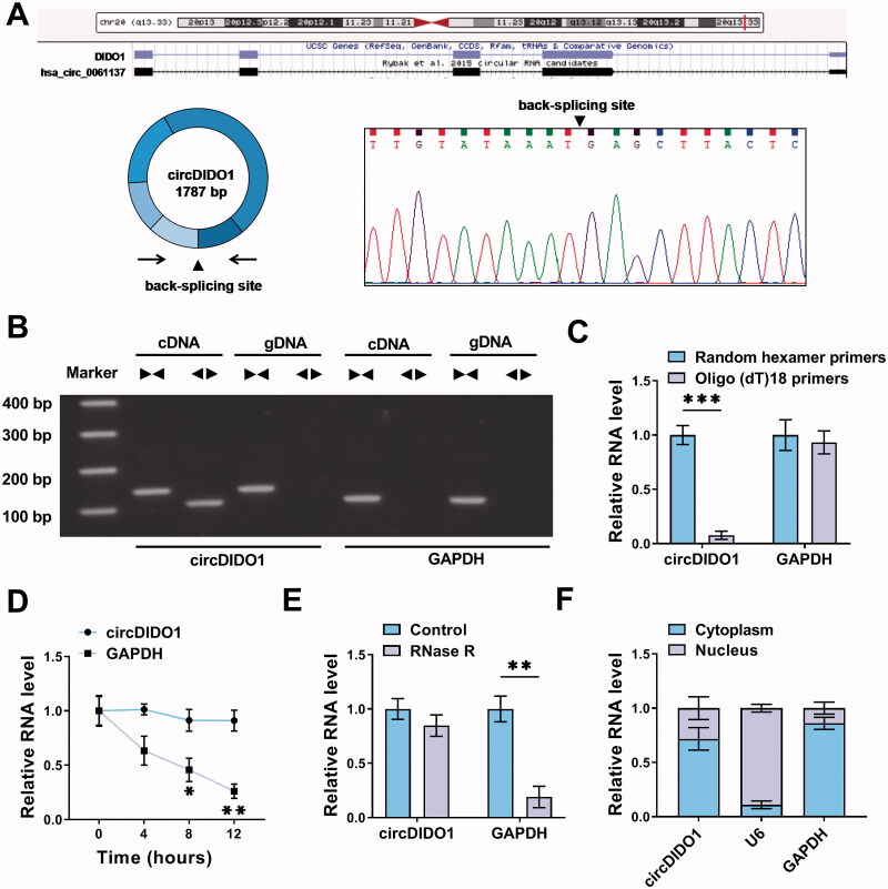
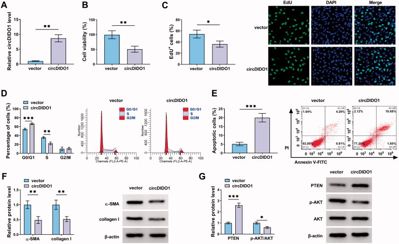
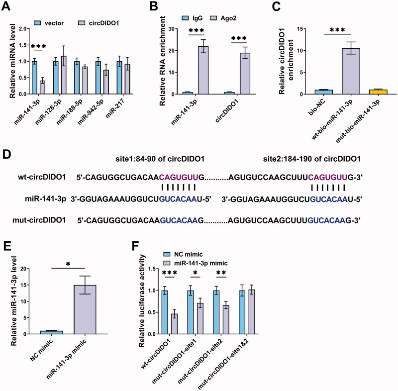
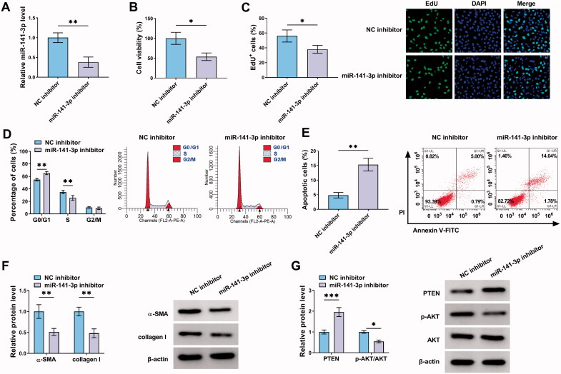
, thus enabling personalized treatment. This study aimed to investigate the role of exosome-based delivery of circDIDO1 in liver fibrosis. Levels of genes and proteins were examined by qRT-PCR and Western blot. Cell proliferation, apoptosis, and cell cycle were analyzed by using cell counting kit-8 (CCK-8) assay, EdU assay, and flow cytometry, respectively. The binding between circDIDO1 and miR-141-3p was confirmed by dual-luciferase reporter, RNA pull-down and RIP assays. Exosomes were isolated by ultracentrifugation, and qualified by transmission electron microscopy (TEM), nanoparticle tracking analysis (NTA) and Western blot. CircDIDO1 overexpression or miR-141-3p inhibition suppressed the proliferation, reduced pro-fibrotic markers, and induced apoptosis as well as cell cycle arrest in hepatic stellate cells (HSCs) by blocking PTEN/AKT pathway. Mechanistically, circDIDO1 acted as an endogenous sponge for miR-141-3p, further rescue experiments showed that circDIDO1 suppressed HSC activation by targeting miR-141-3p. Extracellular circDIDO1 could be incorporated into exosomes isolated from mesenchymal stem cells (MSCs), and transmitted to HSCs to restrain HSC activation. Clinically, low levels of serum circDIDO1 in exosome were correlated with liver failure, and serum exosomal circDIDO1 had a well diagnostic value for liver fibrosis in liver failure patients. Transfer of circDIDO1 mediated by MSC-isolated exosomes suppressed HSC activation through the miR-141-3p/PTEN/AKT pathway, gaining a new insight into the prevention of liver fibrosis in liver failure patients.
Overview
The claims and statistics of this paper haven’t been checked yet.
Run the check — free, no account needed. It reads the open-access text and checks the sources it cites.
Integrity signals
Registry
Cites
Statistics
Figures
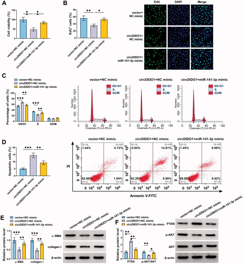
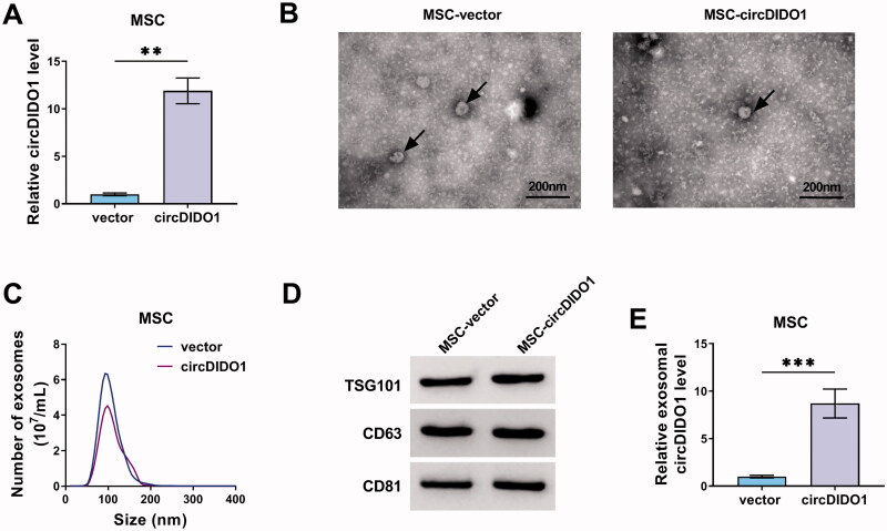
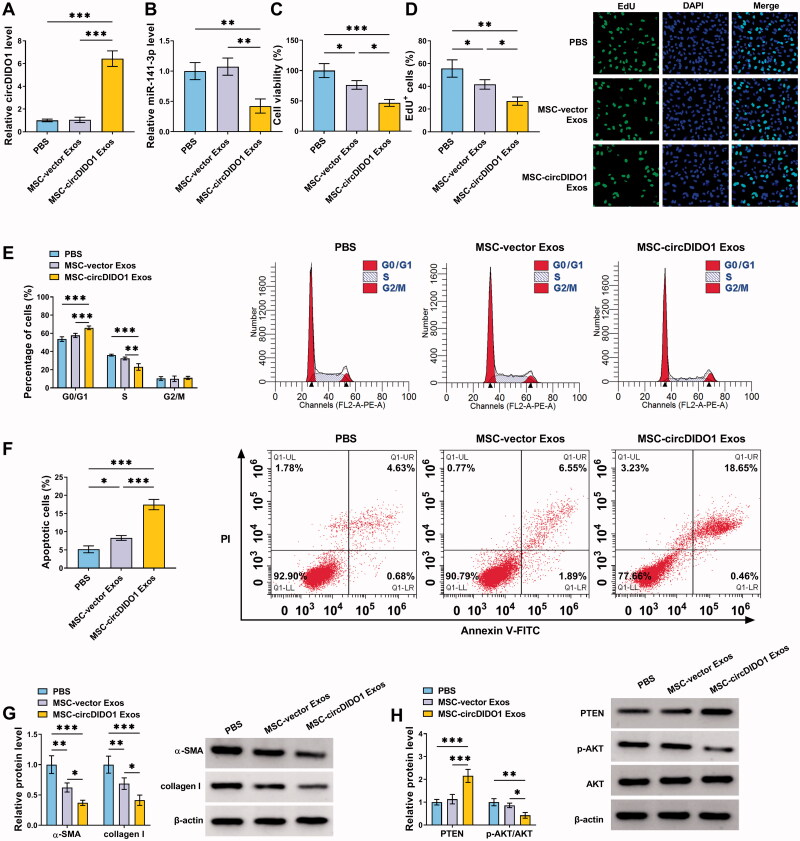
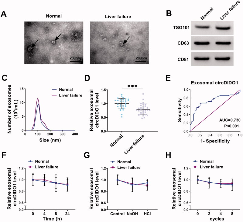
Verification
Claims the paper makes
The claims and statistics of this paper haven’t been checked yet.
Run the check — free, no account needed. It reads the open-access text and checks the sources it cites.
Claims other papers make about this one
Each is checked against this paper’s own text: does it say what the citing paper says it does?
The cited source says what the claim attributes to it.
In the citing paper
“In another study, Ma et al. reported that MSC-derived exosomal circDIDO1 suppressed HSC activation by targeting miR-141-3p/PTEN/AKT pathway [215].”
From Mechanisms, efficacy, and future perspectives of cellular-based therapies for liver fibrosis/cirrhosis: focusing on mesenchymal stromal cells (2025) · cites this paper as [215]
Self-check
Percentages, sums and reported differences re-computed from the paper’s own numbers.
The self-check needs the paper’s full text.
Citations
Cites
The biology , function , and biomedical applications of exosomes
Kalluri, R., & LeBleu, V. S. (2020) · Science · doi:10.1126/science.aau6977 · Cited by 10,770
The biogenesis, biology and characterization of circular RNAs
Kristensen, L. S., Andersen, M. S., Stagsted, L. V. W., Ebbesen, K. K., Hansen, T. B., & Kjems, J. (2019) · Nature Reviews Genetics · doi:10.1038/s41576-019-0158-7 · Cited by 4,823
Mechanisms of hepatic stellate cell activation
Tsuchida, T., & Friedman, S. L. (2017) · Nature Reviews Gastroenterology & Hepatology · doi:10.1038/nrgastro.2017.38 · Cited by 2,909
Horwitz, E. M., Blanc, K. L., Dominici, M., Mueller, I., Slaper‐Cortenbach, I., Marini, F. C., Deans, R. J., Krause, D. S., & Keating, A. (2005) · Cytotherapy · doi:10.1080/14653240500319234 · Cited by 2,071
The Landscape of Circular RNA in Cancer
Vo, J. N., Cieslik, M. P., Zhang, Y., Shukla, S. K., Xiao, L., Zhang, Y., Wu, Y., Dhanasekaran, S. M., Engelke, C. G., Cao, X., et al. (2019) · Cell · doi:10.1016/j.cell.2018.12.021 · Cited by 1,745
Hepatic stellate cells as key target in liver fibrosis
Higashi, T., Friedman, S. L., & Hoshida, Y. (2017) · Advanced Drug Delivery Reviews · doi:10.1016/j.addr.2017.05.007 · Cited by 1,485
Using exosomes, naturally-equipped nanocarriers, for drug delivery
Batrakova, E. V., & Kim, M. S. (2015) · Journal of Controlled Release · doi:10.1016/j.jconrel.2015.07.030 · Cited by 1,041
The PTEN/PI3K/AKT Signalling Pathway in Cancer, Therapeutic Implications
Carnero, A., Blanco‐Aparicio, C., Renner, O., Link, W., & Martínez-Leal, J. F. (2008) · Current Cancer Drug Targets · doi:10.2174/156800908784293659 · Cited by 889
Circular RNA cSMARCA5 inhibits growth and metastasis in hepatocellular carcinoma
Yu, J., Xu, Q., Wang, Z., Yang, Y., Zhang, L., Ma, J., Sun, S., Yang, F., & Zhou, W. (2018) · Journal of Hepatology · doi:10.1016/j.jhep.2018.01.012 · Cited by 764
Samsonraj, R. M., Raghunath, M., Nurcombe, V., Hui, J. H. P., van Wijnen, A. J., & Cool, S. M. (2017) · Stem Cells Translational Medicine · doi:10.1002/sctm.17-0129 · Cited by 732
Mesenchymal stem cell-derived exosomes as a new therapeutic strategy for liver diseases
Lou, G., Chen, Z., Zheng, M., & Liu, Y. (2017) · Experimental & Molecular Medicine · doi:10.1038/emm.2017.63 · Cited by 615
Liver fibrosis and hepatic stellate cells: Etiology, pathological hallmarks and therapeutic targets
Zhang, C.-Y., Yuan, W.-G., He, P., Lei, J.-H., & Wang, C.-X. (2016) · World Journal of Gastroenterology · doi:10.3748/wjg.v22.i48.10512 · Cited by 594
Mutant p53 cancers reprogram macrophages to tumor supporting macrophages via exosomal miR-1246
Cooks, T., Pateras, I. S., Jenkins, L. M. M., Patel, K., Robles, A. I., Morris, J., Forshew, T., Appella, E., Gorgoulis, V. G., & Harris, C. C. (2018) · Nature Communications · doi:10.1038/s41467-018-03224-w · Cited by 497
Exosomes: Mediators of Neurodegeneration, Neuroprotection and Therapeutics
Kalani, A., Tyagi, A. A., & Tyagi, N. (2013) · Molecular Neurobiology · doi:10.1007/s12035-013-8544-1 · Cited by 341
Mesenchymal stem cells secretome: a new paradigm for central nervous system regeneration?
Teixeira, F. G., Carvalho, M. M., Sousa, N., & Salgado, A. J. (2013) · Cellular and Molecular Life Sciences · doi:10.1007/s00018-013-1290-8 · Cited by 337
Rong, X., Liu, J., Yao, X., Jiang, T., Wang, Y., & Xie, F. (2019) · Stem Cell Research & Therapy · doi:10.1186/s13287-019-1204-2 · Cited by 331
Self-citationReif, S. S., Lang, A., Lindquist, J. N., Yata, Y., Gäbele, E., Scanga, A. E., Brenner, D. A., & Rippe, R. A. (2003) · Journal of Biological Chemistry · doi:10.1074/jbc.m212927200 · Cited by 298
González–Begne, M., Lu, B., Han, X., Hagen, F. K., Hand, A. R., Melvin, J. E., & Yates, J. R. (2009) · Journal of Proteome Research · doi:10.1021/pr800658c · Cited by 290
Ma, P.-F., Gao, C.-C., Yi, J., Zhao, J.-L., Liang, S.-Q., Zhao, Y., Ye, Y.-C., Bai, J., Zheng, Q.-J., Dou, K.-F., et al. (2017) · Journal of Hepatology · doi:10.1016/j.jhep.2017.05.022 · Cited by 284
Exosome-transmitted circular RNA hsa_circ_0051443 suppresses hepatocellular carcinoma progression
Chen, W., Quan, Y., Fan, S., Wang, H., Liang, J., Huang, L. T., Chen, L., Liu, Q., He, P., & Ye, Y. (2020) · Cancer Letters · doi:10.1016/j.canlet.2020.01.022 · Cited by 268
Macrophage autophagy protects against liver fibrosis in mice
Lodder, J., Denaës, T., Chobert, M.-N., Wan, J., El-Benna, J., Pawlotsky, J.-M., Lotersztajn, S., & Teixeira-Clerc, F. (2015) · Autophagy · doi:10.1080/15548627.2015.1058473 · Cited by 266
MSCs-Derived Exosomes and Neuroinflammation, Neurogenesis and Therapy of Traumatic Brain Injury
Yang, Y., Ye, Y., Su, X., He, J., Bai, W., & He, X. (2017) · Frontiers in Cellular Neuroscience · doi:10.3389/fncel.2017.00055 · Cited by 237
Emerging Epigenetic Regulation of Circular RNAs in Human Cancer
Wu, J., Qi, X., Liu, L., Hu, X. T., Liu, J., Yang, J., Yang, J., Lu, L., Zhang, Z., Ma, S., et al. (2019) · Molecular Therapy — Nucleic Acids · doi:10.1016/j.omtn.2019.04.011 · Cited by 221
Self-citationThe decade of exosomal long RNA species: an emerging cancer antagonist
Zhou, R., Chen, K. K., Zhang, J., Xiao, B., Huang, Z., Ju, C., Sun, J., Zhang, F., Lv, X.-B., & Huang, G. (2018) · Molecular Cancer · doi:10.1186/s12943-018-0823-z · Cited by 206
Insights into the regulatory role of circRNA in angiogenesis and clinical implications
Liu, Y., Yang, Y., Wang, Z., Fu, X., Chu, X., Li, Y., Wang, Q., He, X., Li, M., Wang, K., et al. (2020) · Atherosclerosis · doi:10.1016/j.atherosclerosis.2020.02.017 · Cited by 102
Chen, Y., Yuan, B., Wu, Z., Dong, Y., Zhang, L., & Zeng, Z. (2017) · Gene · doi:10.1016/j.gene.2017.07.078 · Cited by 90
Circular RNAs in drug resistant tumors
Hua, X., Sun, Y., Chen, J., Wu, Y., Sha, J., Han, S., & Zhu, X. (2019) · Biomedicine & Pharmacotherapy · doi:10.1016/j.biopha.2019.109233 · Cited by 90
Shi, J., Zhao, J., Zhang, X., Cheng, Y., Hu, J., Li, Y., Zhao, X., Shang, Q., Sun, Y., Tu, B., et al. (2017) · Scientific Reports · doi:10.1038/srep44544 · Cited by 66
CircMTO1 inhibits liver fibrosis via regulation of miR‐17‐5p and Smad7
Wang, W., Dong, R., Guo, Y. G., He, J., Shao, C., Yi, P., Yu, F., Gu, D., & Zheng, J. (2019) · Journal of Cellular and Molecular Medicine · doi:10.1111/jcmm.14432 · Cited by 66
Wang, S., Zhan, J., Lin, X., Wang, Y., Wang, Y., & Liu, Y. (2020) · Cell Biochemistry and Function · doi:10.1002/cbf.3543 · Cited by 64
Cao, G., Meng, X., Han, X., & Li, J. (2020) · Bioscience Reports · doi:10.1042/bsr20193436 · Cited by 54
MicroRNA-141 Targets Sirt1 and Inhibits Autophagy to Reduce HBV Replication
Yang, Y., Liu, Y., Xue, J., Yang, Z., Shi, Y. H., Shi, Y., Lou, G., Wu, S., Qi, J., Liu, W., et al. (2017) · Cellular Physiology and Biochemistry · doi:10.1159/000456162 · Cited by 52
Hsa_circ_0070963 inhibits liver fibrosis via regulation of miR-223-3p and LEMD3
Ji, D., Chen, G., Wang, J., Ji, S., Wu, X., Lu, X., Chen, J., & Li, J.-T. (2020) · Aging · doi:10.18632/aging.102705 · Cited by 45
Activation of hepatic stellate cell in Pten null liver injury model
He, L., Gubbins, J., Peng, Z., Medina, V., Fei, F., Asahina, K., Wang, J., Kahn, M., Rountree, C. B., & Stiles, B. L. (2016) · Fibrogenesis & Tissue Repair · doi:10.1186/s13069-016-0045-1 · Cited by 44
Pedrosa, M. F., Gomes, J., Laranjeira, P., Duarte, C. C. M., Pedreiro, S., Antunes, B., Ribeiro, T., dos Santos, F., Martinho, A., Fardilha, M., et al. (2019) · Journal of Tissue Engineering and Regenerative Medicine · doi:10.1002/term.2958 · Cited by 39
HAO, L. S., Zhang, X. L., AN, J. Y., Karlin, J. N., TIAN, X. P., Dun, Z.-N., XIE, S. R., & CHEN, S. (2009) · Apmis · doi:10.1111/j.1600-0463.2009.02515.x · Cited by 34
Li, S., Song, F., Xu, L., Li, J., Fang, L., & Tan, H. (2020) · Aging · doi:10.18632/aging.103257 · Cited by 31
Downregulation of miR‑141 deactivates hepatic stellate�cells by targeting the PTEN/AKT/mTOR pathway
Liang, H., Wang, X., Si, C., Duan, Y., Chen, B., Liang, H., & Yang, D. (2020) · International Journal of Molecular Medicine · doi:10.3892/ijmm.2020.4578 · Cited by 17
[Progress of the application of stem cell therapy for end-stage liver disease].
Duan, X., Wang, Y., He, J., Peng, Y., Yu, J., & Sun, Y. J. (2017) · PubMed · doi:10.11817/j.issn.1672-7347.2017.04.015 · Cited by 2
Cited by
Sorted by citations
Clinical applications of stem cell-derived exosomes
Tan, F., Li, X., Wang, Z., Li, J., Shahzad, K., & Zheng, J. (2024) · Signal Transduction and Targeted Therapy · doi:10.1038/s41392-023-01704-0 · Cited by 708
Zhang, Z., Shang, J., Yang, Q., Dai, Z., Liang, Y., Lai, C., Feng, T., Zhong, D., Zou, H., Ji, G., et al. (2023) · Journal of Nanobiotechnology · doi:10.1186/s12951-023-01788-4 · Cited by 131
Liver fibrosis resolution: from molecular mechanisms to therapeutic opportunities
Pei, Q., Yi, Q., & Tang, L. (2023) · International Journal of Molecular Sciences · doi:10.3390/ijms24119671 · Cited by 121
Mesenchymal stromal cells in hepatic fibrosis/cirrhosis: from pathogenesis to treatment
Yang, X., Li, Q., Liu, W., Zong, C., Wei, L., Shi, Y., & Han, Z. (2023) · Cellular & Molecular Immunology · doi:10.1038/s41423-023-00983-5 · Cited by 120
The potential role and mechanism of circRNA/miRNA axis in cholesterol synthesis
Chen, W., Xu, J., Wu, Y., Liang, B., Yan, M., Sun, C., Wang, D., Hu, X., Liu, L.-Z., Hu, W., et al. (2023) · International Journal of Biological Sciences · doi:10.7150/ijbs.84994 · Cited by 107
Metadata
- Authors
- Ma, L., Wei, J., Zeng, Y., Liu, J., Xiao, E., Kang, Y., & Kang, Y.
- Affiliations
- Zhengzhou University (CN)
- Corresponding author
- Kang, Yi
- Venue
- Drug Delivery
- Publisher
- Informa UK Limited
- Published
- 2022-01-31
- Volume
- 29
- Issue
- 1
- Pages
- 440-453
- ISSN
- 1071-7544 (ISSN-L)
- 1521-0464
- PMC
- PMC8812765
- OpenAlex
- W4210563368
- Cited by
- 109
- References
- 39
- Citations by year
- 2026: 14 · 2025: 22 · 2024: 26 · 2023: 30 · 2022: 17
- FWCI
- 7.76
- Type
- journal article
- Language
- en
- Open access
- gold
- doi.org
- Licence
- Locations
- Drug Delivery (publishedVersion, cc-by-nc)
- PubMed Central (publishedVersion, cc-by-nc)
- PubMed (publishedVersion)
- DOAJ (DOAJ: Directory of Open Access Journals) (submittedVersion, cc-by-sa)
- Topics
- Life Sciences › Biochemistry, Genetics and Molecular Biology › Molecular Biology › Circular RNAs in diseases
- Life Sciences › Biochemistry, Genetics and Molecular Biology › Molecular Biology › Extracellular vesicles in disease
- Health Sciences › Medicine › Hepatology › Liver physiology and pathology
- Keywords
- liver fibrosis, hepatic stellate cell activation, mesenchymal stem cell-derived exosomes, miR-141-3p, PTEN/AKT pathway, microRNA sponge, exosome therapy, liver failure, serum biomarkers, cell cycle arrest
- MeSH terms
- RNA, Circular, Cell Line, DNA-Binding Proteins, Humans, Liver Cirrhosis, Signal Transduction, Apoptosis, MicroRNAs, Cell Proliferation, Proto-Oncogene Proteins c-akt, PTEN Phosphohydrolase, Hepatic Stellate Cells, Exosomes, Mesenchymal Stem Cells
About these checks
Registry, retraction, Cites and statistics results are computed, not judged, with one exception: a printed reference that no index lists is read once by a model before it counts as a citation error. Claims are extracted by a model and kept only when their quote appears verbatim in the text; each cited source is then checked, with a model’s help, for whether it says what the claim relies on. Those verdicts can be wrong — read the claim’s quote and the cited source before relying on one.
Statistics and arithmetic are recomputed, not judged.
Confidence bands are shown only for fields calibrated on our annotated test set. None is calibrated yet, so no band is shown.
What counts as a claim ›How we measure confidence ›How we check this ›
Are you an author, or do you see a problem with this page? Request a review
