

The DOI matches its Crossref record; 49 of the 49 references it cites were found in a registry or an index.
Schwann cell autophagy, myelinophagy, initiates myelin clearance from injured nerves
Open accessAbstract
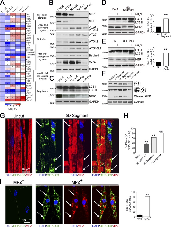
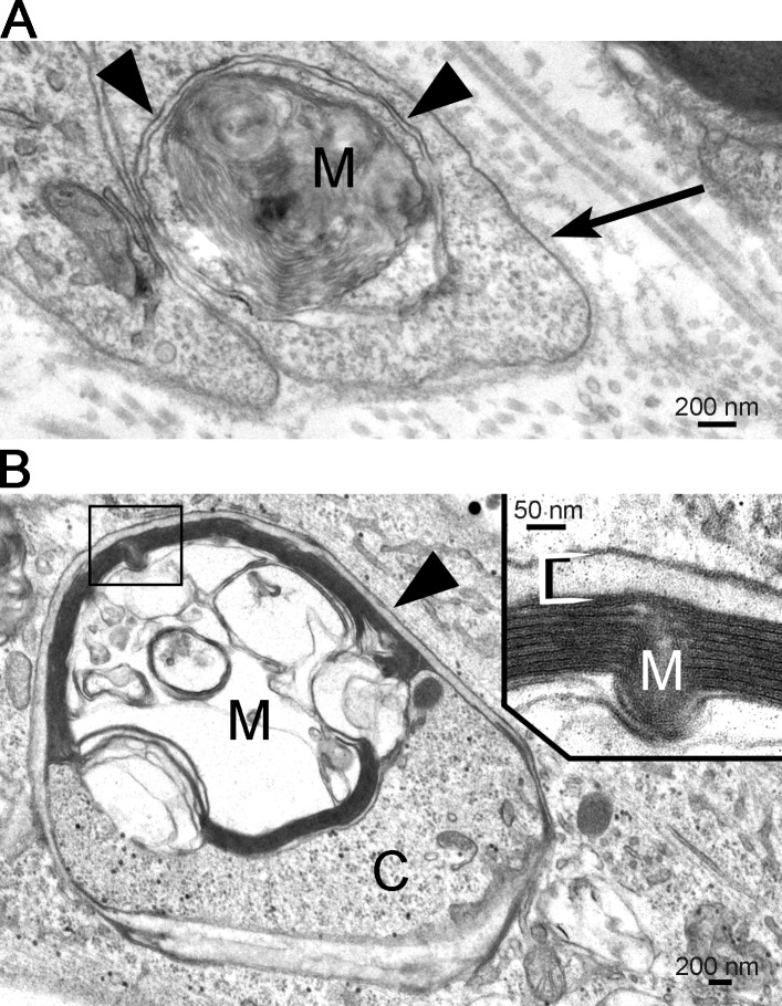
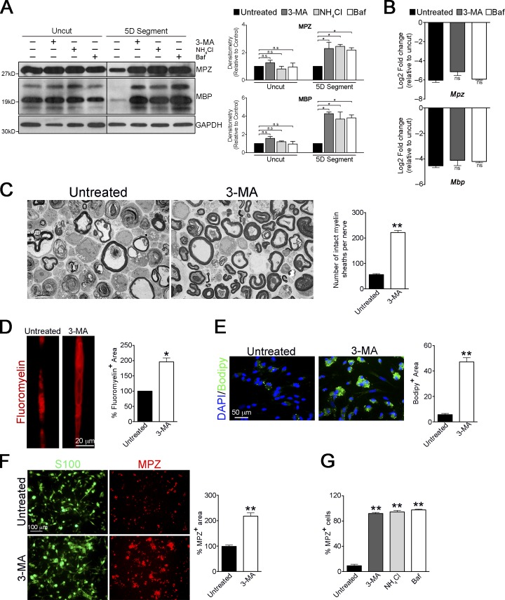
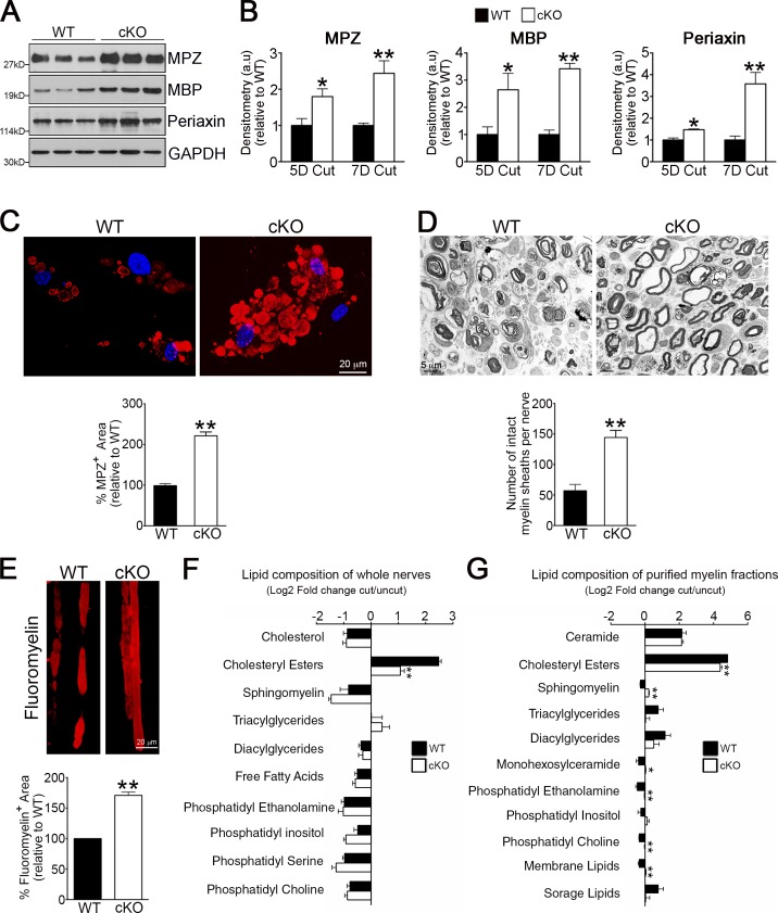
Although Schwann cell myelin breakdown is the universal outcome of a remarkably wide range of conditions that cause disease or injury to peripheral nerves, the cellular and molecular mechanisms that make Schwann cell–mediated myelin digestion possible have not been established. We report that Schwann cells degrade myelin after injury by a novel form of selective autophagy, myelinophagy. Autophagy was up-regulated by myelinating Schwann cells after nerve injury, myelin debris was present in autophagosomes, and pharmacological and genetic inhibition of autophagy impaired myelin clearance. Myelinophagy was positively regulated by the Schwann cell JNK/c-Jun pathway, a central regulator of the Schwann cell reprogramming induced by nerve injury. We also present evidence that myelinophagy is defective in the injured central nervous system. These results reveal an important role for inductive autophagy during Wallerian degeneration, and point to potential mechanistic targets for accelerating myelin clearance and improving demyelinating disease.
Overview
The claims and statistics of this paper haven’t been checked yet.
Run the check — free, no account needed. It reads the open-access text and checks the sources it cites.
Integrity signals
Registry
Cites
Statistics
Figures
Verification
Claims the paper makes
The claims and statistics of this paper haven’t been checked yet.
Run the check — free, no account needed. It reads the open-access text and checks the sources it cites.
Claims other papers make about this one
Each is checked against this paper’s own text: does it say what the citing paper says it does?
In the citing paper
“After nerve injury, Schwann cells oversee the capture of myelin debris in autophagosomes in a process termed “myelinophagy” [459,462,463],”
From Autophagy and ALS: mechanistic insights and therapeutic implications (2022) · cites this paper as [463]
Self-check
Percentages, sums and reported differences re-computed from the paper’s own numbers.
The self-check needs the paper’s full text.
Citations
Cites
A RAPID METHOD OF TOTAL LIPID EXTRACTION AND PURIFICATION
Bligh, E. G., & Dyer, W. J. (1959) · Canadian Journal of Biochemistry and Physiology · doi:10.1139/o59-099 · Cited by 54,751
Repertoires of Autophagy in the Pathogenesis of Ocular Diseases
Li, Y., Jiang, Q. L., Cao, G.-F., Yao, J., & Yan, B. (2015) · Cellular Physiology and Biochemistry · doi:10.1159/000373980 · Cited by 16,351
Calamita, E., Schmid, M., Kunz, M., Ndebele-Murisa, M. R., Magadza, C. H. D., Nyambe, I., & Wehrli, B. (2019) · Figshare · doi:10.26181/6153922fe919c · Cited by 4,239
Guidelines for the use and interpretation of assays for monitoring autophagy
Klionsky, D. J., Abdalla, F. C., Abeliovich, H., Abraham, R. T., Acevedo-Arozena, A., Adeli, K., Agholme, L., Agnello, M., Agostinis, P., Aguirre-Ghiso, J. A., et al. (2012) · Autophagy · doi:10.4161/auto.19496 · Cited by 3,996 · cited 15×
“Conversely, levels of the autophagy substrate NBR1 (Klionsky et al., 2012) showed a clear decrease in injured nerves (Fig.”
InfluentialAutophagy regulates lipid metabolism
Singh, R., Kaushik, S., Wang, Y., Xiang, Y., Novak, I., Komatsu, M., Tanaka, K., Cuervo, A. M., & Czaja, M. J. (2009) · Nature · doi:10.1038/nature07976 · Cited by 3,975
Autophagy and the Integrated Stress Response
Kroemer, G., Mariño, G., & Levine, B. (2010) · Molecular Cell · doi:10.1016/j.molcel.2010.09.023 · Cited by 3,506
Autophagy in immunity and inflammation
Levine, B., Mizushima, N., & Virgin, H. W. (2011) · Nature · doi:10.1038/nature09782 · Cited by 3,289
Impairment of starvation-induced and constitutive autophagy in Atg7 -deficient mice
Komatsu, M., Waguri, S., Ueno, T., Iwata, J., Murata, S., Tanida, I., Ezaki, J., Mizushima, N., Ohsumi, Y., Uchiyama, Y., et al. (2005) · The Journal of Cell Biology · doi:10.1083/jcb.200412022 · Cited by 2,331
Mizushima, N., Yamamoto, A., Matsui, M., Yoshimori, T., & Ohsumi, Y. (2004) · Molecular Biology of the Cell · doi:10.1091/mbc.e03-09-0704 · Cited by 2,105
The machinery of macroautophagy
Feng, Y., He, D., Yao, Z., & Klionsky, D. J. (2014) · Cell Research · doi:10.1038/cr.2013.168 · Cited by 2,083
Cited by
Sorted by citations
Items 101–200 of 453
Orkwis, J. A., Wolf, A. K., Shahid, S. M., Smith, C. S., Esfandiari, L., & Harris, G. M. (2020) · Macromolecular Bioscience · doi:10.1002/mabi.202000197 · Cited by 52
Reed, C. B., Feltri, M. L., & Wilson, E. R. (2021) · Journal of Anatomy · doi:10.1111/joa.13484 · Cited by 59
Role of adipose mesenchymal stem cells and secretome in peripheral nerve regeneration
Sumarwoto, T., Suroto, H., Mahyudin, F., Utomo, D. N., Romaniyanto, Tinduh, D., Notobroto, H. B., Prakoeswa, C. R. S., Rantam, F. A., & Rhatomy, S. (2021) · Annals of Medicine and Surgery · doi:10.1016/j.amsu.2021.102482 · Cited by 50
Haertinger, M., Weiss, T., Mann, A., Tabi, A., Brandel, V., & Radtke, C. (2020) · Cells · doi:10.3390/cells9010163 · Cited by 49
The role of autophagy in axonal degeneration of the optic nerve
Koch, J. C., & Lingor, P. (2016) · Experimental Eye Research · doi:10.1016/j.exer.2015.08.016 · Cited by 46
Jang, S. Y., Yoon, B.-A., Shin, Y. K., Yun, S. H., Jo, Y. R., Choi, Y. Y., Ahn, M., Shin, T., Park, J. I., Kim, J. K., et al. (2017) · Glia · doi:10.1002/glia.23200 · Cited by 46
Aging Schwann cells: mechanisms, implications, future directions
Painter, M. W. (2017) · Current Opinion in Neurobiology · doi:10.1016/j.conb.2017.10.022 · Cited by 49
Cai, Z., Yao, H., Chen, J., Ahmed, A. A., Li, C., Hu, X., Tang, X., & Jiang, C. (2024) · Cancer Letters · doi:10.1016/j.canlet.2024.216689 · Cited by 44
Ye, Z., Wei, J., Zhan, C., & Hou, J. L. (2022) · Frontiers in Neuroscience · doi:10.3389/fnins.2022.917587 · Cited by 43
Review: Myelin clearance is critical for regeneration after peripheral nerve injury
Yuan, Y., Wang, Y., Wu, S., & Zhao, M. Y. (2022) · Frontiers in Neurology · doi:10.3389/fneur.2022.908148 · Cited by 49
Coccurello, R., Nazio, F., Rossi, C., Angelis, F. D., Vacca, V., Giacovazzo, G., Procacci, P., Magnaghi, V., Ciavardelli, D., & Marinelli, S. (2018) · PLoS ONE · doi:10.1371/journal.pone.0208596 · Cited by 46
The role of adherens junction proteins in the regulation of insulin secretion
Dissanayake, W. C., Sorrenson, B., & Shepherd, P. R. (2018) · Bioscience Reports · doi:10.1042/bsr20170989 · Cited by 42
Lipid metabolism adaptations are reduced in human compared to murine Schwann cells following injury
zu Reckendorf, C. M., Brand, C., Pedro, M. T., Hegler, J., Schilling, C., Lerner, R., Bîndilă, L., Antoniadis, G., & Knöll, B. (2020) · Nature Communications · doi:10.1038/s41467-020-15915-4 · Cited by 42
Grove, M., Lee, H., Zhao, H., & Son, Y. (2020) · eLife · doi:10.7554/elife.50138 · Cited by 42
Park, H. T., Kim, J. K., & Tricaud, N. (2018) · Glia · doi:10.1002/glia.23509 · Cited by 41
Electrical Muscle Stimulation Accelerates Functional Recovery After Nerve Injury
Fu, T., Jiang, L., Peng, Y., Li, Z., Liu, S., Lu, J., Zhang, F., & Zhang, J. (2019) · Neuroscience · doi:10.1016/j.neuroscience.2019.10.052 · Cited by 41
Engineered Schwann Cell-Based Therapies for Injury Peripheral Nerve Reconstruction
Su, Q.-S., Nasser, M. I., He, J., Deng, G., Ouyang, Q., Zhuang, D., Deng, Y., Hu, H., Liu, N., Li, Z., et al. (2022) · Frontiers in Cellular Neuroscience · doi:10.3389/fncel.2022.865266 · Cited by 42
Zhou, L., Dong, M., Hu, Z., Tang, Y., Chu, Y., Chen, M., Yang, S., Chen, Z., Wu, L., Wang, W., et al. (2022) · Proceedings of the National Academy of Sciences · doi:10.1073/pnas.2209990120 · Cited by 42
Primary Cilia in Glial Cells: An Oasis in the Journey to Overcoming Neurodegenerative Diseases
Ki, S. M., Jeong, H. S., & Lee, J. E. (2021) · Frontiers in Neuroscience · doi:10.3389/fnins.2021.736888 · Cited by 40
Teo, J. D., Marian, O. C., Spiteri, A. G., Nicholson, M., Song, H., Khor, J. X. Y., McEwen, H. P., Ge, A., Sen, M. K., Piccio, L. M., et al. (2022) · Glia · doi:10.1002/glia.24329 · Cited by 41
Mechanisms and Treatments of Peripheral Nerve Injury
Liu, X., & Duan, X. (2023) · Annals of Plastic Surgery · doi:10.1097/sap.0000000000003480 · Cited by 40
Schwann-Cell Autophagy, Functional Recovery, and Scar Reduction After Peripheral Nerve Repair
Ko, P., Yang, C.-C., Kuo, Y., Su, F., Hsu, T., Tu, Y., & Jou, I. (2018) · Journal of Molecular Neuroscience · doi:10.1007/s12031-018-1056-8 · Cited by 41
Polycomb repression regulates Schwann cell proliferation and axon regeneration after nerve injury
Ma, K. H., Duong, P., Moran, J. J. M., Junaidi, N., & Svaren, J. P. (2018) · Glia · doi:10.1002/glia.23500 · Cited by 41
Epigenetic Control of Schwann Cells
Ma, K. H., & Svaren, J. P. (2018) · The Neuroscientist · doi:10.1177/1073858417751112 · Cited by 39
Millesi, F., Weiss, T., Mann, A., Haertinger, M., Semmler, L., Supper, P., Pils, D., Naghilou, A., & Radtke, C. (2020) · The FASEB Journal · doi:10.1096/fj.202001447r · Cited by 39
Nazareth, L., John, J. A. S., Murtaza, M., & Ekberg, J. A. K. (2021) · Frontiers in Cell and Developmental Biology · doi:10.3389/fcell.2021.660259 · Cited by 42
Gomez-Sanchez, J. A., Patel, N., Martirena, F., Fazal, S. V., Mutschler, C., & Cabedo, H. (2022) · International Journal of Molecular Sciences · doi:10.3390/ijms23062996 · Cited by 39
Self-citationAxonal damage in central and peripheral nervous system inflammatory demyelinating diseases
Barnett, M. H., Mathey, E. K., Kiernan, M. C., & Pollard, J. D. (2016) · Current Opinion in Neurology · doi:10.1097/wco.0000000000000334 · Cited by 40
Calabrò, S., Kankowski, S., Cescon, M., Gambarotta, G., Raimondo, S., Haastert‐Talini, K., & Ronchi, G. (2023) · International Journal of Molecular Sciences · doi:10.3390/ijms24098061 · Cited by 40
Elberg, G., Zaltsman, S. L., Reichert, F., Matozaki, T., Tal, M., & Rotshenker, S. (2019) · Journal of Neuroinflammation · doi:10.1186/s12974-019-1679-x · Cited by 38
How autophagy can restore proteostasis defects in multiple diseases?
Joshi, V., Upadhyay, A., Prajapati, V. K., & Mishra, A. (2020) · Medicinal Research Reviews · doi:10.1002/med.21662 · Cited by 40
Unveiling the Role of Schwann Cell Plasticity in the Pathogenesis of Diabetic Peripheral Neuropathy
Razak, N. H. A., Idris, J., Hassan, N. H., Zaini, F., Muhamad, N., & Daud, M. (2024) · International Journal of Molecular Sciences · doi:10.3390/ijms251910785 · Cited by 37
Chaudhry, P. M., & Piao, X. (2016) · Developmental Dynamics · doi:10.1002/dvdy.24473 · Cited by 37
Modeling the Pathogenesis of Charcot-Marie-Tooth Disease Type 1A Using Patient-Specific iPSCs
Shi, L., Huang, L., He, R., Huang, W., Wang, H., Lai, X., Zou, Z., Sun, J., Ke, Q., Zheng, M., et al. (2017) · Stem Cell Reports · doi:10.1016/j.stemcr.2017.11.013 · Cited by 38
Repair of the Injured Spinal Cord by Schwann Cell Transplantation
Fu, H., Hu, D., Chen, J., Wang, Q., Zhang, Y., Qi, C., & Yu, T. (2022) · Frontiers in Neuroscience · doi:10.3389/fnins.2022.800513 · Cited by 37
Reed, C. B., Frick, L. R., Weaver, A., Sidoli, M., Schlant, E., Feltri, M. L., & Wrabetz, L. G. (2020) · Journal of Neuroscience · doi:10.1523/jneurosci.0951-20.2020 · Cited by 36
Wise, A. K., Pujol, R., Landry, T., Fallon, J. B., & Shepherd, R. K. (2017) · Journal of the Association for Research in Otolaryngology · doi:10.1007/s10162-017-0631-y · Cited by 36
Rotshenker, S. (2022) · Frontiers in Cellular Neuroscience · doi:10.3389/fncel.2022.949079 · Cited by 35
André‐Lévigne, D., Pignel, R., Boet, S., Jaquet, V., Kalbermatten, D. F., & Madduri, S. (2024) · International Journal of Molecular Sciences · doi:10.3390/ijms25042030 · Cited by 35
Injury and stress responses of adult neural crest-derived cells
Parfejevs, V., Antunes, A. T., & Sommer, L. (2018) · Developmental Biology · doi:10.1016/j.ydbio.2018.05.011 · Cited by 35
Cheng, W., Xiang, W., Wang, S., & Xu, K. L. (2019) · PubMed Central · Cited by 35
The properties of human Schwann cells: Lessons from in vitro culture and transplantation studies
Monje, P. V. (2020) · Glia · doi:10.1002/glia.23793 · Cited by 35
Welleford, A. S., Quintero, J. E., Seblani, N. E., Blalock, E. M., Gunewardena, S., Shapiro, S. M., Riordan, S. M., Huettl, P. F., Guduru, Z., Stanford, J. A., et al. (2020) · Cell Transplantation · doi:10.1177/0963689720926157 · Cited by 35
Imaging resident and recruited macrophage contribution to Wallerian degeneration
Boissonnas, A., Louboutin, F., Laviron, M., Loyher, P.-L., Reboussin, É., Barthélémy, S., Goazigo, A. R.-L., Lobsiger, C. S., Combadière, B., Parsadaniantz, S. M., et al. (2020) · The Journal of Experimental Medicine · doi:10.1084/jem.20200471 · Cited by 35
Schwann cells and trigeminal neuralgia
Liao, J.-Y., Zhou, T.-H., Chen, B.-K., & Liu, Z.-X. (2020) · Molecular Pain · doi:10.1177/1744806920963809 · Cited by 35
Calcitonin gene-related peptide (CGRP): role in peripheral nerve regeneration
Jung, A. M. (2017) · Reviews in the Neurosciences · doi:10.1515/revneuro-2017-0060 · Cited by 36
Zhang, J., Ren, J., Liu, Y., Huang, D., & Lu, L. (2020) · Life Sciences · doi:10.1016/j.lfs.2020.117959 · Cited by 34
Schwann cell‐derived exosomes: Janus‐faced mediators of regeneration and disease
Wong, F. C., Ye, L., Demir, İ. E., & Kahlert, C. (2021) · Glia · doi:10.1002/glia.24087 · Cited by 34
Chromatin-remodeling enzymes in control of Schwann cell development, maintenance and plasticity
Jacob, C. (2017) · Current Opinion in Neurobiology · doi:10.1016/j.conb.2017.08.007 · Cited by 34
The Sonic Hedgehog signaling pathway regulates inferior alveolar nerve regeneration
Yamada, Y., Ohazama, A., Maeda, T., & Seo, K. (2018) · Neuroscience Letters · doi:10.1016/j.neulet.2017.12.051 · Cited by 33
Brief Electrical Stimulation Promotes Recovery after Surgical Repair of Injured Peripheral Nerves
Gordon, T. (2024) · International Journal of Molecular Sciences · doi:10.3390/ijms25010665 · Cited by 32
Jiang, Y., Liang, J., Li, R., Peng, Y., Huang, J., & Huang, L. (2021) · Journal of Cellular and Molecular Medicine · doi:10.1111/jcmm.16274 · Cited by 33
Florio, F., Ferri, C., Scapin, C., Feltri, M. L., Wrabetz, L. G., & D’Antonio, M. (2018) · Journal of Neuroscience · doi:10.1523/jneurosci.0201-18.2018 · Cited by 32
Schwann Cells as Orchestrators of Nerve Repair: Implications for Tissue Regeneration and PathologiesPreprint
Stassart, R. M., Gomez-Sanchez, J. A., & Lloyd, A. C. (2024) · Cold Spring Harbor Perspectives in Biology · doi:10.1101/cshperspect.a041363 · Cited by 32
Self-citationMohseni, S., Badii, M. O., Kylhammar, A., Thomsen, N., Eriksson, K., Malik, R. A., Rosén, I., & Dahlin, L. B. (2017) · Brain and Behavior · doi:10.1002/brb3.763 · Cited by 32
Autophagy and lysosomal pathways in nervous system disorders
Bingol, B. (2018) · Molecular and Cellular Neuroscience · doi:10.1016/j.mcn.2018.04.009 · Cited by 32
The molecular and cellular insight into the toxicology of bortezomib-induced peripheral neuropathy
Yan, W., Wu, Z., Zhang, Y., Hong, D., Dong, X., Liu, L., Rao, Y., Huang, L., Zhang, X., & Wu, J. (2021) · Biomedicine & Pharmacotherapy · doi:10.1016/j.biopha.2021.112068 · Cited by 32
Myelin debris phagocytosis in demyelinating disease
Gao, R., Song, S., Tian, M., Wang, L., Zhang, Y., & Li, X. (2024) · Glia · doi:10.1002/glia.24602 · Cited by 31
The Complex Work of Proteases and Secretases in Wallerian Degeneration: Beyond Neuregulin-1
Pellegatta, M., & Taveggia, C. (2019) · Frontiers in Cellular Neuroscience · doi:10.3389/fncel.2019.00093 · Cited by 30
Axo‐glial interaction in the injured PNS
Stassart, R. M., & Woodhoo, A. (2020) · Developmental Neurobiology · doi:10.1002/dneu.22771 · Cited by 30
Self-citationLiu, J., Ma, X., Hu, X., Wen, J., Zhang, H., Xu, J., He, Y., Wang, X., & Guo, J. (2023) · Glia · doi:10.1002/glia.24365 · Cited by 30
Lipin1 Alleviates Autophagy Disorder in Sciatic Nerve and Improves Diabetic Peripheral Neuropathy
Wang, M., Xie, M., Yu, S., Shang, P. F., Zhang, C., Han, X., Fan, C., Chen, L., Zhuang, X., & Chen, S. (2021) · Molecular Neurobiology · doi:10.1007/s12035-021-02540-5 · Cited by 31
Transcriptomic Characterization of the Human Insular Cortex and Claustrum
Ibrahim, C., Foll, B. L., & French, L. (2019) · Frontiers in Neuroanatomy · doi:10.3389/fnana.2019.00094 · Cited by 31
Hedging against Neuropathic Pain: Role of Hedgehog Signaling in Pathological Nerve Healing
Moreau, N., & Boucher, Y. (2020) · International Journal of Molecular Sciences · doi:10.3390/ijms21239115 · Cited by 32
Ascorbic acid accelerates Wallerian degeneration after peripheral nerve injury
Guo, J., Li, L., Xu, Y., Wang, X., Liu, J., Hu, X., Tan, D., & Li, Z. (2020) · Neural Regeneration Research · doi:10.4103/1673-5374.300459 · Cited by 29
Zhu, Y., Peng, N., Wang, J., Jin, Z., Zhu, L., Wang, Y., Chen, S., Hu, Y., Zhang, T., Song, Q., et al. (2022) · Biomaterials Research · doi:10.1186/s40824-022-00264-8 · Cited by 29
Patzig, J., Kusch, K., Fledrich, R., Eichel‐Vogel, M. A., Lüders, K. A., Möbius, W., Sereda, M. W., Nave, K., Martini, R., & Werner, H. B. (2015) · Glia · doi:10.1002/glia.22922 · Cited by 29
Schwann Cell Responses and Plasticity in Different Dental Pulp Scenarios
Couve, E., & Schmachtenberg, O. (2018) · Frontiers in Cellular Neuroscience · doi:10.3389/fncel.2018.00299 · Cited by 28
Feng, X., Takayama, Y., Ohno, N., Kanda, H., Dai, Y., Sokabe, T., & Tominaga, M. (2020) · Communications Biology · doi:10.1038/s42003-020-01444-9 · Cited by 28
Hybrid material mimics a hypoxic environment to promote regeneration of peripheral nerves
Quan, Q., Hong, L., Wang, Y., Li, R., Yin, X. H., Cheng, X., Liu, G., Tang, H., Meng, H., Liu, S., et al. (2021) · Biomaterials · doi:10.1016/j.biomaterials.2021.121068 · Cited by 28
Rahman, M. A., Rahman, M. H., Mamun-Or-Rashid, A. N. M., Hwang, H., Chung, S., Kim, B., & Rhim, H. (2022) · Biomedicines · doi:10.3390/biomedicines10051027 · Cited by 31
Weiss, T., Taschner‐Mandl, S., Ambros, P. F., & Ambros, I. M. (2018) · Methods in molecular biology · doi:10.1007/978-1-4939-7649-2_5 · Cited by 28
Logan, A. M., Mammel, A. E., Robinson, D. C., Chin, A. L., Condon, A. F., & Robinson, F. L. (2017) · Glia · doi:10.1002/glia.23173 · Cited by 27
Forese, M. G., Pellegatta, M., Canevazzi, P., Gullotta, G. S., Podini, P., Rivellini, C., Previtali, S. C., Bacigaluppi, M., Quattrini, A., & Taveggia, C. (2019) · Glia · doi:10.1002/glia.23705 · Cited by 27
Sardella-Silva, G., Mietto, B. S., & Ribeiro‐Resende, V. T. (2021) · Biomolecules · doi:10.3390/biom11121887 · Cited by 27
Haddad, M. E., Eid, S., Harb, F., Massry, M. E., Azar, S. T., Sauleau, E., & Eid, A. A. (2022) · Journal of Pain · doi:10.1016/j.jpain.2022.02.011 · Cited by 28
Human repair‐related Schwann cells adopt functions of antigen‐presenting cells in vitro
Berner, J., Weiss, T., Sorger, H., Rifatbegovic, F., Kauer, M., Windhager, R. H., Dohnal, A. M., Ambros, P. F., Ambros, I. M., Boztuǧ, K., et al. (2022) · Glia · doi:10.1002/glia.24257 · Cited by 27
Bombeiro, A. L., Pereira, B. T. N., & de Oliveira, A. L. R. (2018) · European Journal of Neuroscience · doi:10.1111/ejn.14106 · Cited by 28
Powell, R., Eleftheriadou, D., Kellaway, S. C., & Phillips, J. B. (2021) · Frontiers in Bioengineering and Biotechnology · doi:10.3389/fbioe.2021.674473 · Cited by 27
Autophagic degradation of CNS myelin maintains axon integrity
Ktena, N., Kaplanis, S. I., Kolotuev, I., Georgilis, A., Stavroulaki, V., Kallergi, E., Nikoletopoulou, V., Karagogeos, D., & Savvaki, M. (2022) · Cell Stress · doi:10.15698/cst2022.12.274 · Cited by 28
Induction of Autophagy and Its Role in Peripheral Nerve Regeneration after Peripheral Nerve Injury
Yon, D. K., Kim, Y. J., Park, D. C., Jung, S. Y., Kim, S. S., Yeo, J. H., Lee, J., Lee, J. M., & Yeo, S. G. (2023) · International Journal of Molecular Sciences · doi:10.3390/ijms242216219 · Cited by 26
Kim, Y. H., Kim, Y. H., Shin, Y. K., Jo, Y. R., Park, D. K., Song, M., Yoon, B., Nam, S. H., Kim, J. H., Choi, B., et al. (2019) · Annals of Clinical and Translational Neurology · doi:10.1002/acn3.50828 · Cited by 26
Park, H. T., Kim, Y. H., Lee, K. E., & Kim, J. K. (2019) · Cellular and Molecular Life Sciences · doi:10.1007/s00018-019-03431-8 · Cited by 26
Transcriptional inhibition in Schwann cell development and nerve regeneration
Brinkmann, B., & Quintes, S. (2017) · Neural Regeneration Research · doi:10.4103/1673-5374.213537 · Cited by 26
Yang, K., Yang, S., Teng, X., He, X., Sun, T., & Chen, H. (2025) · npj Regenerative Medicine · doi:10.1038/s41536-025-00443-w · Cited by 25
Role of autophagy in the pathogenesis and regulation of pain
Zheng, G., Ren, J., Shang, L., & Bao, Y. (2023) · European Journal of Pharmacology · doi:10.1016/j.ejphar.2023.175859 · Cited by 24
Innovations in Peripheral Nerve Regeneration
Lam, T. C., & Leung, Y. Y. (2024) · Bioengineering · doi:10.3390/bioengineering11050444 · Cited by 24
Advances and challenges in modeling inherited peripheral neuropathies using iPSCs
Lent, J. V., Prior, R. C., Pérez‐Siles, G., Cutrupi, A. N., Kennerson, M. L., Vangansewinkel, T., Wolfs, E., Mukherjee-Clavin, B., Nevin, Z. S., Judge, L. M., et al. (2024) · Experimental & Molecular Medicine · doi:10.1038/s12276-024-01250-x · Cited by 24
Nunes, G. D., Wilson, E. R., Hurley, E. L., He, B., O’Malley, B. W., Poitelon, Y., Wrabetz, L. G., & Feltri, M. L. (2021) · eLife · doi:10.7554/elife.66278 · Cited by 24
The phenotypic changes of Schwann cells promote the functional repair of nerve injury
Liao, S., Chen, Y., Luo, Y., Zhang, M., & Min, J. (2024) · Neuropeptides · doi:10.1016/j.npep.2024.102438 · Cited by 23
Kitada, M., Murakami, T., Wakao, S., Li, G., & Dezawa, M. (2019) · Glia · doi:10.1002/glia.23582 · Cited by 21
Klein, D., Groh, J., Yuan, X., Berve, K., Stassart, R. M., Fledrich, R., & Martini, R. (2022) · Glia · doi:10.1002/glia.24158 · Cited by 23
Tesařová, M., Peterková, L., Šťastná, M., Kolář, M., Lacina, L., Smetana, K., Hynek, R., Betka, J., Vlasák, A., Lukeš, P., et al. (2022) · Biomedicines · doi:10.3390/biomedicines11010032 · Cited by 23
Li, R., Ma, K., Zhao, H., Feng, Z., Yang, Y., Ge, H., Zhang, X., Tang, J., Yin, Y., Liu, X., et al. (2016) · Scientific Reports · doi:10.1038/srep35923 · Cited by 22
Myelin breakdown favours Mycobacterium leprae survival in Schwann cells
Mietto, B. S., de Souza, B. J., Rosa, P. S., Pessolani, M. C. V., Lara, F. A., & Sarno, E. N. (2019) · Cellular Microbiology · doi:10.1111/cmi.13128 · Cited by 22
Wu, L., He, J., Shen, N., & Chen, S. (2024) · IBRO Neuroscience Reports · doi:10.1016/j.ibneur.2024.12.013 · Cited by 22
Adler, M., Pellett, S., Sharma, S. K., Lebeda, F. J., Dembek, Z. F., & Mahan, M. A. (2022) · Microorganisms · doi:10.3390/microorganisms10050886 · Cited by 21
Research progress on the reduced neural repair ability of aging Schwann cells
Zhang, H., Zhang, Z., & Lin, H. (2023) · Frontiers in Cellular Neuroscience · doi:10.3389/fncel.2023.1228282 · Cited by 21
Gitik, M., Elberg, G., Reichert, F., Tal, M., & Rotshenker, S. (2023) · Journal of Neuroinflammation · doi:10.1186/s12974-023-02929-0 · Cited by 21
Runx2 regulates peripheral nerve regeneration to promote Schwann cell migration and re-myelination
Hu, R., Dun, X., Singh, L., & Banton, M. C. (2023) · Neural Regeneration Research · doi:10.4103/1673-5374.387977 · Cited by 21
Metadata
- Authors
- Gomez-Sanchez, J. A., Carty, L., Iruarrizaga-Lejarreta, M., Palomo-Irigoyen, M., Varela-Rey, M., Griffith, M., Hantke, J., Macias-Camara, N., Azkargorta, M., Aurrekoetxea, I., De Juan, V. G., Jefferies, H. B. J., Aspichueta, P., Elortza, F., Aransay, A. M., Martínez-Chantar, M. L., Baas, F., Mato, J. M., Mirsky, R., Woodhoo, A., & Jessen, K. R.
- Affiliations
- University College London (GB)
- CIC bioGUNE, Centro de Investigación Biomédica en Red de Enfermedades Hepáticas y Digestivas (CIBERehd), 48160 Derio, Bizkaia, Spain 2
- CIC bioGUNE (ES)
- Instituto de Salud Carlos III (ES)
- University of the Basque Country (ES)
- The Francis Crick Institute (GB)
- Amsterdam UMC Location University of Amsterdam (NL)
- Ikerbasque (ES)
- Corresponding author
- Woodhoo, Ashwin
- ORCID
- Venue
- Journal of Cell Biology
- Publisher
- Rockefeller University Press
- Published
- 2015-07-06
- Volume
- 210
- Issue
- 1
- Pages
- 153-168
- ISSN
- 0021-9525 (ISSN-L)
- 1540-8140
- PMC
- PMC4494002
- OpenAlex
- W1655540037
- Cited by
- 453
- References
- 49
- Citations by year
- 2026: 33 · 2025: 47 · 2024: 40 · 2023: 45 · 2022: 52 · 2021: 55 · 2020: 43 · 2019: 38 · 2018: 36 · 2017: 32 · 2016: 22 · 2015: 8
- FWCI
- 25.61
- Type
- journal article
- Language
- en
- Open access
- hybrid
- doi.org
- Licence
- cc-by-nc-sa
- Locations
- The Journal of Cell Biology (publishedVersion, cc-by-nc-sa)
- Europe PMC (PubMed Central) (publishedVersion, cc-by-nc-sa)
- PubMed Central (publishedVersion, cc-by-nc-sa)
- PubMed (publishedVersion)
- Pure Amsterdam UMC (submittedVersion, other-oa)
- Data Archiving and Networked Services (DANS) (submittedVersion)
- UCL Discovery (University College London) (submittedVersion)
- Figshare (submittedVersion, cc-by-nc-sa)
- Funders
- Wellcome Trust (FP7/2007–2013)
- Francis Crick Institute (10187)
- Cancer Research UK (15153)
- Instituto de Salud Carlos III
- National Center for Complementary and Integrative Health (R01AT001576)
- Topics
- Life Sciences › Biochemistry, Genetics and Molecular Biology › Cell Biology › Autophagy in Disease and Therapy
- Life Sciences › Neuroscience › Cellular and Molecular Neuroscience › Nerve injury and regeneration
- Life Sciences › Neuroscience › Cellular and Molecular Neuroscience › Hereditary Neurological Disorders
- Keywords
- Schwann cells, selective autophagy, myelin debris clearance, nerve injury, Wallerian degeneration, JNK/c-Jun signaling, demyelinating disease, autophagosomes, myelin debris, central nervous system
- MeSH terms
- Animals, Autophagy, Cells, Cultured, Mice, Transgenic, Myelin Sheath, Sciatic Nerve, Wallerian Degeneration, Proto-Oncogene Proteins c-jun, JNK Mitogen-Activated Protein Kinases, Lipid Metabolism, TOR Serine-Threonine Kinases, Peripheral Nerve Injuries
About these checks
Registry, retraction, Cites and statistics results are computed, not judged, with one exception: a printed reference that no index lists is read once by a model before it counts as a citation error. Claims are extracted by a model and kept only when their quote appears verbatim in the text; each cited source is then checked, with a model’s help, for whether it says what the claim relies on. Those verdicts can be wrong — read the claim’s quote and the cited source before relying on one.
Statistics and arithmetic are recomputed, not judged.
Confidence bands are shown only for fields calibrated on our annotated test set. None is calibrated yet, so no band is shown.
What counts as a claim ›How we measure confidence ›How we check this ›
Are you an author, or do you see a problem with this page? Request a review
