

The DOI matches its Crossref record; 49 of the 49 references it cites were found in a registry or an index.
Schwann cell autophagy, myelinophagy, initiates myelin clearance from injured nerves
Open accessAbstract
Although Schwann cell myelin breakdown is the universal outcome of a remarkably wide range of conditions that cause disease or injury to peripheral nerves, the cellular and molecular mechanisms that make Schwann cell–mediated myelin digestion possible have not been established. We report that Schwann cells degrade myelin after injury by a novel form of selective autophagy, myelinophagy. Autophagy was up-regulated by myelinating Schwann cells after nerve injury, myelin debris was present in autophagosomes, and pharmacological and genetic inhibition of autophagy impaired myelin clearance. Myelinophagy was positively regulated by the Schwann cell JNK/c-Jun pathway, a central regulator of the Schwann cell reprogramming induced by nerve injury. We also present evidence that myelinophagy is defective in the injured central nervous system. These results reveal an important role for inductive autophagy during Wallerian degeneration, and point to potential mechanistic targets for accelerating myelin clearance and improving demyelinating disease.
Overview
The claims and statistics of this paper haven’t been checked yet.
Run the check — free, no account needed. It reads the open-access text and checks the sources it cites.
Integrity signals
Registry
Cites
Statistics
Figures
Verification
Claims the paper makes
The claims and statistics of this paper haven’t been checked yet.
Run the check — free, no account needed. It reads the open-access text and checks the sources it cites.
Claims other papers make about this one
Each is checked against this paper’s own text: does it say what the citing paper says it does?
In the citing paper
“After nerve injury, Schwann cells oversee the capture of myelin debris in autophagosomes in a process termed “myelinophagy” [459,462,463],”
From Autophagy and ALS: mechanistic insights and therapeutic implications (2022) · cites this paper as [463]
Self-check
Percentages, sums and reported differences re-computed from the paper’s own numbers.
The self-check needs the paper’s full text.
Citations
Cites
A RAPID METHOD OF TOTAL LIPID EXTRACTION AND PURIFICATION
Bligh, E. G., & Dyer, W. J. (1959) · Canadian Journal of Biochemistry and Physiology · doi:10.1139/o59-099 · Cited by 54,751
Repertoires of Autophagy in the Pathogenesis of Ocular Diseases
Li, Y., Jiang, Q. L., Cao, G.-F., Yao, J., & Yan, B. (2015) · Cellular Physiology and Biochemistry · doi:10.1159/000373980 · Cited by 16,351
Calamita, E., Schmid, M., Kunz, M., Ndebele-Murisa, M. R., Magadza, C. H. D., Nyambe, I., & Wehrli, B. (2019) · Figshare · doi:10.26181/6153922fe919c · Cited by 4,239
Guidelines for the use and interpretation of assays for monitoring autophagy
Klionsky, D. J., Abdalla, F. C., Abeliovich, H., Abraham, R. T., Acevedo-Arozena, A., Adeli, K., Agholme, L., Agnello, M., Agostinis, P., Aguirre-Ghiso, J. A., et al. (2012) · Autophagy · doi:10.4161/auto.19496 · Cited by 3,996 · cited 15×
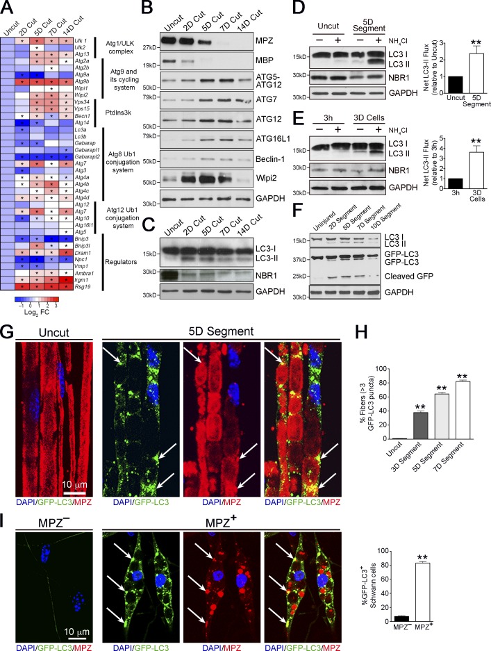
“Conversely, levels of the autophagy substrate NBR1 (Klionsky et al., 2012) showed a clear decrease in injured nerves (Fig.”
InfluentialAutophagy regulates lipid metabolism
Singh, R., Kaushik, S., Wang, Y., Xiang, Y., Novak, I., Komatsu, M., Tanaka, K., Cuervo, A. M., & Czaja, M. J. (2009) · Nature · doi:10.1038/nature07976 · Cited by 3,975
Autophagy and the Integrated Stress Response
Kroemer, G., Mariño, G., & Levine, B. (2010) · Molecular Cell · doi:10.1016/j.molcel.2010.09.023 · Cited by 3,506
Autophagy in immunity and inflammation
Levine, B., Mizushima, N., & Virgin, H. W. (2011) · Nature · doi:10.1038/nature09782 · Cited by 3,289
Impairment of starvation-induced and constitutive autophagy in Atg7 -deficient mice
Komatsu, M., Waguri, S., Ueno, T., Iwata, J., Murata, S., Tanida, I., Ezaki, J., Mizushima, N., Ohsumi, Y., Uchiyama, Y., et al. (2005) · The Journal of Cell Biology · doi:10.1083/jcb.200412022 · Cited by 2,331
Mizushima, N., Yamamoto, A., Matsui, M., Yoshimori, T., & Ohsumi, Y. (2004) · Molecular Biology of the Cell · doi:10.1091/mbc.e03-09-0704 · Cited by 2,105
The machinery of macroautophagy
Feng, Y., He, D., Yao, Z., & Klionsky, D. J. (2014) · Cell Research · doi:10.1038/cr.2013.168 · Cited by 2,083
Regulation of Mammalian Autophagy in Physiology and Pathophysiology
Ravikumar, B., Sarkar, S., Davies, J. E., Futter, M., García-Arencibia, M., Green-Thompson, Z. W., Jiménez-Sánchez, M., Korolchuk, V. I., Lichtenberg, M., Luo, S., et al. (2010) · Physiological Reviews · doi:10.1152/physrev.00030.2009 · Cited by 1,789
Preparation of PCR-Quality Mouse Genomic DNA with Hot Sodium Hydroxide and Tris (HotSHOT)
Truett, G. E., Heeger, P. S., Mynatt, R. L., Truett, A. A., Walker, J. A., & Warman, M. L. (2000) · BioTechniques · doi:10.2144/00291bm09 · Cited by 1,748
Arthur‐Farraj, P., Latouche, M., Wilton, D. K., Quintes, S., Chabrol, E., Banerjee, A., Woodhoo, A., Jenkins, B., Rahman, M., Turmaine, M., et al. (2012) · Neuron · doi:10.1016/j.neuron.2012.06.021 · Cited by 866
Self-citationSchwann Cells: Development and Role in Nerve RepairPreprint
Jessen, K. R., Mirsky, R., & Lloyd, A. C. (2015) · Cold Spring Harbor Perspectives in Biology · doi:10.1101/cshperspect.a020487 · Cited by 759
Self-citationMechanisms of Autophagosome Biogenesis
Rubinsztein, D. C., Shpilka, T., & Elazar, Z. (2012) · Current Biology · doi:10.1016/j.cub.2011.11.034 · Cited by 517
c-Jun is a negative regulator of myelination
Parkinson, D. B., Bhaskaran, A., Arthur‐Farraj, P., Noon, L. A., Woodhoo, A., Lloyd, A. C., Feltri, M. L., Wrabetz, L. G., Behrens, A., Mirsky, R., et al. (2008) · The Journal of Cell Biology · doi:10.1083/jcb.200803013 · Cited by 414
Self-citationNotch controls embryonic Schwann cell differentiation, postnatal myelination and adult plasticity
Woodhoo, A., Alonso, M. B. D., Droggiti, A., Turmaine, M., D’Antonio, M., Parkinson, D. B., Wilton, D. K., Al‐Shawi, R., Simons, J. P., Shen, J., et al. (2009) · Nature Neuroscience · doi:10.1038/nn.2323 · Cited by 364
Self-citationConditional disruption of β1 integrin in Schwann cells impedes interactions with axons
Feltri, M. L., Porta, D. G., Previtali, S. C., Nodari, A., Migliavacca, B., Cassetti, A., Littlewood-Evans, A., Reichardt, L. F., Messing, A., Quattrini, A., et al. (2002) · The Journal of Cell Biology · doi:10.1083/jcb.200109021 · Cited by 325
LYSOSOMES IN THE RAT SCIATIC NERVE FOLLOWING CRUSH
Holtzman, E., & Novikoff, A. B. (1965) · The Journal of Cell Biology · doi:10.1083/jcb.27.3.651 · Cited by 323
Lipid metabolism in myelinating glial cells: lessons from human inherited disorders and mouse models
Chrast, R., Saher, G., Nave, K.-A., & Verheijen, M. H. G. (2011) · Journal of Lipid Research · doi:10.1194/jlr.r009761 · Cited by 314
Contrasting the Glial Response to Axon Injury in the Central and Peripheral Nervous Systems
Lutz, A. B., & Barres, B. A. (2014) · Developmental Cell · doi:10.1016/j.devcel.2013.12.002 · Cited by 233
Novel signals controlling embryonic Schwann cell development, myelination and dedifferentiation
Mirsky, R., Woodhoo, A., Parkinson, D. B., Arthur‐Farraj, P., Bhaskaran, A., & Jessen, K. R. (2008) · Journal of the Peripheral Nervous System · doi:10.1111/j.1529-8027.2008.00168.x · Cited by 229
Self-citationObesity-Dependent Metabolic Signatures Associated with Nonalcoholic Fatty Liver Disease Progression
Barr, J., Caballería, J., Martínez‐Arranz, I., Domínguez‐Díez, A., Alonso, C., Muntané, J., Pérez-Cormenzana, M., García‐Monzón, C., Mayo, R., Martín‐Duce, A., et al. (2012) · Journal of Proteome Research · doi:10.1021/pr201223p · Cited by 226
Self-citationFunctional indices for sciatic, peroneal, and posterior tibial nerve lesions in the mouse
Inserra, M. M., Bloch, D. A., & Terris, D. J. (1998) · Microsurgery · doi:10.1002/(sici)1098-2752(1998)18:2 · Cited by 222
Myelin phagocytosis by macrophages and nonmacrophages during Wallerian degeneration
Hirata, K., & Kawabuchi, M. (2002) · Microscopy Research and Technique · doi:10.1002/jemt.10108 · Cited by 202
Ruiz-Sanz, J. I., & Ochoa, B. (1997) · Journal of Lipid Research · doi:10.1016/s0022-2275(20)37430-7 · Cited by 198
Niemi, J. P., Oranburg, T. D., Roldán-Hernández, L., Lindborg, J. A., Mandell, D., & Zigmond, R. E. (2013) · Journal of Neuroscience · doi:10.1523/jneurosci.3319-12.2013 · Cited by 193
Vargas, M. E., Watanabe, J., Singh, S. J., Robinson, W. H., & Barres, B. A. (2010) · Proceedings of the National Academy of Sciences · doi:10.1073/pnas.1001948107 · Cited by 168
Periaxin, a novel protein of myelinating schwann cells with a possible role in axonal ensheathment
Gillespie, C. S., Sherman, D. L., Blair, G. E., & Brophy, P. (1994) · Neuron · doi:10.1016/0896-6273(94)90208-9 · Cited by 166
Rapamycin Activates Autophagy and Improves Myelination in Explant Cultures from Neuropathic Mice
Rangaraju, S., Verrier, J. D., Madorsky, I., Nicks, J., Dunn, W. A., & Notterpek, L. M. (2010) · Journal of Neuroscience · doi:10.1523/jneurosci.1356-10.2010 · Cited by 153
Fernández‐Valle, C., Bunge, R. P., & Bunge, M. B. (1995) · Journal of Neurocytology · doi:10.1007/bf01179817 · Cited by 134
Perry, V. H., Tsao, J. W., Feam, S., & Brown, M. C. (1995) · European Journal of Neuroscience · doi:10.1111/j.1460-9568.1995.tb01063.x · Cited by 126
Role of Inflammation and Cytokines in Peripheral Nerve Regeneration
Dubový, P., Jančálek, R., & Kubek, T. (2013) · International review of neurobiology · doi:10.1016/b978-0-12-410499-0.00007-1 · Cited by 123
Actin Polymerization Is Essential for Myelin Sheath Fragmentation during Wallerian Degeneration
Jung, J., Cai, W., Lee, H. K., Pellegatta, M., Shin, Y. K., Jang, S. Y., Suh, D. J., Wrabetz, L. G., Feltri, M. L., & Park, H. T. (2011) · Journal of Neuroscience · doi:10.1523/jneurosci.4537-10.2011 · Cited by 106
Larocca, J. N., & Norton, W. T. (2006) · Current Protocols in Cell Biology · doi:10.1002/0471143030.cb0325s33 · Cited by 105
Ceramide-induced autophagy: To junk or to protect cells?
Pattingre, S., Bauvy, C., Levade, T., Levine, B., & Codogno, P. (2009) · Autophagy · doi:10.4161/auto.5.4.8390 · Cited by 105
Conditional Disruption Of Beta 1 Integrin In Schwann Cells Impedes Interactions With Axons
Feltri, M., Porta, D., Previtali, S. C., Nodari, A., Migliavacca, B., Cassetti, A., Littlewood-Evans, A. J., Reichardt, L. F., Messing, A., Quattrini, A., et al. (2002) · Journal of the Peripheral Nervous System · doi:10.1046/j.1529-8027.2002.02026_1.x · Cited by 90
S-adenosylmethionine Levels Regulate the Schwann Cell DNA Methylome
Varela‐Rey, M., Iruarrizaga‐Lejarreta, M., Lozano, J. J., Aransay, A. M., Fernández, A. F., Lavín, J. L., Mosén-Ansorena, D., Berdasco, M., Turmaine, M., Luka, Z., et al. (2014) · Neuron · doi:10.1016/j.neuron.2014.01.037 · Cited by 78
Self-citationAutophagy in unicellular eukaryotes
Kiel, J. A. K. W. (2010) · Philosophical Transactions of the Royal Society B Biological Sciences · doi:10.1098/rstb.2009.0237 · Cited by 78
c-Jun activation in Schwann cells protects against loss of sensory axons in inherited neuropathy
Hantke, J., Carty, L., Wagstaff, L. J., Turmaine, M., Wilton, D. K., Quintes, S., Koltzenburg, M., Baas, F., Mirsky, R., & Jessen, K. R. (2014) · Brain · doi:10.1093/brain/awu257 · Cited by 76
Self-citationReverter, G. P., Agudo‐Barriuso, M., Nadal-Nicolás, F. M., Alarcón-Martínez, L., Jiménez-López, M., Salinas‐Navarro, M., Sobrado‐Calvo, P., Bernal-Garro, J. M., Villegas‐Pérez, M. P., & Vidal‐Sanz, M. (2009) · Vision Research · doi:10.1016/j.visres.2009.08.020 · Cited by 75
Proteomics of the Injured Rat Sciatic Nerve Reveals Protein Expression Dynamics During Regeneration
Jiménez, C. R., Stam, F. J., Li, K. W., Gouwenberg, Y., Hornshaw, M. P., de Winter, F., Verhaagen, J., & Smit, A. B. (2004) · Molecular & Cellular Proteomics · doi:10.1074/mcp.m400076-mcp200 · Cited by 70
Myelin and Axon Pathology in a Long-Term Study ofPMP22-Overexpressing Mice
Verhamme, C., King, R. H. M., ten Asbroek, A. L. M. A., Muddle, J. R., Nourallah, M., Wolterman, R. A., Baas, F., & van Schaik, I. N. (2011) · Journal of Neuropathology & Experimental Neurology · doi:10.1097/nen.0b013e318217eba0 · Cited by 63
Self-citationc-Jun expression in human neuropathies: a pilot study
Hutton, E. J., Carty, L., Laurá, M., Houlden, H., Lunn, M. P. T., Brandner, S., Mirsky, R., Jessen, K., & Reilly, M. M. (2011) · Journal of the Peripheral Nervous System · doi:10.1111/j.1529-8027.2011.00360.x · Cited by 63
Self-citationJang, S. Y., Shin, Y. K., Park, S. Y., Park, J. Y., Rha, S.-H., Kim, J. K., Lee, H. J., & Park, H. T. (2015) · PLOS ONE · doi:10.1371/journal.pone.0116624 · Cited by 61 · cited 2×
“G; see, however, Jang et al., 2015).”
Goodrum, J. F., Earnhardt, T. S., Goines, N. D., & Bouldin, T. W. (1994) · Journal of Neuroscience · doi:10.1523/jneurosci.14-01-00357.1994 · Cited by 59
Soluble Complement Receptor 1 Protects the Peripheral Nerve from Early Axon Loss after Injury
Ramaglia, V., Wolterman, R. A., de Kok, M., Vigar, M. A., Bos, I. G. A. C., King, R. H. M., Morgan, B. P., & Baas, F. (2008) · American Journal Of Pathology · doi:10.2353/ajpath.2008.070660 · Cited by 49
Self-citationJung, J., Cai, W., Jang, S. Y., Shin, Y. K., Suh, D. J., Kim, J. K., & Park, H. T. (2011) · Anatomy & Cell Biology · doi:10.5115/acb.2011.44.1.41 · Cited by 33
Vaudano, E., Campbell, G., & Hunt, S. P. (1996) · Neuroscience · doi:10.1016/0306-4522(96)00199-6 · Cited by 10
Cited by
Sorted by citations
Guidelines for the use and interpretation of assays for monitoring autophagy (4th edition) 1
Klionsky, D. J., Abdel-Aziz, A. K., Abdelfatah, S., Abdellatif, M., Abdoli, A., Abel, S., Abeliovich, H., Abildgaard, M. H., Abudu, Y. P., Acevedo-Arozena, A., et al. (2021) · Autophagy · doi:10.1080/15548627.2020.1797280 · Cited by 2,897
Molecular definitions of autophagy and related processes
Galluzzi, L., Baehrecke, E. H., Ballabio, A., Boya, P., Bravo‐San Pedro, J. M., Cecconi, F., Choi, A. M., Chu, C. T., Codogno, P., Colombo, M. I., et al. (2017) · The EMBO Journal · doi:10.15252/embj.201796697 · Cited by 1,646
Autophagy in major human diseases
Klionsky, D. J., Petroni, G., Amaravadi, R. K., Baehrecke, E. H., Ballabio, A., Boya, P., Bravo‐San Pedro, J. M., Cadwell, K., Cecconi, F., Choi, A. M. K., et al. (2021) · The EMBO Journal · doi:10.15252/embj.2021108863 · Cited by 1,585
The repair Schwann cell and its function in regenerating nerves
Jessen, K. R., & Mirsky, R. (2016) · The Journal of Physiology · doi:10.1113/jp270874 · Cited by 1,178
Self-citationAutophagy and Neurodegeneration: Pathogenic Mechanisms and Therapeutic Opportunities
Menzies, F. M., Fleming, A., Caricasole, A., Bento, C. F., Andrews, S. P., Ashkenazi, A., Füllgrabe, J., Jackson, A., Jimenez Sanchez, M., Karabiyik, C., et al. (2017) · Neuron · doi:10.1016/j.neuron.2017.01.022 · Cited by 1,134
Metadata
- Authors
- Gomez-Sanchez, J. A., Carty, L., Iruarrizaga-Lejarreta, M., Palomo-Irigoyen, M., Varela-Rey, M., Griffith, M., Hantke, J., Macias-Camara, N., Azkargorta, M., Aurrekoetxea, I., De Juan, V. G., Jefferies, H. B. J., Aspichueta, P., Elortza, F., Aransay, A. M., Martínez-Chantar, M. L., Baas, F., Mato, J. M., Mirsky, R., Woodhoo, A., & Jessen, K. R.
- Affiliations
- University College London (GB)
- CIC bioGUNE, Centro de Investigación Biomédica en Red de Enfermedades Hepáticas y Digestivas (CIBERehd), 48160 Derio, Bizkaia, Spain 2
- CIC bioGUNE (ES)
- Instituto de Salud Carlos III (ES)
- University of the Basque Country (ES)
- The Francis Crick Institute (GB)
- Amsterdam UMC Location University of Amsterdam (NL)
- Ikerbasque (ES)
- Corresponding author
- Woodhoo, Ashwin
- ORCID
- Venue
- Journal of Cell Biology
- Publisher
- Rockefeller University Press
- Published
- 2015-07-06
- Volume
- 210
- Issue
- 1
- Pages
- 153-168
- ISSN
- 0021-9525 (ISSN-L)
- 1540-8140
- PMC
- PMC4494002
- OpenAlex
- W1655540037
- Cited by
- 453
- References
- 49
- Citations by year
- 2026: 33 · 2025: 47 · 2024: 40 · 2023: 45 · 2022: 52 · 2021: 55 · 2020: 43 · 2019: 38 · 2018: 36 · 2017: 32 · 2016: 22 · 2015: 8
- FWCI
- 25.61
- Type
- journal article
- Language
- en
- Open access
- hybrid
- doi.org
- Licence
- cc-by-nc-sa
- Locations
- The Journal of Cell Biology (publishedVersion, cc-by-nc-sa)
- Europe PMC (PubMed Central) (publishedVersion, cc-by-nc-sa)
- PubMed Central (publishedVersion, cc-by-nc-sa)
- PubMed (publishedVersion)
- Pure Amsterdam UMC (submittedVersion, other-oa)
- Data Archiving and Networked Services (DANS) (submittedVersion)
- UCL Discovery (University College London) (submittedVersion)
- Figshare (submittedVersion, cc-by-nc-sa)
- Funders
- Wellcome Trust (FP7/2007–2013)
- Francis Crick Institute (10187)
- Cancer Research UK (15153)
- Instituto de Salud Carlos III
- National Center for Complementary and Integrative Health (R01AT001576)
- Topics
- Life Sciences › Biochemistry, Genetics and Molecular Biology › Cell Biology › Autophagy in Disease and Therapy
- Life Sciences › Neuroscience › Cellular and Molecular Neuroscience › Nerve injury and regeneration
- Life Sciences › Neuroscience › Cellular and Molecular Neuroscience › Hereditary Neurological Disorders
- Keywords
- Schwann cells, selective autophagy, myelin debris clearance, nerve injury, Wallerian degeneration, JNK/c-Jun signaling, demyelinating disease, autophagosomes, myelin debris, central nervous system
- MeSH terms
- Animals, Autophagy, Cells, Cultured, Mice, Transgenic, Myelin Sheath, Sciatic Nerve, Wallerian Degeneration, Proto-Oncogene Proteins c-jun, JNK Mitogen-Activated Protein Kinases, Lipid Metabolism, TOR Serine-Threonine Kinases, Peripheral Nerve Injuries
About these checks
Registry, retraction, Cites and statistics results are computed, not judged, with one exception: a printed reference that no index lists is read once by a model before it counts as a citation error. Claims are extracted by a model and kept only when their quote appears verbatim in the text; each cited source is then checked, with a model’s help, for whether it says what the claim relies on. Those verdicts can be wrong — read the claim’s quote and the cited source before relying on one.
Statistics and arithmetic are recomputed, not judged.
Confidence bands are shown only for fields calibrated on our annotated test set. None is calibrated yet, so no band is shown.
What counts as a claim ›How we measure confidence ›How we check this ›
Are you an author, or do you see a problem with this page? Request a review
