Cited by 154Lipid metabolism and biosynthesisCellular transport and secretionMitochondrial Function and Pathology

The DOI matches its Crossref record; 58 of the 58 references it cites were found in a registry or an index.

Structure–function insights into direct lipid transfer between membranes by Mmm1–Mdm12 of ERMES

Open access

Abstract

The endoplasmic reticulum (ER)–mitochondrial encounter structure (ERMES) physically links the membranes of the ER and mitochondria in yeast. Although the ER and mitochondria cooperate to synthesize glycerophospholipids, whether ERMES directly facilitates the lipid exchange between the two organelles remains controversial. Here, we compared the x-ray structures of an ERMES subunit Mdm12 from Kluyveromyces lactis with that of Mdm12 from Saccharomyces cerevisiae and found that both Mdm12 proteins possess a hydrophobic pocket for phospholipid binding. However in vitro lipid transfer assays showed that Mdm12 alone or an Mmm1 (another ERMES subunit) fusion protein exhibited only a weak lipid transfer activity between liposomes. In contrast, Mdm12 in a complex with Mmm1 mediated efficient lipid transfer between liposomes. Mutations in Mmm1 or Mdm12 impaired the lipid transfer activities of the Mdm12–Mmm1 complex and furthermore caused defective phosphatidylserine transport from the ER to mitochondrial membranes via ERMES in vitro. Therefore, the Mmm1–Mdm12 complex functions as a minimal unit that mediates lipid transfer between membranes.

Overview

The claims and statistics of this paper haven’t been checked yet.

Run the check — free, no account needed. It reads the open-access text and checks the sources it cites.

A few free checks a day · papers already verified open instantly · results are public

Integrity signals

Registry

Crossref · 10.1083/jcb.201704119

Cites

58 of 58 references verified0 citation errors · 0 not checkable

Statistics

Figures

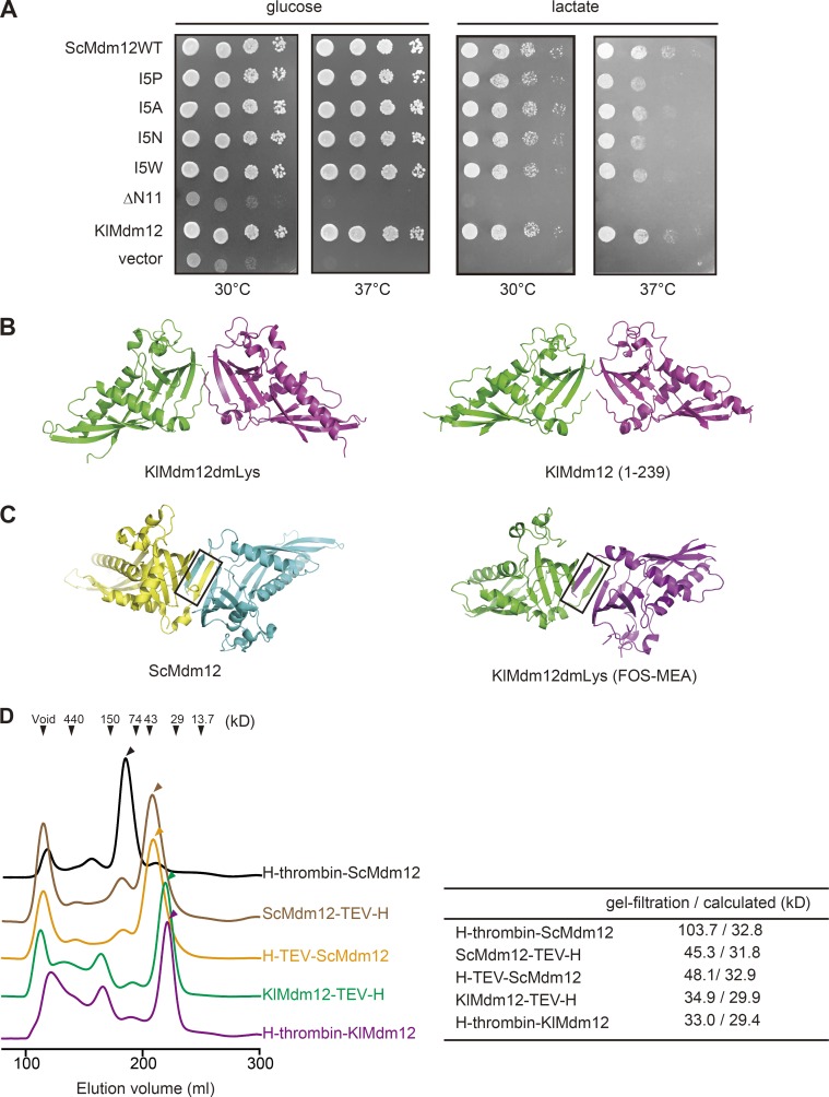
Figure 1. Oligomer formation and lipid binding of Mdm12. (A) A pRS314 vector containing the gene for ScMdm12 (ScMdm12WT); ScMdm12 with the mutation of I5P, I5A, I5N, or I5W; ScMdm12 lacking the N-terminal 11 residues (ΔN11); or KlMdm12(1–239) was introduced into the S. cerevisiae mdm12Δ strain by plasmid shuffling. An empty pR314 vector was introduced into the mdm12Δ strain as a control (vector). Saturated cultures of the indicated cells were diluted with 10-fold increments, spotted on SCD (−Trp, +FOA) or SCLac (−Trp, +FOA) plates, and incubated at 30°C or 37°C for 2 or 3 d, respectively. (B) Dimeric arrangement found in the crystal structures of KlMdm12. Structures of KlMdm12(1–239), residues 1–239 of KlMdm12 without modification (left), and KlMdm12dmLys, dimethyl-lysine–modified KlMdm12(1–239) (right) are shown in ribbon form. (C) Interlocking dimers found in ScMdm12 (left; Jeong et al., 2016) and KlMdm12dmLys crystallized in the presence of FOS-MEA-10 (right). The domain-swapped regions are shown by black squares. (D) The indicated proteins were purified from E. coli cells and subjected to gel-filtration analyses using a HiLoad26/600 Superdex 200 pg column. Absorption at 280 nm is plotted against elution volume, and positions of molecular mass (kilodalton) marker proteins are indicated. Apparent molecular masses of the Mdm12 derivatives estimated from the peak tops (indicated with arrowheads) of the elution profiles (gel filtration) and calculated molecular masses for the monomeric derivatives (calculated) are shown in the right table. H-TEV–ScMdm12, ScMdm12 with the N-terminally attached His10 tag followed by the TEV protease recognition sequence; H-thrombin-KlMdm12, KlMdm12 with the N-terminally attached His6 tag followed by the thrombin protease recognition sequence; H-thrombin–ScMdm12, ScMdm12 with the N-terminally attached His6 tag followed by the thrombin protease recognition sequence; KlMdm12TEV-H, KlMdm12 followed by the TEV protease recognition sequence and the C-terminally attached His6 tag; ScMdm12TEV-H, ScMdm12 followed by the TEV protease recognition sequence and the C-terminally attached His6 tag.
Figure 1. Oligomer formation and lipid binding of Mdm12. (A) A pRS314 vector containing the gene for ScMdm12 (ScMdm12WT); ScMdm12 with the mutation of I5P, I5A, I5N, or I5W; ScMdm12 lacking the N-terminal 11 residues (ΔN11); or KlMdm12(1–239) was introduced into the S. cerevisiae mdm12Δ strain by plasmid shuffling. An empty pR314 vector was introduced into the mdm12Δ strain as a control (vector). Saturated cultures of the indicated cells were diluted with 10-fold increments, spotted on SCD (−Trp, +FOA) or SCLac (−Trp, +FOA) plates, and incubated at 30°C or 37°C for 2 or 3 d, respectively. (B) Dimeric arrangement found in the crystal structures of KlMdm12. Structures of KlMdm12(1–239), residues 1–239 of KlMdm12 without modification (left), and KlMdm12dmLys, dimethyl-lysine–modified KlMdm12(1–239) (right) are shown in ribbon form. (C) Interlocking dimers found in ScMdm12 (left; Jeong et al., 2016) and KlMdm12dmLys crystallized in the presence of FOS-MEA-10 (right). The domain-swapped regions are shown by black squares. (D) The indicated proteins were purified from E. coli cells and subjected to gel-filtration analyses using a HiLoad26/600 Superdex 200 pg column. Absorption at 280 nm is plotted against elution volume, and positions of molecular mass (kilodalton) marker proteins are indicated. Apparent molecular masses of the Mdm12 derivatives estimated from the peak tops (indicated with arrowheads) of the elution profiles (gel filtration) and calculated molecular masses for the monomeric derivatives (calculated) are shown in the right table. H-TEV–ScMdm12, ScMdm12 with the N-terminally attached His10 tag followed by the TEV protease recognition sequence; H-thrombin-KlMdm12, KlMdm12 with the N-terminally attached His6 tag followed by the thrombin protease recognition sequence; H-thrombin–ScMdm12, ScMdm12 with the N-terminally attached His6 tag followed by the thrombin protease recognition sequence; KlMdm12TEV-H, KlMdm12 followed by the TEV protease recognition sequence and the C-terminally attached His6 tag; ScMdm12TEV-H, ScMdm12 followed by the TEV protease recognition sequence and the C-terminally attached His6 tag.CC BY-NC-SA · PubMed Central
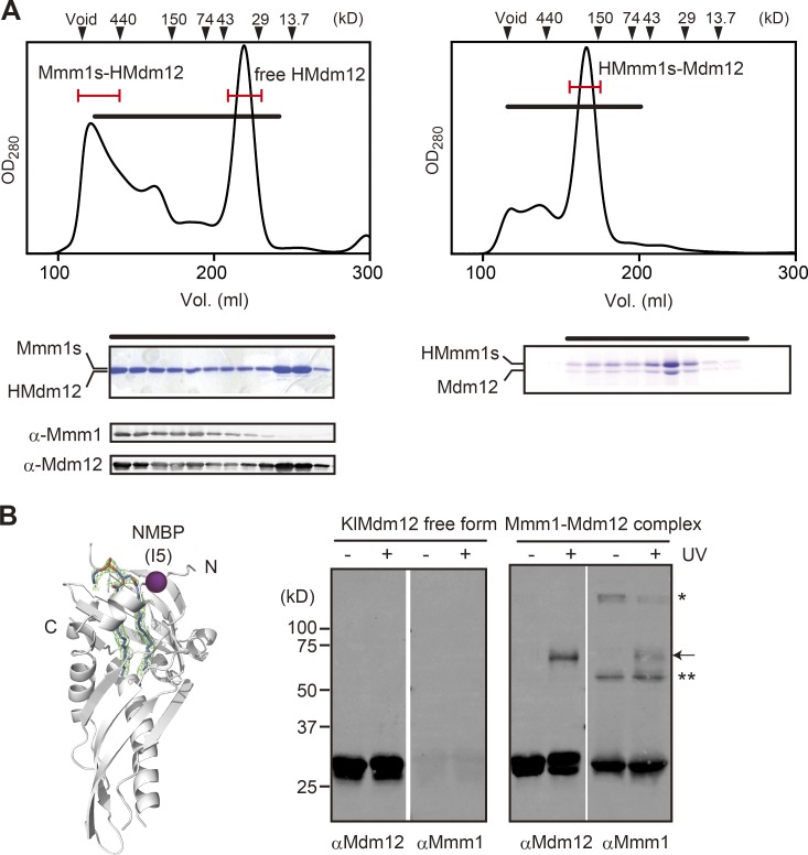
Figure 2. Hetero-oligomer formation of K. lactis Mdm12 and Mmm1. (A) KlMdm12 and Mmm1s form hetero-oligomeric complexes when expressed in E. coli cells. Mmm1s and N-terminally His6-tagged KlMdm12 (HMdm12) and N-terminally His6-tagged K. lactis Mmm1s (HMmm1s) and KlMdm12 were expressed in E. coli cells and affinity purified using a Ni-NTA column. Eluted fractions were then pooled and subjected to gel filtration by a Superdex 200 column (left: Mmm1s-HMdm12; right: HMmm1s-Mdm12). Fractions indicated with black bars in the elution profiles (top) were fractionated and analyzed by SDS-PAGE and Coomassie staining (bottom) or immunoblotting with the indicated antibodies. Fractions indicated with red bars are collected for further analyses for lipid transfer. Positions of molecular mass (kilodalton) marker proteins are indicated. The presence of the His6 tag at the N terminus of either Mmm1 or Mdm12 affects the gel-filtration elution profiles of the Mmm1s–Mdm12 hetero-oligomers. Apparent sizes of the HMmm1s–Mdm12 and Mmm1s–HMdm12 complexes are ∼200 kD and ∼500–600 kD, respectively. (B) Left: The position of I5 in KlMdm12 is shown. Two forms (free KlMdm12 and KlMdm12 complexed with Mmm1s) of I5C mutant KlMdm12 were purified and modified with NMBP to introduce a photoreactive benzophenone group into the sulfhydryl group of C5. Samples were then treated with or without UV irradiation. Proteins were analyzed by SDS-PAGE followed by immunoblotting with the indicated antibodies. The arrow points to cross-linked product; asterisks indicate nonspecific signals.
Figure 2. Hetero-oligomer formation of K. lactis Mdm12 and Mmm1. (A) KlMdm12 and Mmm1s form hetero-oligomeric complexes when expressed in E. coli cells. Mmm1s and N-terminally His6-tagged KlMdm12 (HMdm12) and N-terminally His6-tagged K. lactis Mmm1s (HMmm1s) and KlMdm12 were expressed in E. coli cells and affinity purified using a Ni-NTA column. Eluted fractions were then pooled and subjected to gel filtration by a Superdex 200 column (left: Mmm1s-HMdm12; right: HMmm1s-Mdm12). Fractions indicated with black bars in the elution profiles (top) were fractionated and analyzed by SDS-PAGE and Coomassie staining (bottom) or immunoblotting with the indicated antibodies. Fractions indicated with red bars are collected for further analyses for lipid transfer. Positions of molecular mass (kilodalton) marker proteins are indicated. The presence of the His6 tag at the N terminus of either Mmm1 or Mdm12 affects the gel-filtration elution profiles of the Mmm1s–Mdm12 hetero-oligomers. Apparent sizes of the HMmm1s–Mdm12 and Mmm1s–HMdm12 complexes are ∼200 kD and ∼500–600 kD, respectively. (B) Left: The position of I5 in KlMdm12 is shown. Two forms (free KlMdm12 and KlMdm12 complexed with Mmm1s) of I5C mutant KlMdm12 were purified and modified with NMBP to introduce a photoreactive benzophenone group into the sulfhydryl group of C5. Samples were then treated with or without UV irradiation. Proteins were analyzed by SDS-PAGE followed by immunoblotting with the indicated antibodies. The arrow points to cross-linked product; asterisks indicate nonspecific signals.CC BY-NC-SA · PubMed Central
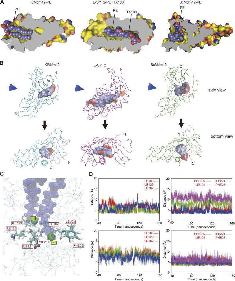
Figure 3. Bottom of the hydrophobic pocket in KlMdm12 is tightly closed. (A) Structural comparison of the KlMdm12–PE complex (left), the complex of E-SYT2 with PE and Triton X-100 (TX100; middle), and the ScMdm12–PE complex (right). Proteins are represented by the molecular surface (C, yellow; O, red; and N, blue) with cutaway views to show cavities and the bound substrates in space-filling form. (B) Solvent-accessible pockets (light blue surface) and main-chain foldings of KlMdm12 (left), human E-SYT2 (middle), and ScMdm12 (right). The solvent-accessible pockets were searched by query in the POCASSA server (http://altair.sci.hokudai.ac.jp/g6.service/pocassa/; Yu et al., 2010) and visualized. Pockets were defined as the region between the protein surface and the “probe surface,” which is generated by a probe sphere with diameter of 4 Å rolling along the protein surface. Blue cones indicate the directions of the views for the bottom panels. Openings of the pockets are indicated by transparent red areas. Note that the cavity in E-SYT2 has multiple separate openings that make up a continuing cavity through the molecule. The volumes of the cavities are 1,242 Å3, 2,086 Å3, and 2,152 Å3 for KlMdm12, E-SYT2 (monomer), and ScMdm12, respectively. N and C indicate the N and C termini, respectively. (C and D) MD simulation of the PE-bound KlMdm12 structure. MD simulation was used to test whether the bottom of the lipid-binding pocket is stably closed. The distances from the terminal methyl C atoms of the 1-palmitoyl group (C1) and of 2-oleoyl group (C2) to the hydrophobic side chains of the indicated residues (C) at the bottom of the lipid-binding pocket of KlMdm12 were traced down for 160 ns in the top and bottom panels, respectively (D).
Figure 3. Bottom of the hydrophobic pocket in KlMdm12 is tightly closed. (A) Structural comparison of the KlMdm12–PE complex (left), the complex of E-SYT2 with PE and Triton X-100 (TX100; middle), and the ScMdm12–PE complex (right). Proteins are represented by the molecular surface (C, yellow; O, red; and N, blue) with cutaway views to show cavities and the bound substrates in space-filling form. (B) Solvent-accessible pockets (light blue surface) and main-chain foldings of KlMdm12 (left), human E-SYT2 (middle), and ScMdm12 (right). The solvent-accessible pockets were searched by query in the POCASSA server (http://altair.sci.hokudai.ac.jp/g6.service/pocassa/; Yu et al., 2010) and visualized. Pockets were defined as the region between the protein surface and the “probe surface,” which is generated by a probe sphere with diameter of 4 Å rolling along the protein surface. Blue cones indicate the directions of the views for the bottom panels. Openings of the pockets are indicated by transparent red areas. Note that the cavity in E-SYT2 has multiple separate openings that make up a continuing cavity through the molecule. The volumes of the cavities are 1,242 Å3, 2,086 Å3, and 2,152 Å3 for KlMdm12, E-SYT2 (monomer), and ScMdm12, respectively. N and C indicate the N and C termini, respectively. (C and D) MD simulation of the PE-bound KlMdm12 structure. MD simulation was used to test whether the bottom of the lipid-binding pocket is stably closed. The distances from the terminal methyl C atoms of the 1-palmitoyl group (C1) and of 2-oleoyl group (C2) to the hydrophobic side chains of the indicated residues (C) at the bottom of the lipid-binding pocket of KlMdm12 were traced down for 160 ns in the top and bottom panels, respectively (D).CC BY-NC-SA · PubMed Central
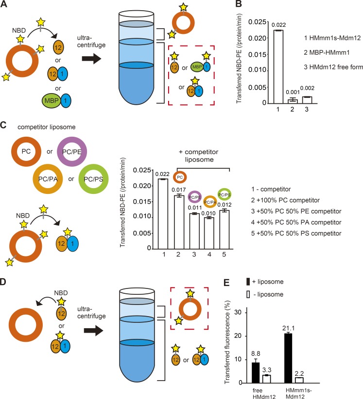
Figure 4. Mmm1–Mdm12 can extract lipids from membranes and insert them into membranes. (A) Fluorescent lipid extraction by a protein from liposomes. Liposomes consisting of PC and NBD-PE (PC/NBD-PE = 80/20) were incubated with free KlMdm12, MBP–HMmm1s, or HMmm1s–Mdm12 and then separated by sucrose step-gradient centrifugation. The orange and blue ovals indicate Mdm12 and Mmm1s, respectively. (B) Lipid-extraction activities as fluorescence intensity in the protein fraction in the assay in A (broken-line square) are plotted by bars and figures above the bar. Bars show means ± SE of three independent experiments. (C) Competition of HMmm1s–Mdm12–mediated fluorescent lipid extraction from liposome by nonfluorescent liposomes. 200 µM liposomes (PC/NBD-PE = 80/20) were incubated with HMmm1s–Mdm12 in the presence or absence of 200 µM PC liposomes (orange), PC/PE (50/50; purple), PC/PA (50/50; yellow orange), or PC/PS (50/50; yellow green) liposomes and separated by sucrose step-gradient centrifugation. The molar ratio of transferred NBD-PE to HMmm1s–Mdm12 was plotted by bars and figures above the bars. Bars show means ± SE of three independent experiments. Total NBD-PE fluorescence intensity was estimated by solubilizing NBD-PE–containing liposomes with Triton X-100. (D) Lipid transfer from a protein preloaded with a fluorescent lipid to liposomes. Free form of Mdm12 (free Mdm12) or HMmm1s–Mdm12 preloaded with NBD-PE was incubated with PC liposomes and then separated by Nycodenz step-gradient centrifugation. (E) Lipid-transfer activities (protein to liposomes) as fluorescence intensity in the floating liposome fraction normalized by the fluorescence intensity in total fractions are plotted by bars and figures above the bars. Bars show means ± SE of three independent experiments. We confirmed that the added proteins did not stably bind to liposomes in these assays.
Figure 4. Mmm1–Mdm12 can extract lipids from membranes and insert them into membranes. (A) Fluorescent lipid extraction by a protein from liposomes. Liposomes consisting of PC and NBD-PE (PC/NBD-PE = 80/20) were incubated with free KlMdm12, MBP–HMmm1s, or HMmm1s–Mdm12 and then separated by sucrose step-gradient centrifugation. The orange and blue ovals indicate Mdm12 and Mmm1s, respectively. (B) Lipid-extraction activities as fluorescence intensity in the protein fraction in the assay in A (broken-line square) are plotted by bars and figures above the bar. Bars show means ± SE of three independent experiments. (C) Competition of HMmm1s–Mdm12–mediated fluorescent lipid extraction from liposome by nonfluorescent liposomes. 200 µM liposomes (PC/NBD-PE = 80/20) were incubated with HMmm1s–Mdm12 in the presence or absence of 200 µM PC liposomes (orange), PC/PE (50/50; purple), PC/PA (50/50; yellow orange), or PC/PS (50/50; yellow green) liposomes and separated by sucrose step-gradient centrifugation. The molar ratio of transferred NBD-PE to HMmm1s–Mdm12 was plotted by bars and figures above the bars. Bars show means ± SE of three independent experiments. Total NBD-PE fluorescence intensity was estimated by solubilizing NBD-PE–containing liposomes with Triton X-100. (D) Lipid transfer from a protein preloaded with a fluorescent lipid to liposomes. Free form of Mdm12 (free Mdm12) or HMmm1s–Mdm12 preloaded with NBD-PE was incubated with PC liposomes and then separated by Nycodenz step-gradient centrifugation. (E) Lipid-transfer activities (protein to liposomes) as fluorescence intensity in the floating liposome fraction normalized by the fluorescence intensity in total fractions are plotted by bars and figures above the bars. Bars show means ± SE of three independent experiments. We confirmed that the added proteins did not stably bind to liposomes in these assays.CC BY-NC-SA · PubMed Central
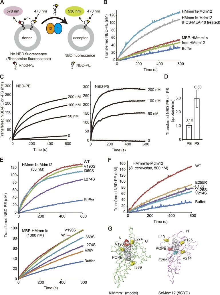
Figure 5. Mmm1–Mdm12 can transfer lipids between lipid membranes. (A) A schematic diagram of the fluorescent-based NBD-PE transfer between liposomes. 12.5 µM donor liposomes contain NBD-PE and Rhod-PE, and NBD fluorescence is quenched by FRET to Rhod. Once NBD-PE is transferred to acceptor liposomes (50 µM), NBD fluorescence increases, which is monitored in B. (B) NBD-PE transfer activities of the 50 nM HMmm1s–Mdm12 complex, the FOS-MEA-10–treated HMmm1s–Mdm12 complex, MBP-HMmm1s, and HMdm12 were measured at 25°C by the assay shown in A. At 0 s, the protein or buffer was added to the reaction mixture, and NBD fluorescence intensities were set to 0 at 0 s. (C) Transfer of NBD-PE and NBD-PS between liposomes by different concentrations of HMmm1s–Mdm12 was compared by the assays as in B. (D) The amounts of NBD-PE and NBD-PS transferred from 12.5 µM donor liposomes containing NBD-PE or NBD-PS with Rhod-PE to 50 µM acceptor liposomes after 10 s reaction. Total NBD-PE or NBD-PS fluorescence intensities were estimated by solubilizing NBD-PE or NBD-PS containing liposomes with Triton X-100. Bars show means ± SD of three independent experiments. (E) NBD-PE transfer activities of the 50 nM HMmm1s–Mdm12 complex and 1,000 nM MBP-HMmm1s containing K. lactis wild-type Mmm1s (WT) or the indicated K. lactis Mmm1s mutants (measured as in A and B). Total NBD-PE fluorescence intensity was estimated by solubilizing NBD-PE–containing liposomes with Triton X-100. (F) NBD-PE transfer activities of the 500 nM ScHMmm1s–ScMdm12 complex analyzed as in E. The curves for the mutations at the same positions between the Mmm1 model structure (E) and Mdm12 x-ray structure (F) are indicated with the same colors. (G) Positions of the mutated hydrophobic residues in the S. cerevisiae Mdm12 structure (Jeong et al., 2016) and in the homology model of K. lactis Mmm1s (KlMmm1s, main-chain folding in green) are shown by space-filling form in yellow. E255 in the S. cerevisiae structure is shown by space-filling form in cyan. The positions of POPE in the ScMdm12 structure and in the model structure of KlMmm1s are shown in space-filling form. Homology modeling of KlMmm1s based on the KlMdm12 structure was performed using Modeller software (Šali and Blundell, 1993). The predicted model structure of KlMmm1s resembles KlMdm12 and possesses a hydrophobic (likely phospholipid-binding) pocket. To assess the significance of the hydrophobic pockets of Mmm1 and Mdm12 in lipid transfer activity by mutational analyses, three residues were chosen according to their positions at the entrance, middle, and bottom part of the pocket: L10, V125, and V214 in ScMdm12 and V190, L274, and I369 (KlMmm1 numbering) in KlMmm1.
Figure 5. Mmm1–Mdm12 can transfer lipids between lipid membranes. (A) A schematic diagram of the fluorescent-based NBD-PE transfer between liposomes. 12.5 µM donor liposomes contain NBD-PE and Rhod-PE, and NBD fluorescence is quenched by FRET to Rhod. Once NBD-PE is transferred to acceptor liposomes (50 µM), NBD fluorescence increases, which is monitored in B. (B) NBD-PE transfer activities of the 50 nM HMmm1s–Mdm12 complex, the FOS-MEA-10–treated HMmm1s–Mdm12 complex, MBP-HMmm1s, and HMdm12 were measured at 25°C by the assay shown in A. At 0 s, the protein or buffer was added to the reaction mixture, and NBD fluorescence intensities were set to 0 at 0 s. (C) Transfer of NBD-PE and NBD-PS between liposomes by different concentrations of HMmm1s–Mdm12 was compared by the assays as in B. (D) The amounts of NBD-PE and NBD-PS transferred from 12.5 µM donor liposomes containing NBD-PE or NBD-PS with Rhod-PE to 50 µM acceptor liposomes after 10 s reaction. Total NBD-PE or NBD-PS fluorescence intensities were estimated by solubilizing NBD-PE or NBD-PS containing liposomes with Triton X-100. Bars show means ± SD of three independent experiments. (E) NBD-PE transfer activities of the 50 nM HMmm1s–Mdm12 complex and 1,000 nM MBP-HMmm1s containing K. lactis wild-type Mmm1s (WT) or the indicated K. lactis Mmm1s mutants (measured as in A and B). Total NBD-PE fluorescence intensity was estimated by solubilizing NBD-PE–containing liposomes with Triton X-100. (F) NBD-PE transfer activities of the 500 nM ScHMmm1s–ScMdm12 complex analyzed as in E. The curves for the mutations at the same positions between the Mmm1 model structure (E) and Mdm12 x-ray structure (F) are indicated with the same colors. (G) Positions of the mutated hydrophobic residues in the S. cerevisiae Mdm12 structure (Jeong et al., 2016) and in the homology model of K. lactis Mmm1s (KlMmm1s, main-chain folding in green) are shown by space-filling form in yellow. E255 in the S. cerevisiae structure is shown by space-filling form in cyan. The positions of POPE in the ScMdm12 structure and in the model structure of KlMmm1s are shown in space-filling form. Homology modeling of KlMmm1s based on the KlMdm12 structure was performed using Modeller software (Šali and Blundell, 1993). The predicted model structure of KlMmm1s resembles KlMdm12 and possesses a hydrophobic (likely phospholipid-binding) pocket. To assess the significance of the hydrophobic pockets of Mmm1 and Mdm12 in lipid transfer activity by mutational analyses, three residues were chosen according to their positions at the entrance, middle, and bottom part of the pocket: L10, V125, and V214 in ScMdm12 and V190, L274, and I369 (KlMmm1 numbering) in KlMmm1.CC BY-NC-SA · PubMed Central
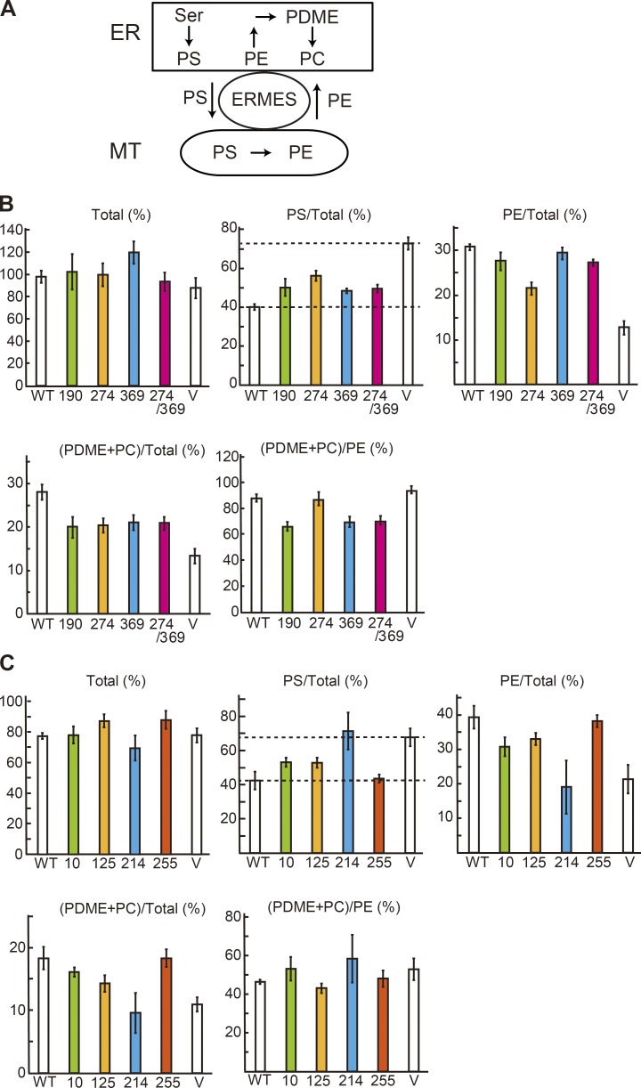
Figure 6. Mmm1 and Mdm12 mediate PS transport from the ER to mitochondria. (A) A schematic diagram of the PS transfer/conversion pathway involving the ER and mitochondria (MT). The indicated phospholipids were analyzed in this assay. PDME, phosphatidyldimethylethanolamine. (B) In vitro PS transport assays were performed using the HMFs isolated from mmm1Δ cells expressing Vps13-D716H with S. cerevisiae wild-type (WT) or mutant Mmm1 (V190S [190], L274S [274], L369S [369], or L274S/L369S [274/369]) or without Mmm1 derivatives (V). Total phospholipids were extracted and analyzed by TLC and radioimaging. Amounts of PS, PE, and PDME+PC relative to total phospholipids and PDME+PC relative to PE were quantified after 30 min of incubation. Bars show mean ± SE of three independent experiments. The amount of total phospholipids synthesized with wild-type cells after 40-min incubation was set to 100% (total). The amounts of PS/total reflect PS transport from the ER to mitochondria. (C) In vitro PS transport assays were performed using the HMFs isolated from mdm12Δ cells expressing Vps13-D716H with S. cerevisiae wild-type (WT) or mutant Mdm12 (L10S [10], V125S [125], V214S [214], or E255R [255]) or without Mdm12 derivatives (V) as in B. The amounts of PS/total reflect PS transport from the ER to mitochondria. The bars for the mutations at the same positions between the Mmm1 model structure (B) and Mdm12 x-ray structure (C) are indicated with the same colors.
Figure 6. Mmm1 and Mdm12 mediate PS transport from the ER to mitochondria. (A) A schematic diagram of the PS transfer/conversion pathway involving the ER and mitochondria (MT). The indicated phospholipids were analyzed in this assay. PDME, phosphatidyldimethylethanolamine. (B) In vitro PS transport assays were performed using the HMFs isolated from mmm1Δ cells expressing Vps13-D716H with S. cerevisiae wild-type (WT) or mutant Mmm1 (V190S [190], L274S [274], L369S [369], or L274S/L369S [274/369]) or without Mmm1 derivatives (V). Total phospholipids were extracted and analyzed by TLC and radioimaging. Amounts of PS, PE, and PDME+PC relative to total phospholipids and PDME+PC relative to PE were quantified after 30 min of incubation. Bars show mean ± SE of three independent experiments. The amount of total phospholipids synthesized with wild-type cells after 40-min incubation was set to 100% (total). The amounts of PS/total reflect PS transport from the ER to mitochondria. (C) In vitro PS transport assays were performed using the HMFs isolated from mdm12Δ cells expressing Vps13-D716H with S. cerevisiae wild-type (WT) or mutant Mdm12 (L10S [10], V125S [125], V214S [214], or E255R [255]) or without Mdm12 derivatives (V) as in B. The amounts of PS/total reflect PS transport from the ER to mitochondria. The bars for the mutations at the same positions between the Mmm1 model structure (B) and Mdm12 x-ray structure (C) are indicated with the same colors.CC BY-NC-SA · PubMed Central
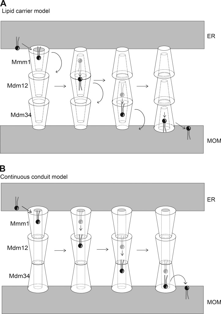
Figure 7. Models of phospholipid transfer by ERMES between the ER and mitochondrial OMs. (A) Lipid carrier model assumes that Mmm1, Mdm12, and Mdm34 have a hydrophobic lipid-binding pocket with an outlet only at the head of the molecules for binding to a phospholipid molecule. They pass the lipid molecule sequentially from Mmm1 at the ER membrane to Mdm34 at the mitochondrial OM via Mdm12 for the lipid transport from the ER to mitochondria. To achieve efficient transfer of lipid molecules between each ERMES component, they have to change their orientations and positions relative to the other components to put the outlet of the lipid-binding pocket of one component close to the one of the other component. (B) The continuous conduit model assumes the presence of the hydrophobic lipid-binding tunnels with two outlets at the head and tail of the molecules of Mmm1, Mdm12, and Mdm34. These tunnels are connected with each other to form a continuous lipid-moving conduit running from Mmm1 to Mdm34 via Mdm12. For lipid transport from the ER to mitochondria, a lipid molecule extracted from the ER membrane by Mmm1 will enter the hydrophobic tunnel in Mmm1 and diffuse through the tunnel to reach the one in Mdm12 and then the one in Mdm34. Finally, the lipid molecule will be released from the other outlet of the tunnel of Mdm34 for insertion into the mitochondrial OM.
Figure 7. Models of phospholipid transfer by ERMES between the ER and mitochondrial OMs. (A) Lipid carrier model assumes that Mmm1, Mdm12, and Mdm34 have a hydrophobic lipid-binding pocket with an outlet only at the head of the molecules for binding to a phospholipid molecule. They pass the lipid molecule sequentially from Mmm1 at the ER membrane to Mdm34 at the mitochondrial OM via Mdm12 for the lipid transport from the ER to mitochondria. To achieve efficient transfer of lipid molecules between each ERMES component, they have to change their orientations and positions relative to the other components to put the outlet of the lipid-binding pocket of one component close to the one of the other component. (B) The continuous conduit model assumes the presence of the hydrophobic lipid-binding tunnels with two outlets at the head and tail of the molecules of Mmm1, Mdm12, and Mdm34. These tunnels are connected with each other to form a continuous lipid-moving conduit running from Mmm1 to Mdm34 via Mdm12. For lipid transport from the ER to mitochondria, a lipid molecule extracted from the ER membrane by Mmm1 will enter the hydrophobic tunnel in Mmm1 and diffuse through the tunnel to reach the one in Mdm12 and then the one in Mdm34. Finally, the lipid molecule will be released from the other outlet of the tunnel of Mdm34 for insertion into the mitochondrial OM.CC BY-NC-SA · PubMed Central

Verification

Claims the paper makes

The claims and statistics of this paper haven’t been checked yet.

Run the check — free, no account needed. It reads the open-access text and checks the sources it cites.

A few free checks a day · papers already verified open instantly · results are public

Claims other papers make about this one

Each is checked against this paper’s own text: does it say what the citing paper says it does?

Self-check

Percentages, sums and reported differences re-computed from the paper’s own numbers.

The self-check needs the paper’s full text.

Citations

Cites

Show all 58 references

Cited by

Sorted by citations

See all 154

Metadata

Authors
Kawano, S., Tamura, Y., Kojima, R., Bala, S., Asai, E., Michel, A. H., Kornmann, B., Riezman, I., Riezman, H., Sakae, Y., Okamoto, Y., & Endo, T.
Affiliations
  • Kyoto Sangyo University (JP)
  • Yamagata University (JP)
  • Nagoya University (JP)
  • ETH Zurich (CH)
  • Institute of Biochemistry, ETH Zürich, Zürich, Switzerland 9
  • University of Geneva (CH)
Corresponding author
Endo, Toshiya
Venue
Journal of Cell Biology
Published
2017-12-26
Volume
217
Issue
3
Pages
959-974
ISSN
  • 0021-9525 (ISSN-L)
  • 1540-8140
OpenAlex
W2777157902
Cited by
148
References
58
Citations by year
2026: 12 · 2025: 11 · 2024: 20 · 2023: 11 · 2022: 14 · 2021: 17 · 2020: 23 · 2019: 26 · 2018: 14
FWCI
8.72
Type
journal article
Language
en
Open access
Locations
  • The Journal of Cell Biology (publishedVersion, cc-by-nc-sa)
  • Europe PMC (PubMed Central) (publishedVersion, cc-by-nc-sa)
  • PubMed Central (publishedVersion, cc-by-nc-sa)
  • PubMed (publishedVersion)
  • Archive ouverte UNIGE (University of Geneva) (publishedVersion, other-oa)
  • Archive ouverte UNIGE (University of Geneva) (submittedVersion, cc-by)
  • Repository for Publications and Research Data (ETH Zurich) (submittedVersion, cc-by-nc-sa)
  • ETH Zürich Research Collection
Funders
  • Japan Society of the Promotion of Science (25840020, 17K18230, 15H05595, 15H05705, 2222703)
  • Japan Science and Technology Agency
  • Core Research for Evolutional Science and Technology (JPMJCR12M1)
  • Swiss National Science Foundation
  • National Centre of Competence in Research
Topics
  • Life Sciences › Biochemistry, Genetics and Molecular Biology › Biochemistry › Lipid metabolism and biosynthesis
  • Life Sciences › Biochemistry, Genetics and Molecular Biology › Cell Biology › Cellular transport and secretion
  • Life Sciences › Biochemistry, Genetics and Molecular Biology › Molecular Biology › Mitochondrial Function and Pathology
Keywords
ERMES, lipid transfer, endoplasmic reticulum-mitochondria contacts, phosphatidylserine transport, glycerophospholipid biosynthesis, Kluyveromyces lactis, Saccharomyces cerevisiae, protein structure, membrane lipid exchange
MeSH terms
Biological Transport, Active, Endoplasmic Reticulum, Kluyveromyces, Membrane Proteins, Phospholipids, Saccharomyces cerevisiae, Structure-Activity Relationship, Mitochondrial Proteins, Saccharomyces cerevisiae Proteins, Multiprotein Complexes, Mitochondrial Membranes

About these checks

Registry, retraction, Cites and statistics results are computed, not judged, with one exception: a printed reference that no index lists is read once by a model before it counts as a citation error. Claims are extracted by a model and kept only when their quote appears verbatim in the text; each cited source is then checked, with a model’s help, for whether it says what the claim relies on. Those verdicts can be wrong — read the claim’s quote and the cited source before relying on one.

Statistics and arithmetic are recomputed, not judged.

Confidence bands are shown only for fields calibrated on our annotated test set. None is calibrated yet, so no band is shown.

What counts as a claim ›How we measure confidence ›How we check this ›

Are you an author, or do you see a problem with this page? Request a review