

The DOI matches its Crossref record; 32 of the 33 references it cites were found in a registry or an index.
Characterization of Human CD8+ T Cells Reactive with Mycobacterium tuberculosis–infected Antigen-presenting Cells
Open accessAbstract
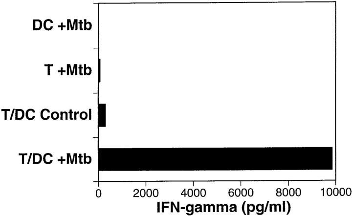
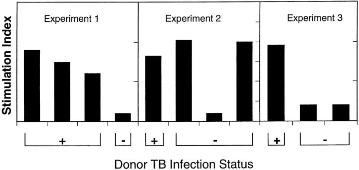
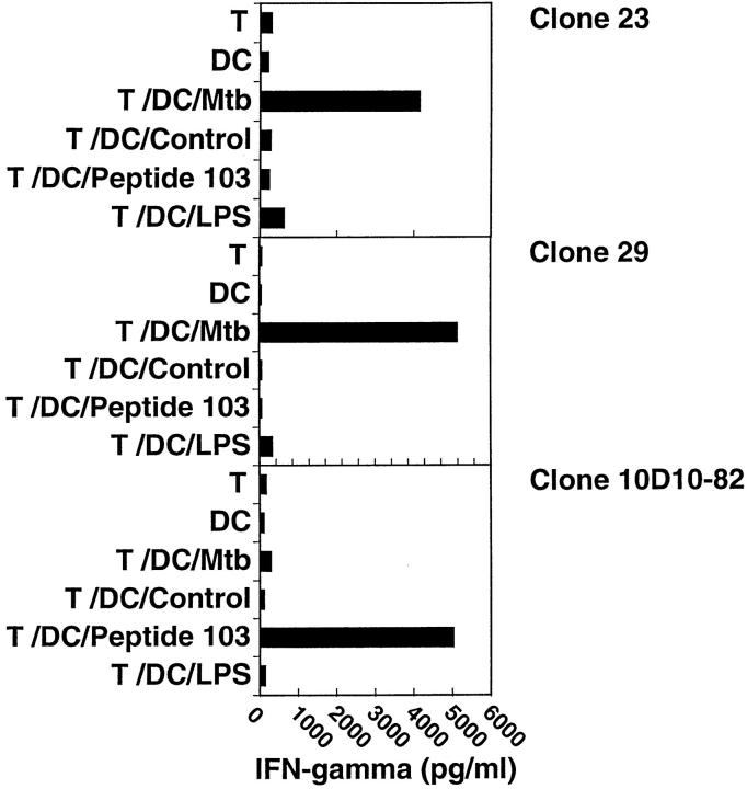
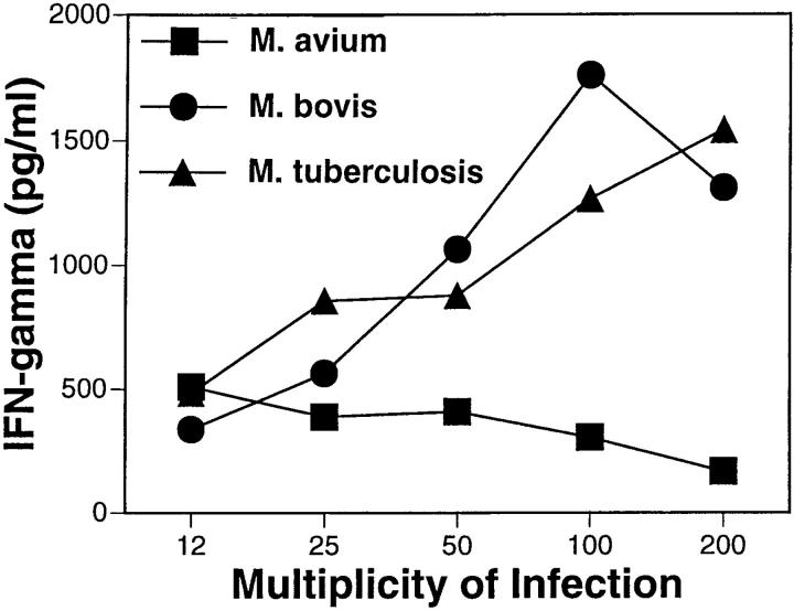
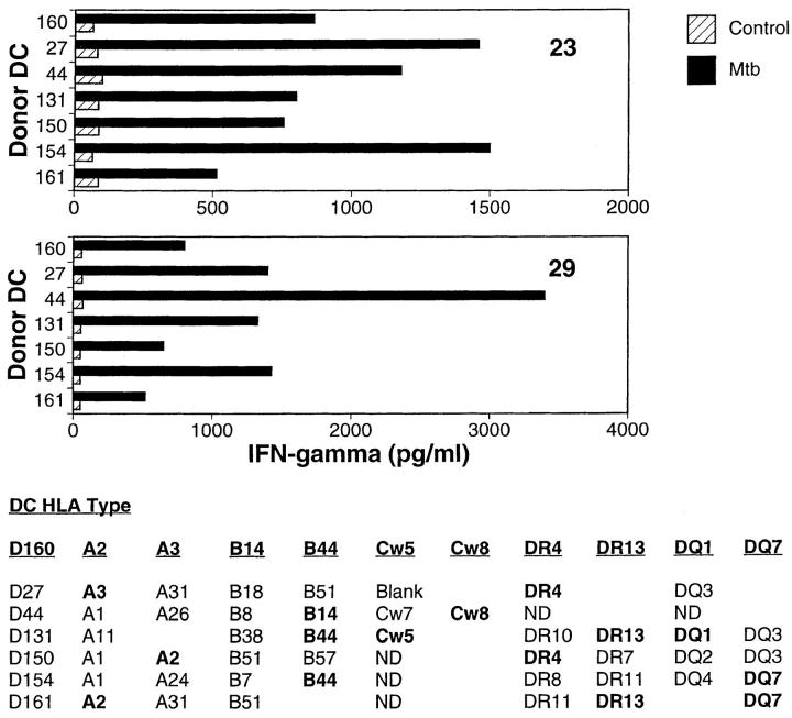
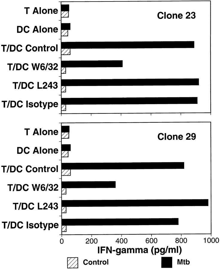
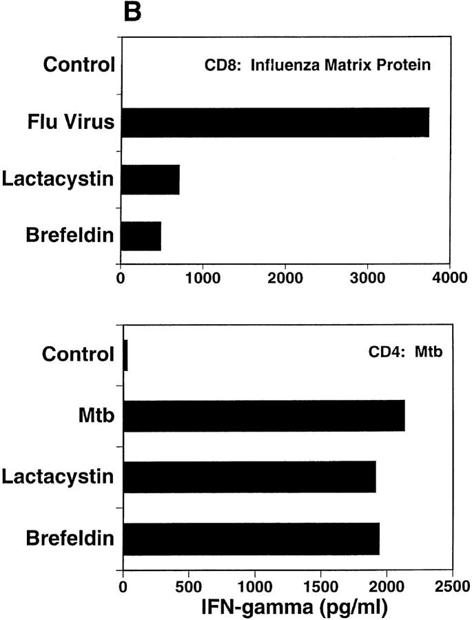
Previous studies in murine models, including those using the β2 microglobulin knockout mouse, have suggested an important role for CD8+ T cells in host defense to Mycobacterium tuberculosis (Mtb). At present, little is understood about these cells in the human immune response to tuberculosis. This report demonstrates the existence of human Mtb-reactive CD8+ T cells. These cells are present preferentially in persons infected with Mtb and produce interferon γ in response to stimulation with Mtb-infected target cells. Recognition of Mtb-infected cells by these CD8+ T cells is restricted neither by the major histocompatibility complex (MHC) class I A, B, or C alleles nor by CD1, although it is inhibited by anti–MHC class I antibody. The Mtb-specific CD8+ T cells recognize an antigen which is generated in the proteasome, but which does not require transport through the Golgi-ER. The data suggest the possible use of nonpolymorphic MHC class Ib antigen presenting structures other than CD1.
Overview
The claims and statistics of this paper haven’t been checked yet.
Run the check — free, no account needed. It reads the open-access text and checks the sources it cites.
Integrity signals
Registry
Cites
Statistics
Figures
Verification
Claims the paper makes
The claims and statistics of this paper haven’t been checked yet.
Run the check — free, no account needed. It reads the open-access text and checks the sources it cites.
Claims other papers make about this one
Each is checked against this paper’s own text: does it say what the citing paper says it does?
In the citing paper
“We demonstrate that bacterial HLA-I and HLA-II peptides can be presented by both antigen-presenting cells (as has previously been shown25–28) and by melanoma cells”
From Identification of bacteria-derived HLA-bound peptides in melanoma (2021) · cites this paper as [28]
Self-check
Percentages, sums and reported differences re-computed from the paper’s own numbers.
The self-check needs the paper’s full text.
Citations
Cites
An essential role for interferon gamma in resistance to Mycobacterium tuberculosis infection.
Flynn, J. L., Chan, J., Triebold, K. J., Dalton, D. K., Stewart, T. A., & Bloom, B. R. (1993) · The Journal of Experimental Medicine · doi:10.1084/jem.178.6.2249 · Cited by 2,468
Proliferating dendritic cell progenitors in human blood.
Romani, N., Grüner, S., Brang, D., Kämpgen, E., Lenz, A., Trockenbacher, B., Konwalinka, G., Fritsch, P. O., Steinman, R. M., & Schuler, G. (1994) · The Journal of Experimental Medicine · doi:10.1084/jem.180.1.83 · Cited by 1,841
Flynn, J. L., Goldstein, M. M., Chan, J., Triebold, K. J., Pfeffer, K., Lowenstein, C. J., Schrelber, R., Mak, T. W., & Bloom, B. R. (1995) · Immunity · doi:10.1016/1074-7613(95)90001-2 · Cited by 1,678
Recognition of a lipid antigen by CD1-restricted αβ+ T cells
Beckman, E. M., Porcelli, S. A., Morita, C. T., Behar, S. M., Furlong, S. T., & Brenner, M. B. (1994) · Nature · doi:10.1038/372691a0 · Cited by 977
The global tuberculosis situation and the new control strategy of the World Health Organization
Kochi, A. (1991) · Tubercle · doi:10.1016/0041-3879(91)90017-m · Cited by 849
Flynn, J. L., Goldstein, M. M., Triebold, K. J., Koller, B. H., & Bloom, B. R. (1992) · Proceedings of the National Academy of Sciences · doi:10.1073/pnas.89.24.12013 · Cited by 793
Immunogenicity and protective efficacy of a tuberculosis DNA vaccine
Huygen, K., Content, J., Denis, O., Montgomery, D. L., Yawman, A. M., Deck, R. R., DeWitt, C. M., Orme, I. M., Baldwin, S. L., D'Souza, C. D., et al. (1996) · Nature Medicine · doi:10.1038/nm0896-893 · Cited by 628
Phagocytic processing of bacterial antigens for class I MHC presentation to T cells
Pfeifer, J. D., Wick, M. J., Roberts, R. L., Findlay, K., Normark, S. J., & Harding, C. V. (1993) · Nature · doi:10.1038/361359a0 · Cited by 615 · cited 2×
“However, particulate antigens have been shown to gain access to the MHC class I pathway (18, 19), although the efficiency of these pathways remains controversial (20).”
BackgroundKovacsovics-Bankowski, M., Clark, K., Benacerraf, B., & Rock, K. L. (1993) · Proceedings of the National Academy of Sciences · doi:10.1073/pnas.90.11.4942 · Cited by 601
CDlb restricts the response of human CD4−8−T lymphocytes to a microbial antigen
Porcelli, S. A., Morita, C. T., & Brenner, M. B. (1992) · Nature · doi:10.1038/360593a0 · Cited by 572
Cited by
Sorted by citations
Flynn, J., & Chan, J. (2003) · Annual Review of Immunology · doi:10.1146/annurev.immunol.19.1.93 · Cited by 1,144
How can immunology contribute to the control of tuberculosis?
Kaufmann, S. H. E. (2001) · Nature reviews. Immunology · doi:10.1038/35095558 · Cited by 727
Innate Immunity toMycobacterium tuberculosis
van Crevel, R., Ottenhoff, T. H. M., & van der Meer, J. W. M. (2002) · Clinical Microbiology Reviews · doi:10.1128/cmr.15.2.294-309.2002 · Cited by 602
Guinn, K. M., Hickey, M. J., Mathur, S. K., Zakel, K. L., Grotzke, J. E., Lewinsohn, D. M., Smith, S., & Sherman, D. R. (2004) · Molecular Microbiology · doi:10.1046/j.1365-2958.2003.03844.x · Cited by 508
Self-citationCaruso, A. M., Serbina, N. V., Klein, E. C., Triebold, K. J., Bloom, B. R., & Flynn, J. L. (1999) · PubMed · Cited by 489
Metadata
- Authors
- Lewinsohn, D. M., Alderson, M. R., Briden, A. L., Riddell, S. R., Reed, S. G., & Grabstein, K. H.
- Affiliations
- Infectious Disease Research Institute (US)
- Corresponding author
- Lewinsohn, David M.
- Venue
- The Journal of Experimental Medicine
- Publisher
- Rockefeller University Press
- Published
- 1998-05-18
- Volume
- 187
- Issue
- 10
- Pages
- 1633-1640
- ISSN
- 0022-1007 (ISSN-L)
- 1540-9538
- PMC
- PMC2212289
- OpenAlex
- W2111354206
- Cited by
- 165
- References
- 33
- Citations by year
- 2026: 2 · 2025: 3 · 2022: 3 · 2021: 1 · 2020: 2 · 2019: 2 · 2018: 3 · 2017: 2 · 2016: 6 · 2015: 1 · 2014: 2 · 2013: 1 · 2012: 6
- FWCI
- 16.05
- Type
- journal article
- Language
- en
- Open access
- green
- www.ncbi.nlm.nih.gov
- Licence
- cc-by-nc-sa
- Locations
- The Journal of Experimental Medicine (publishedVersion)
- PubMed Central (publishedVersion, cc-by-nc-sa)
- PubMed (publishedVersion)
- Funders
- National Heart, Lung, and Blood Institute (5T32 HL-07287, T32HL007287)
- Topics
- Health Sciences › Medicine › Infectious Diseases › Tuberculosis Research and Epidemiology
- Life Sciences › Immunology and Microbiology › Immunology › Immunotherapy and Immune Responses
- Life Sciences › Immunology and Microbiology › Immunology › T-cell and B-cell Immunology
- Keywords
- CD8+ T cells, Mycobacterium tuberculosis, tuberculosis, interferon-gamma, MHC class I restriction, MHC class Ib, antigen presentation, human immune response
- MeSH terms
- Acetylcysteine, Amino Acid Sequence, Animals, Chloroquine, Humans, Lymphocyte Cooperation, Molecular Sequence Data, Mycobacterium tuberculosis, Tuberculosis, Cysteine Proteinase Inhibitors, Antigen Presentation, CD8-Positive T-Lymphocytes, Mice
About these checks
Registry, retraction, Cites and statistics results are computed, not judged, with one exception: a printed reference that no index lists is read once by a model before it counts as a citation error. Claims are extracted by a model and kept only when their quote appears verbatim in the text; each cited source is then checked, with a model’s help, for whether it says what the claim relies on. Those verdicts can be wrong — read the claim’s quote and the cited source before relying on one.
Statistics and arithmetic are recomputed, not judged.
Confidence bands are shown only for fields calibrated on our annotated test set. None is calibrated yet, so no band is shown.
What counts as a claim ›How we measure confidence ›How we check this ›
Are you an author, or do you see a problem with this page? Request a review
