

The DOI matches its Crossref record; 67 of the 68 references it cites were found in a registry or an index.
Involvement of Guanosine Triphosphatases and Phospholipase C-γ2 in Extracellular Signal–regulated Kinase, c-Jun NH2-terminal Kinase, and p38 Mitogen-activated Protein Kinase Activation by the B Cell Antigen Receptor
Open accessAbstract
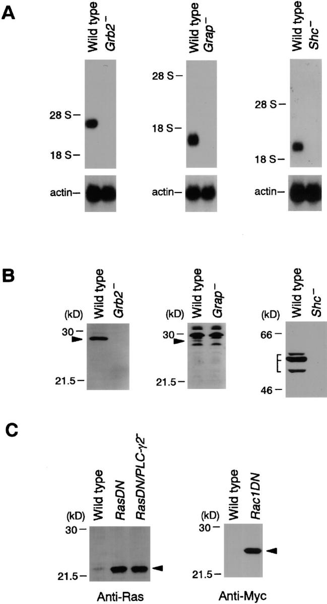
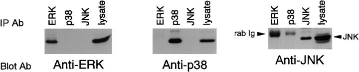
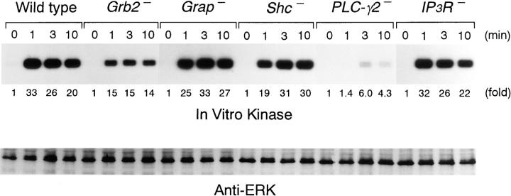
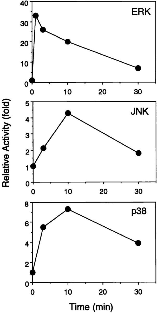
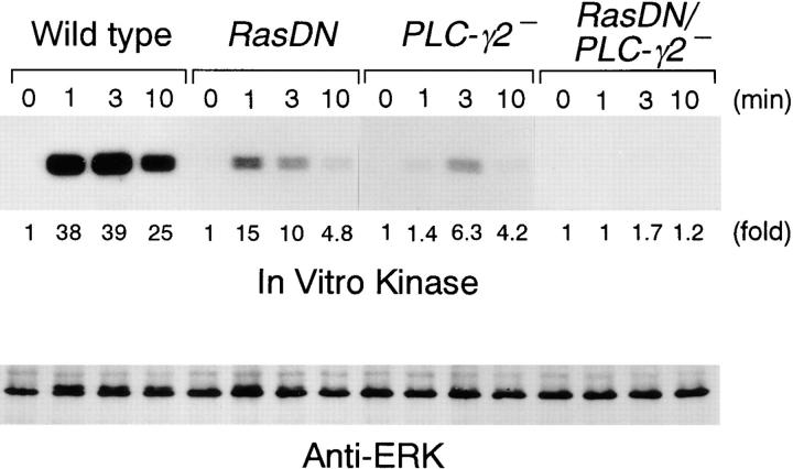
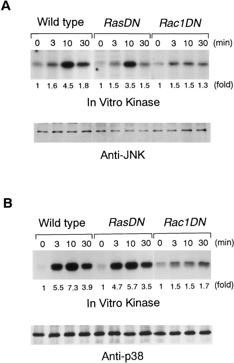
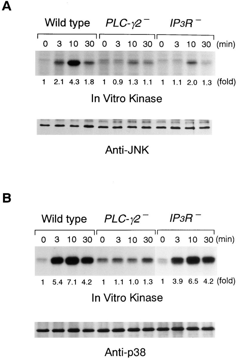
Mitogen-activated protein (MAP) kinase family members, including extracellular signal–regulated kinase (ERK), c-Jun NH2-terminal kinase ( JNK), and p38 MAP kinase, have been implicated in coupling the B cell antigen receptor (BCR) to transcriptional responses. However, the mechanisms that lead to the activation of these MAP kinase family members have been poorly elucidated. Here we demonstrate that the BCR-induced ERK activation is reduced by loss of Grb2 or expression of a dominant-negative form of Ras, RasN17, whereas this response is not affected by loss of Shc. The inhibition of the ERK response was also observed in phospholipase C (PLC)-γ2–deficient DT40 B cells, and expression of RasN17 in the PLC-γ2–deficient cells completely abrogated the ERK activation. The PLC-γ2 dependency of ERK activation was most likely due to protein kinase C (PKC) activation rather than calcium mobilization, since loss of inositol 1,4,5-trisphosphate receptors did not affect ERK activation. Similar to cooperation of Ras with PKC activation in ERK response, both PLC-γ2–dependent signal and GTPase are required for BCR-induced JNK and p38 responses. JNK response is dependent on Rac1 and calcium mobilization, whereas p38 response requires Rac1 and PKC activation.
Overview
The claims and statistics of this paper haven’t been checked yet.
Run the check — free, no account needed. It reads the open-access text and checks the sources it cites.
Integrity signals
Registry
Cites
Statistics
Figures
Verification
Claims the paper makes
The claims and statistics of this paper haven’t been checked yet.
Run the check — free, no account needed. It reads the open-access text and checks the sources it cites.
Claims other papers make about this one
Each is checked against this paper’s own text: does it say what the citing paper says it does?
The cited source says what the claim attributes to it.
In the citing paper
“In addition to the production of IP3, DAG, and calcium flux, PLCγ2 activation results in activation of phospholipase D and cell spreading (12, 13, 14, 15, 16, 17), bringing on other vital cell signaling processes such as Erk (18).”
From The role of PLCγ2 in immunological disorders, cancer, and neurodegeneration (2021) · cites this paper as [18]
Self-check
Percentages, sums and reported differences re-computed from the paper’s own numbers.
The self-check needs the paper’s full text.
Citations
Cites
Calcium's Role in Mechanotransduction during Muscle Development
Damm, T. B., & Egli, M. (2014) · Cellular Physiology and Biochemistry · doi:10.1159/000356667 · Cited by 14,552
Differential activation of transcription factors induced by Ca2+ response amplitude and duration
Dolmetsch, R. E., Lewis, R. S., Goodnow, C. C., & Healy, J. I. (1997) · Nature · doi:10.1038/386855a0 · Cited by 1,763
The SH2 and SH3 domain-containing protein GRB2 links receptor tyrosine kinases to ras signaling
Lowenstein, E. J., Daly, R. J., Batzer, A., Li, W., Margolis, B. L., Lammers, R., Ullrich, A., Skolnik, E. Y., Bar‐Sagi, D., & Schlessinger, J. (1992) · Cell · doi:10.1016/0092-8674(92)90167-b · Cited by 1,632
The small GTP-binding proteins Rac1 and Cdc42regulate the activity of the JNK/SAPK signaling pathway
Coso, O. A., Chiariello, M., Yu, J., Teramoto, H., Crespo, P., Xu, N., Miki, T., & Gutkind, J. S. (1995) · Cell · doi:10.1016/s0092-8674(05)80018-2 · Cited by 1,629
Differential activation of transcription factors induced by Ca2+ response amplitude and duration
Dolmetsch, R. E., Lewis, R. S., Goodnow, C. C., & Healy, J. I. (1997) · Nature · doi:10.1038/40912 · Cited by 1,521
Minden, A., Lin, A., Claret, F.-X., Abo, A., & Karin, M. (1995) · Cell · doi:10.1016/s0092-8674(05)80019-4 · Cited by 1,481
A novel transforming protein (SHC) with an SH2 domain is implicated in mitogenic signal transduction
Pelicci, G., Lanfrancone, L., Grignani, F., McGlade, J. C., Cavallo, F., Forni, G., Nicoletti, I., Grignani, F., Pawson, T. J., & Pelicci, P. G. (1992) · Cell · doi:10.1016/0092-8674(92)90536-l · Cited by 1,326
Zhang, W., Sloan‐Lancaster, J., Kitchen, J., Trible, R. P., & Samelson, L. E. (1998) · Cell · doi:10.1016/s0092-8674(00)80901-0 · Cited by 1,271
The Rho family GTPases RhoA, Racl , and CDC42Hsregulate transcriptional activation by SRF
Hill, C. S., Wynne, J., & Treisman, R. (1995) · Cell · doi:10.1016/s0092-8674(05)80020-0 · Cited by 1,269
Egan, S. E., Giddings, B. W., Brooks, M. W., Buday, L., Sizeland, A. M., & Weinberg, R. A. (1993) · Nature · doi:10.1038/363045a0 · Cited by 1,200
Cited by
Sorted by citations
The p38 signal transduction pathway Activation and function
Ono, K., & Han, J. (2000) · Cellular Signalling · doi:10.1016/s0898-6568(99)00071-6 · Cited by 1,571
Role of Bruton’s tyrosine kinase in B cells and malignancies
Pal Singh, S., Dammeijer, F., & Hendriks, R. W. (2018) · Molecular Cancer · doi:10.1186/s12943-018-0779-z · Cited by 726
Roles of Fc receptors in autoimmunity
Takai, T. (2002) · Nature reviews. Immunology · doi:10.1038/nri856 · Cited by 627
Regulation of B-cell fate by antigen-receptor signals
Niiro, H., & Clark, E. A. (2002) · Nature reviews. Immunology · doi:10.1038/nri955 · Cited by 654
Self-citationTargeting Bruton's tyrosine kinase in B cell malignancies
Hendriks, R. W., Yuvaraj, S., & Kil, L. P. (2014) · Nature reviews. Cancer · doi:10.1038/nrc3702 · Cited by 580
Metadata
- Authors
- Hashimoto, A., Okada, H., Jiang, A., Kurosaki, M., Greenberg, S., Clark, E. A., & Kurosaki, T.
- Affiliations
- Kansai Medical University (JP)
- Corresponding author
- Kurosaki, Tomohiro
- Venue
- The Journal of Experimental Medicine
- Publisher
- Rockefeller University Press
- Published
- 1998-10-05
- Volume
- 188
- Issue
- 7
- Pages
- 1287-1295
- ISSN
- 0022-1007 (ISSN-L)
- 1540-9538
- PMC
- PMC2212492
- OpenAlex
- W1965678722
- Cited by
- 199
- References
- 68
- Citations by year
- 2026: 1 · 2025: 2 · 2024: 1 · 2021: 4 · 2020: 7 · 2019: 3 · 2018: 8 · 2017: 4 · 2016: 4 · 2015: 8 · 2014: 6 · 2013: 5 · 2012: 2
- FWCI
- 5.59
- Type
- journal article
- Language
- en
- Open access
- green
- www.ncbi.nlm.nih.gov
- Licence
- cc-by-nc-sa
- Locations
- The Journal of Experimental Medicine (publishedVersion)
- PubMed Central (publishedVersion, cc-by-nc-sa)
- PubMed (publishedVersion)
- Funders
- National Institutes of Health (5p01gm042508-09)
- National Institute of General Medical Sciences (GM42508, GM-37905, R01GM037905)
- Topics
- Life Sciences › Biochemistry, Genetics and Molecular Biology › Molecular Biology › Protein Kinase Regulation and GTPase Signaling
- Life Sciences › Biochemistry, Genetics and Molecular Biology › Molecular Biology › Protein Tyrosine Phosphatases
- Life Sciences › Biochemistry, Genetics and Molecular Biology › Cancer Research › NF-κB Signaling Pathways
- Keywords
- B cell receptor, extracellular signal-regulated kinase, c-Jun NH2-terminal kinase, p38 MAPK, PLCγ2, Ras, Grb2, Shc, protein kinase C, Rac1, calcium mobilization, IP3 receptor, MAPK activation, signal transduction, B cell signaling
- MeSH terms
- Animals, Cell Line, Chickens, Enzyme Activation, Isoenzymes, Type C Phospholipases, Protein Kinase C, Proteins, Receptors, Antigen, B-Cell, Signal Transduction, Calcium-Calmodulin-Dependent Protein Kinases, GTP-Binding Proteins, Mitogen-Activated Protein Kinase 1, GTP Phosphohydrolases, rac GTP-Binding Proteins, Mitogen-Activated Protein Kinases, JNK Mitogen-Activated Protein Kinases, p38 Mitogen-Activated Protein Kinases, Adaptor Proteins, Signal Transducing, GRB2 Adaptor Protein, Phospholipase C gamma
About these checks
Registry, retraction, Cites and statistics results are computed, not judged, with one exception: a printed reference that no index lists is read once by a model before it counts as a citation error. Claims are extracted by a model and kept only when their quote appears verbatim in the text; each cited source is then checked, with a model’s help, for whether it says what the claim relies on. Those verdicts can be wrong — read the claim’s quote and the cited source before relying on one.
Statistics and arithmetic are recomputed, not judged.
Confidence bands are shown only for fields calibrated on our annotated test set. None is calibrated yet, so no band is shown.
What counts as a claim ›How we measure confidence ›How we check this ›
Are you an author, or do you see a problem with this page? Request a review
