

The DOI matches its Crossref record; 58 of the 58 references it cites were found in a registry or an index.
The Serpin Proteinase Inhibitor 9 Is an Endogenous Inhibitor of Interleukin 1β–Converting Enzyme (Caspase-1) Activity in Human Vascular Smooth Muscle Cells
Open accessAbstract
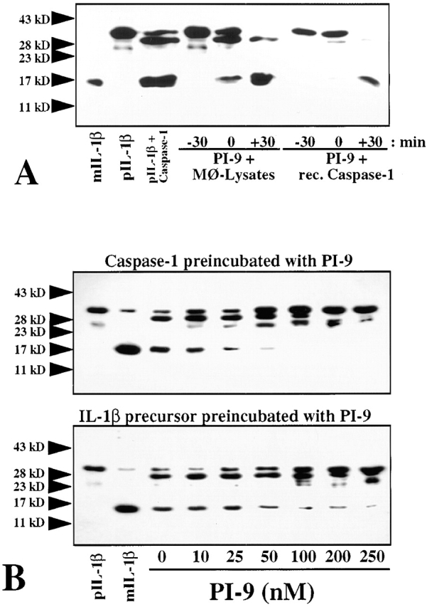
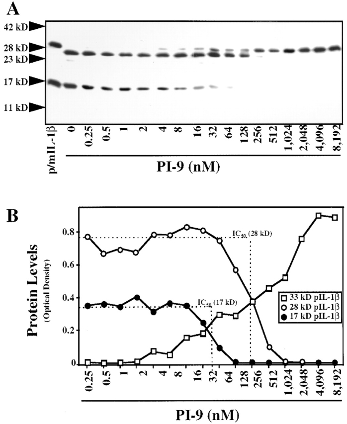
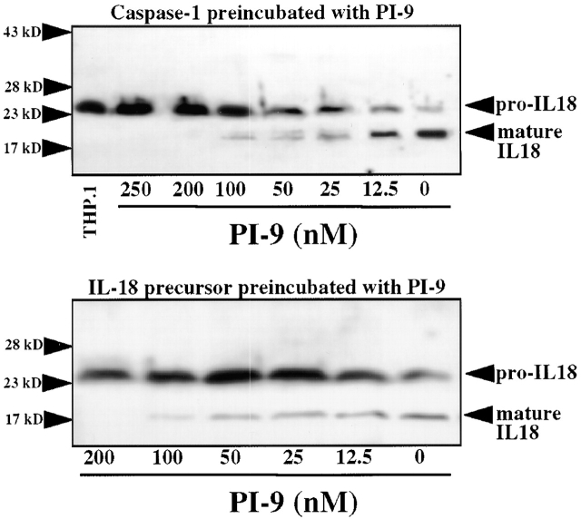
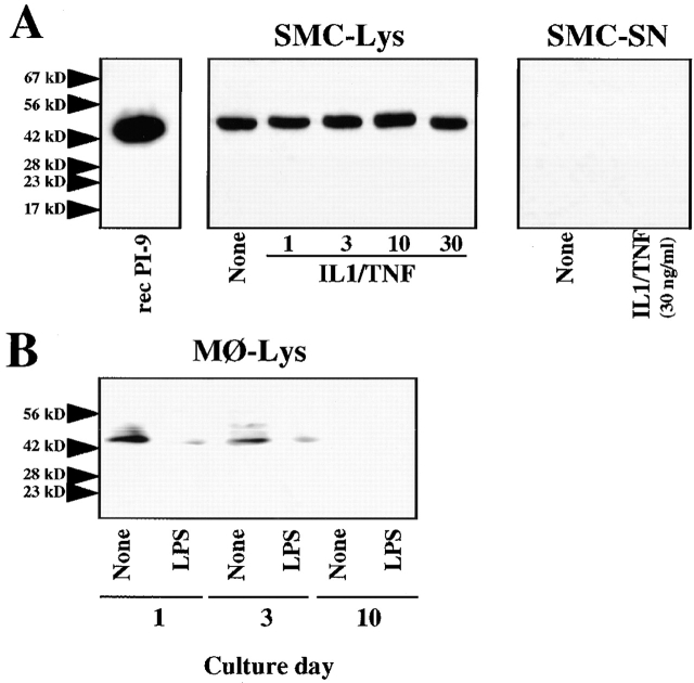
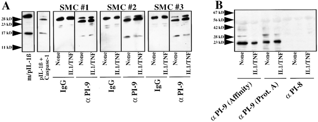
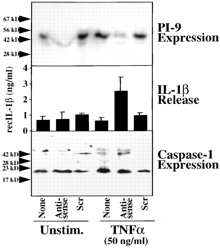
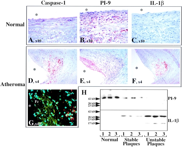
Interleukin-1beta-converting enzyme (ICE, caspase-1) regulates key steps in inflammation and immunity, by activating the proinflammatory cytokines interleukin (IL-)1beta and IL-18, or mediating apoptotic processes. We recently provided evidence for the regulation of caspase-1 activity via an endogenous inhibitor expressed by human vascular smooth muscle cells (SMCs) (Schönbeck, U., M. Herzberg, A. Petersen, C. Wohlenberg, J. Gerdes, H.-D. Flad, and H. Loppnow. 1997. J. Exp. Med. 185:1287-1294). However, the molecular identity of this endogenous inhibitor remained undefined. We report here that the serine proteinase inhibitor (serpin) PI-9 accounts for the endogenous caspase-1 inhibitory activity in human SMCs and prevents processing of the enzyme's natural substrates, IL-1beta and IL-18 precursor. Treatment of SMC lysates with anti-PI-9 antibody abrogated the caspase-1 inhibitory activity and coprecipitated the enzyme, demonstrating protein-protein interaction. Furthermore, PI-9 antisense oligonucleotides coordinately reduced PI-9 expression and promoted IL-1beta release. Since SMCs comprise the majority of cells in the vascular wall, and because IL-1 is implicated in atherogenesis, we tested the biological validity of our in vitro findings within human atheroma in situ. The unaffected arterial wall contains abundant and homogeneously distributed PI-9. In human atherosclerotic lesions, however, PI-9 expression correlated inversely with immunoreactive IL-1beta, supporting a potential role of the endogenous caspase-1 inhibitor in this chronic inflammatory disease. Thus, our results provide new insights into the regulation of this enzyme involved in immune and inflammatory processes of chronic inflammatory diseases, and point to an endogenous antiinflammatory action of PI-9, dysregulated in a prevalent human disease.
Overview
The claims and statistics of this paper haven’t been checked yet.
Run the check — free, no account needed. It reads the open-access text and checks the sources it cites.
Integrity signals
Registry
Cites
Statistics
Figures
Verification
Claims the paper makes
The claims and statistics of this paper haven’t been checked yet.
Run the check — free, no account needed. It reads the open-access text and checks the sources it cites.
Claims other papers make about this one
Each is checked against this paper’s own text: does it say what the citing paper says it does?
In the citing paper
“In addition, other inhibitors such as PI-9 act by directly binding and inhibiting caspase-1 (40,41).”
From Sustained Inflammasome Activity in Macrophages Impairs Wound Healing in Type 2 Diabetic Humans and Mice (2014) · cites this paper as [40]
Self-check
Percentages, sums and reported differences re-computed from the paper’s own numbers.
The self-check needs the paper’s full text.
Citations
Cites
Atherosclerosis--an inflammatory disease
Ross, R. (1999) · New England Journal of Medicine · doi:10.1056/nejm199901143400207 · Cited by 21,725
The pathogenesis of atherosclerosis: a perspective for the 1990s
Ross, R. (1993) · Nature · doi:10.1038/362801a0 · Cited by 10,618
Isolation of mononuclear cells and granulocytes from human blood.
Bøyum, A. (1968) · Scandinavian Journal of Clinical and Laboratory Investigation · Cited by 9,585
Apoptosis in the Pathogenesis and Treatment of Disease
Thompson, C. B. (1995) · Science · doi:10.1126/science.7878464 · Cited by 6,524
Biologic basis for interleukin-1 in disease.
Dinarello, C. (1996) · Blood · doi:10.1182/blood.v87.6.2095.bloodjournal8762095 · Cited by 4,344
The Glomerular Permeability Determined by Dextran Clearance Using Sephadex Gel Filtration
Mogensen, C. E. (1968) · Scandinavian Journal of Clinical and Laboratory Investigation · doi:10.3109/00365516809076979 · Cited by 2,654
A novel heterodimeric cysteine protease is required for interleukin-1βprocessing in monocytes
Thornberry, N. A., Bull, H. G., Calaycay, J. R., Chapman, K. T., Howard, A. D., Kostura, M. J., Miller, D. K., Molineaux, S. M., Weidner, J. R., Aunins, J. G., et al. (1992) · Nature · doi:10.1038/356768a0 · Cited by 2,613
Human ICE/CED-3 Protease Nomenclature
Alnemri, E. S., Livingston, D. J., Nicholson, D. W., Salvesen, G. S., Thornberry, N. A., Wong, W. W., & Yuan, J. (1996) · Cell · doi:10.1016/s0092-8674(00)81334-3 · Cited by 2,371
Cloning, sequence and expression of two distinct human interleukin-1 complementary DNAs
March, C. J., Mosley, B., Larsen, A., Cerretti, D. P., Braedt, G., Price, V. L., Gillis, S. A., Henney, C. S., Kronheim, S. R., Grabstein, K. H., et al. (1985) · Nature · doi:10.1038/315641a0 · Cited by 1,559
Research works printed but not linked: 2
- Ross, 1980, Morphogenesis of vascular smooth muscle in atherosclerosis and cell structure, 66
- Howard, 1991, IL-1-converting enzyme requires aspartic acid residues for processing of the IL-1 beta precursor at two distinct sites and does not cleave 31-kDa IL-1 alpha, J. Immunol., 147, 2964, 10.4049/jimmunol.147.9.2964 · DOI
Cited by
Sorted by citations
Items 101–133 of 133
Dicer regulates activation of the NLRP3 inflammasome
Ojcius, D. M., Jafari, A., Yeruva, L., Schindler, C., & Sater, A. A. A. (2019) · PLoS ONE · doi:10.1371/journal.pone.0215689 · Cited by 15
Virome and Inflammasomes, a Finely Tuned Balance with Important Consequences for the Host Health
Freer, G., Maggi, F., & Pistello, M. (2019) · Current Medicinal Chemistry · doi:10.2174/0929867324666171005112921 · Cited by 10
Pohjanen, V., Kokkonen, T. S., Arvonen, M., Augustin, M. A., Patankar, M., Turunen, S., Vähäsalo, P., & Karttunen, T. J. (2013) · International Journal of Immunopathology and Pharmacology · doi:10.1177/039463201302600408 · Cited by 10
Ogbourne, S. M., & Antalis, T. (2001) · Nucleic Acids Research · doi:10.1093/nar/29.19.3919 · Cited by 10
The Regulation and Consequences of Immune-Mediated Cell Death in Atheromatous Diseases
Choy, J. C., Podor, T. J., Yanagawa, B., Lai, J. C. K., Granville, D. J., Walker, D. C., & McManus, B. M. (2003) · Cardiovascular Toxicology · doi:10.1385/ct:3:3:269 · Cited by 9
Activación y regulación del inflamasoma NLRP3 en las enfermedades infecciosas
López, J. C. H., & Inchima, S. U. (2012) · IATREIA · doi:10.17533/udea.iatreia.13134 · Cited by 8
Tanaka, K., Harashima, N., Niiya, F., Miyagi, Y., Hida, N., Ochi, M., Imai, N., Harada, M., Itoh, K., & Shichijo, S. (2002) · Japanese Journal of Cancer Research · doi:10.1111/j.1349-7006.2002.tb01259.x · Cited by 8
Wågsäter, D., Jatta, K., Ocaya, P., Dimberg, J., & Sirsjö, A. (2006) · Journal of Vascular Research · doi:10.1159/000094258 · Cited by 8
Activation and Regulation of the NLRP3 Inflammasome
Hernández, J. C., Sirois, C. M., & Latz, E. (2011) · Springer eBooks · doi:10.1007/978-3-0348-0148-5_13 · Cited by 8
Huang, J., Zhu, L., Zhao, X., Wu, X., Yang, J., Xu, B., Zhao, Z. G., Gu, L., & Su, L. (2021) · Neurological Sciences · doi:10.1007/s10072-021-05418-5 · Cited by 7
Orikaza, C. M., Pessoa, C. C., Paladino, F. V., Florentino, P. T. V., Barbiéri, C. L., Goto, H., Ramos-Sanchez, E. M., da Silveira, J. F., Rabinovitch, M., Mortara, R. A., et al. (2020) · Infection and Immunity · doi:10.1128/iai.00261-20 · Cited by 7
Morales-Maldonado, A., Humphry, M., Figg, N. L., & Clarke, M. C. H. (2023) · Atherosclerosis · doi:10.1016/j.atherosclerosis.2023.117308 · Cited by 6
An Emerging Role for Serine Protease Inhibitors in T Lymphocyte Immunity and Beyond
Ashton‐Rickardt, P. G. (2012) · ISRN Immunology · doi:10.5402/2012/354365 · Cited by 5
Role for IL‐4 nonproducing NKT cells in CC‐chemokine ligand 2‐induced Th2 cell generation
Fujita, K., Kobayashi, M., Brutkiewicz, R. R., Hanafusa, T., Herndon, D. N., & Suzuki, F. (2005) · Immunology and Cell Biology · doi:10.1111/j.1440-1711.2005.01400.x · Cited by 4
Serpins in T Lymphocyte Immunity and Blood Development
Ashton‐Rickardt, P. G. (2015) · Springer eBooks · doi:10.1007/978-3-319-22711-5_7 · Cited by 3
Caspases: Activation, Regulation, and Function
Riedl, S. J., & Scott, F. L. (2009) · Humana Press eBooks · doi:10.1007/978-1-60327-381-7_1 · Cited by 2
Defining the Function of Pyrin, the Familial Mediterranean Fever-associated Protein, in Inflammation
Hesker, P. R. (2019) · Carolina Digital Repository (University of North Carolina at Chapel Hill) · doi:10.17615/z8zz-6k39 · Cited by 1
Gerdes, N. (2005) · Kölner Universitäts PublikationsServer (Universität zu Köln) · Cited by 1
Inflammation, Infection, and Homocysteine in Atherosclerosis
DeGraba, T. J. (2002) · CRC Press eBooks · doi:10.1201/noe1842141151.ch5 · Cited by 1
Roles in innate immune receptors: Host : pathogen interactions and strengthening vaccines
Nyström, S. (2013) · Figshare
IL-18 and IL-18 Receptor Knockout Mice
Tsutsui, H., Yoshimoto, T., Okamura, H., Akira, S., & Nakanishi, K. (2003) · doi:10.1007/978-1-59259-405-4_18
Mechanisms of inflammasome activation and inhibition during Yersinia infection
LaRock, C. N. (2012) · ResearchWorks at the University of Washington (University of Washington)
The extracellular role of granzyme B in abdominal aortic aneurysm
Ang, L. S. (2014) · cIRcle (University of British Columbia) · doi:10.14288/1.0165706
Stahnke, B., & Fischer, R. (2011) · Publikationsdatenbank der Fraunhofer-Gesellschaft (Fraunhofer-Gesellschaft)
Huang, M. T.-H. (2019) · Carolina Digital Repository (University of North Carolina at Chapel Hill) · doi:10.17615/cg1d-4t84
Cheema, A. K., Li, Y., Moulton, J., Girgis, M. J., Wise, S. Y., Carpenter, A. D., Fatanmi, O. O., & Singh, V. K. (2021) · bioRxiv (Cold Spring Harbor Laboratory) · doi:10.1101/2021.10.16.464661
Hansson, G. K. (2017) · doi:10.1093/med/9780198755777.003.0004
Established and novel methods for treating inflammatory diseases by inhibition of cytokines
Dinarello, C. A. (2007) · Springer eBooks · doi:10.1007/1-4020-4144-6_28
AlMalki, M., Pierri, C. R. D., Méric, T., Mkaouar, H., Slimani, D., Mariaule, V., Tebassi, S., Akermi, N., Maguin, E., Hernandez, J. L., et al. (2026) · Gut Microbes · doi:10.1080/19490976.2026.2726641
Young, J. L. (2008) · The Journal of the American Medical Association (JAMA) Network (American Medical Association) · doi:10.13028/4kma-sa11
Self-citation
Metadata
- Authors
- Young, J. L., Sukhova, G. K., Foster, D., Kisiel, W., Libby, P., & Schönbeck, U.
- Affiliations
- Brigham and Women's Hospital (US)
- bZymoGenetics, Incorporated, Seattle, Washington 93102
- University of New Mexico (US)
- Venue
- The Journal of Experimental Medicine
- Publisher
- Rockefeller University Press
- Published
- 2000-05-01
- Volume
- 191
- Issue
- 9
- Pages
- 1535-1544
- ISSN
- 0022-1007 (ISSN-L)
- 1540-9538
- PMC
- PMC2213432
- OpenAlex
- W2171074690
- Cited by
- 133
- References
- 58
- Citations by year
- 2026: 1 · 2024: 1 · 2023: 1 · 2022: 1 · 2021: 4 · 2020: 6 · 2019: 6 · 2018: 1 · 2017: 5 · 2016: 2 · 2015: 3 · 2014: 1 · 2013: 10 · 2012: 7
- FWCI
- 3.62
- Type
- journal article
- Language
- en
- Open access
- green
- www.ncbi.nlm.nih.gov
- Licence
- cc-by-nc-sa
- Locations
- The Journal of Experimental Medicine (publishedVersion)
- PubMed Central (publishedVersion, cc-by-nc-sa)
- PubMed (publishedVersion)
- Funders
- National Institutes of Health (5r01hl034636-23)
- National Heart, Lung, and Blood Institute (R37HL034636, R01-HL034636, HL-34636)
- Topics
- Life Sciences › Biochemistry, Genetics and Molecular Biology › Molecular Biology › Inflammasome and immune disorders
- Life Sciences › Biochemistry, Genetics and Molecular Biology › Cancer Research › Protease and Inhibitor Mechanisms
- Life Sciences › Biochemistry, Genetics and Molecular Biology › Molecular Biology › Cell death mechanisms and regulation
- Keywords
- caspase-1, IL-1β, IL-18, vascular smooth muscle cells, protein-protein interaction, atherosclerosis, inflammation, anti-inflammatory regulation, atherosclerotic plaque
- MeSH terms
- Arteries, Arteriosclerosis, Gene Expression Regulation, Humans, Interleukin-1, Interleukins, Muscle, Smooth, Vascular, Protein Precursors, Protein Processing, Post-Translational, Serpins, Interleukin-18, Caspase Inhibitors
About these checks
Registry, retraction, Cites and statistics results are computed, not judged, with one exception: a printed reference that no index lists is read once by a model before it counts as a citation error. Claims are extracted by a model and kept only when their quote appears verbatim in the text; each cited source is then checked, with a model’s help, for whether it says what the claim relies on. Those verdicts can be wrong — read the claim’s quote and the cited source before relying on one.
Statistics and arithmetic are recomputed, not judged.
Confidence bands are shown only for fields calibrated on our annotated test set. None is calibrated yet, so no band is shown.
What counts as a claim ›How we measure confidence ›How we check this ›
Are you an author, or do you see a problem with this page? Request a review
