The DOI matches its Crossref record; 60 of the 60 references it cites were found in a registry or an index.
Unique pathological tau conformers from Alzheimer’s brains transmit tau pathology in nontransgenic mice
Open accessAbstract
Filamentous tau aggregates are hallmark lesions in numerous neurodegenerative diseases, including Alzheimer’s disease (AD). Cell culture and animal studies showed that tau fibrils can undergo cell-to-cell transmission and seed aggregation of soluble tau, but this phenomenon was only robustly demonstrated in models overexpressing tau. In this study, we found that intracerebral inoculation of tau fibrils purified from AD brains (AD-tau), but not synthetic tau fibrils, resulted in the formation of abundant tau inclusions in anatomically connected brain regions in nontransgenic mice. Recombinant human tau seeded by AD-tau revealed unique conformational features that are distinct from synthetic tau fibrils, which could underlie the differential potency in seeding physiological levels of tau to aggregate. Therefore, our study establishes a mouse model of sporadic tauopathies and points to important differences between tau fibrils that are generated artificially and authentic ones that develop in AD brains.
Overview
The claims and statistics of this paper haven’t been checked yet.
Run the check — free, no account needed. It reads the open-access text and checks the sources it cites.
Integrity signals
Registry
Cites
Statistics
Figures
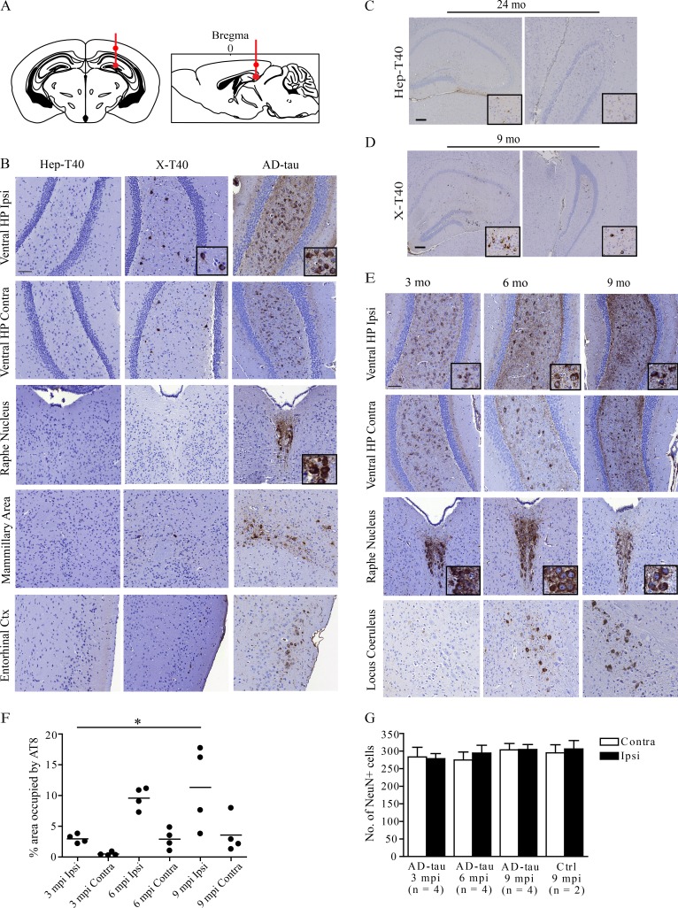
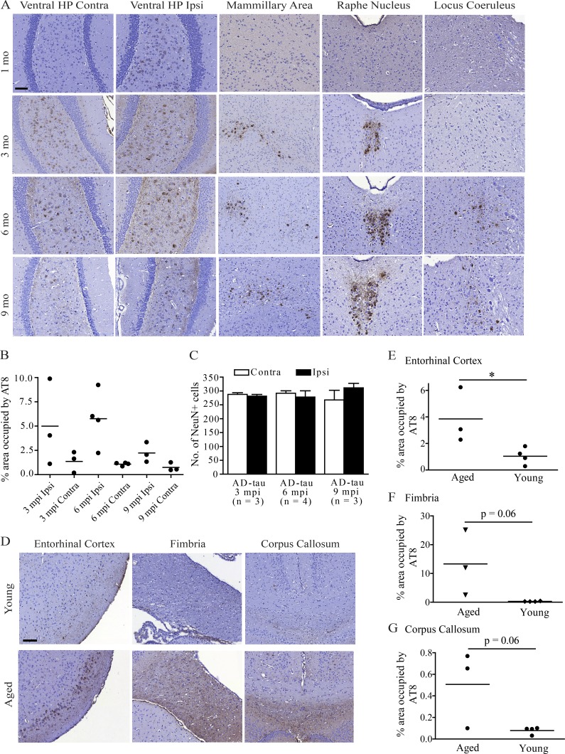
![Figure 3. AD-tau is a more potent seed for tau aggregation in non-Tg mice. (A) Schematics showing injection sites in the dorsal hippocampus and overlying cortex indicated by red dots (Bregma −2.54 mm, 2 mm from midline, and −1.4 mm from skull [for the cortex] and −2.4 mm from skull [for the hippocampus]). (B) Differential induction of tau pathology recognized by AT8 (a monoclonal antibody specific for tau phosphorylated at S202/T205) in non-Tg mice at 3 mo p.i. of different tau fibrils. Amount of tau injected per mouse: 9 µg for Hep-T40 (four mice) and X-T40 fibrils (six mice); 8 µg for AD-tau (four mice). Contra, contralateral; Ctx, cortex; HP, hippocampus; Ipsi, ipsilateral. Bar, 100 µm. (C) AT8 immunostaining at 24 mo p.i. of Hep-T40 fibrils (9 µg/mouse; two mice). (D) AT8 immunostaining at 9 mo p.i. of X-T40 fibrils (9 µg/mouse; three mice). Considerable variability was found among injected animals, with the one mouse developing the most abundant pathology shown here. (C and D) Bars, 200 µm. (E) AT8 immunostaining at 3, 6, and 9 mo p.i. of AD-tau (8 µg/mouse; four mice per time point). Bar, 100 µm. (F) Quantification of percent area occupied by AT8-positive tau pathology developed in the ventral hippocampal hilus with 8 µg AD-tau injection (four mice per time point; each dot represents one mouse). One-way ANOVA was performed across different time points for ipsilateral and contralateral pathology separately. Significant differences were found for ipsilateral pathology, and Tukey’s multiple comparison test identified a significant difference between 3 and 9 mo p.i. *, P < 0.05. mpi, months p.i. (G) AD-tau inoculation (8 µg/mouse) did not lead to significant neuron loss in the ventral hippocampal hilus region as compared with mice injected with control (Ctrl) brain extracts. Data are shown as mean + SEM. One-way ANOVA found no significant differences among the groups. Data shown in this figure were obtained from injections performed all on 2–3-mo-old non-Tg mice (C57BL6 or C57BL6/C3H F1). Refer to Table S4 for the details of animals analyzed per group.](/paper-figures/9aed7d9a91e54302a2ab3c5cf3403995/unique-pathological-tau-conformers-from-alzheimers-brains-transmit-tau-pathology-figure-3.jpg)


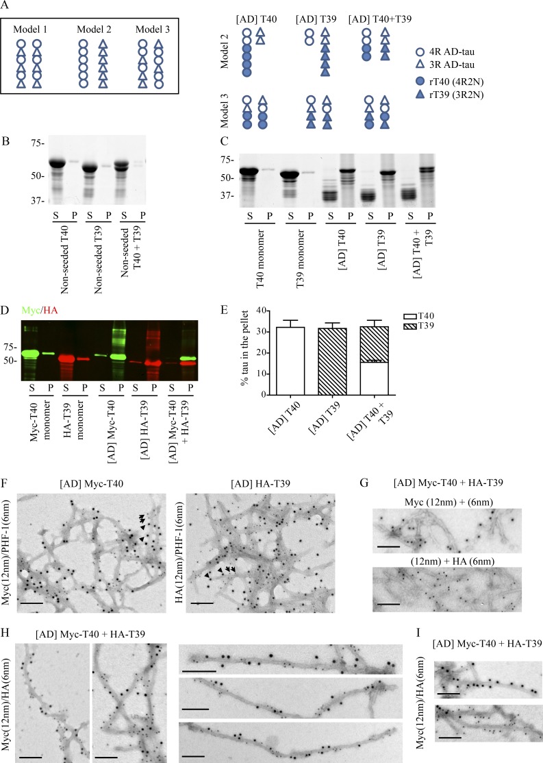
![Figure 7. rTau fibrils templated by AD-tau reveal structural relationship between 4R and 3R tau. (A) Three possible models of 4R/3R tau arrangement in AD PHFs (left) and proposed patterns of recombinant T40/T39 (rT40/rT39) recruitment by AD-tau based on models 2 and 3 (right). (B) Sedimentation test for nonseeded T40 and T39 after 3-d incubation at 37°C with constant agitation at 1,000 rpm. Cofibrillization was conducted at the same total protein concentration as single-isoform fibrillization with T40 and T39 mixed at a 1:1 ratio. S, supernatant; P, pellet. (C) Sedimentation test for singly or cofibrillized T40 and T39 seeded by 10% AD-tau ([AD]T40, [AD]T39, and [AD]T40 + T39) under the same fibrillization condition as in B. Monomers were used as the controls. rTau remaining in the soluble fraction appeared to be truncated. (D) Sedimentation test for singly or cofibrillized Myc-T40 and HA-T39 seeded by 10% AD-tau ([AD]Myc-T40, [AD]HA-T39, and [AD]Myc-T40 + HA-T39) under the same fibrillization condition as in B, with monomers as the controls. Supernatant and pellet fractions were immunoblotted with anti-Myc and anti-HA antibodies. These two antibodies do not label truncated tau in the supernatant as seen in C, suggesting the C terminus of tau was cleaved together with the tag. (B–D) Data shown are representative of at least two independent experiments. (E) Quantification for the percentage of starting tau monomers recruited into the pellet fraction with AD-tau seeding as shown in C and D. Data are displayed as mean + SEM and are based on three independent experiments. (F) Double-labeling immuno-EM images for [AD]Myc-T40 and [AD]HA-T39 fibrils, stained with PHF-1 and anti-Myc or anti-HA antibody. Secondary antibodies are conjugated to colloidal gold particles of two different sizes (6 nm or 12 nm). Arrowheads point to examples of Myc or HA immunoreactivity (12 nm), and arrows point to examples of PHF-1 immunoreactivity (6 nm). (G) Immuno-EM images of [AD]Myc-T40 + HA-T39 fibrils stained with a single primary antibody (anti-Myc alone at the top and anti-HA alone at the bottom) but with both secondary antibodies showed labeling by single-size gold particles, suggesting lack of nonspecific binding by secondary antibodies. (H and I) Double-labeling immuno-EM images for [AD]Myc-T40 + HA-T39 fibrils stained with both anti-Myc and anti-HA antibodies showed frequent intermingling of Myc (12 nm) and HA (6 nm) immunoreactivities on the same filaments (H) and occasional filaments with only Myc or only HA immunoreactivities (I). Bars, 100 nm. (F–I) Images are representative of at least two independent experiments. (B–D) Molecular mass is indicated in kilodaltons.](/paper-figures/8b0959abfdc7490b85c58003b9b9e949/unique-pathological-tau-conformers-from-alzheimers-brains-transmit-tau-pathology-figure-7.jpg)
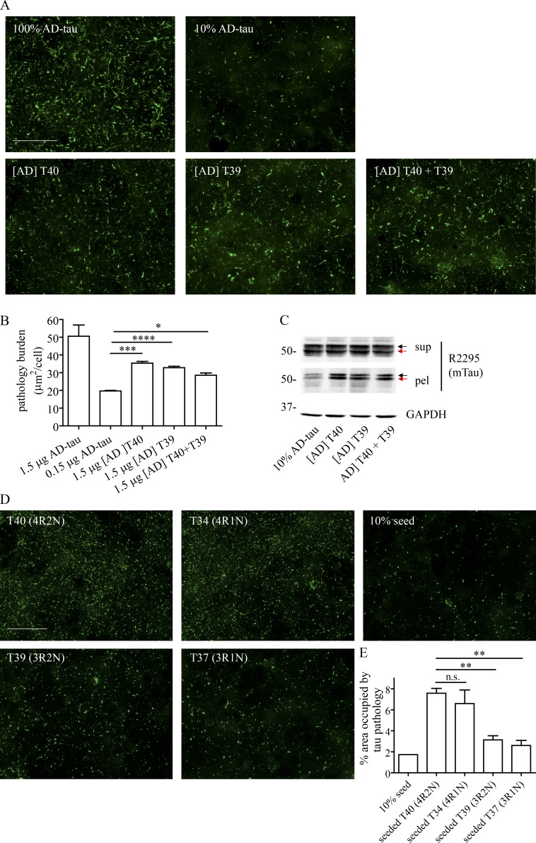
![Figure 8. rTau fibrils templated by AD-tau acquire seeding competency in non-Tg neurons. (A) Mouse tau aggregates (methanol-resistant T49 staining in green) induced in non-Tg neurons by [AD]rTau fibrils compared with the same dose of AD-tau (100% AD-tau) and 10% AD-tau seed control (amount of tau per coverslip: 0.15 µg for 10% AD-tau and 1.5 µg for the rest). (B) Quantification of the area occupied by mouse tau pathology normalized to total cell count for the experiment shown in A, displayed as mean + SEM. Four different sets of [AD]rTau fibrils that were seeded by three different preparations of AD-tau were tested; two independent sets of non-Tg neurons were used. Pairwise Student’s t tests were performed between seeded rTau fibrils and 10% seed control. *, P < 0.05; ***, P < 0.0005; ****, P < 0.00005. (C) Immunoblotting for sarkosyl-soluble (sup) and -insoluble (pel) fractions of non-Tg neurons treated with [AD]rTau fibrils or 10% AD-tau seed control (9 µg [AD]rTau and 0.9 µg AD-tau for each well on a 12-well plate). Black and red arrows indicate 4R and 3R mouse tau, respectively. Data are representative of two independent experiments. mTau, mouse Tau. Molecular mass is indicated in kilodaltons. (D) AT8-positive mouse tau pathology induced by different isoforms of tau seeded by 10% X-T40 fibrils. (E) Quantification of tau pathology for the experiment shown in D. The same preparation of X-T40 fibrils was used to seed different batches of independently purified monomers: four batches of monomers for T40 and two batches of monomers for T34, T39, and T37. Data are shown as mean + SEM. Pairwise Student’s t tests were conducted between seeded T40 and the other isoforms. **, P < 0.005. Bars, 100 µm.](/paper-figures/2e725aabc7ba4827a196043373da3807/unique-pathological-tau-conformers-from-alzheimers-brains-transmit-tau-pathology-figure-8.jpg)

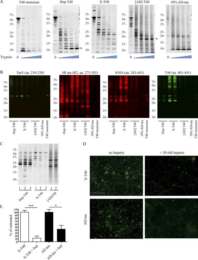
![Figure 10. The differential seeding activities of distinct tau fibril variants are underlain by their conformational differences and influenced by the presence of heparin. (A) The different variants of tau fibrils (Hep-T40, X-T40, and [AD]T40) and control samples (equal amount of T40 monomer; 10% of AD-tau) were incubated with increasing concentrations of trypsin (0%, 0.00125%, 0.0025%, 0.005%, and 0.01%) for 30 min at 37°C. The resulted digestion products were resolved on SDS-PAGE and stained by Coomassie blue. The asterisk indicates dominant trypsin-resistant fragments for Hep-T40 and [AD]T40 fibrils. The 10% AD-tau seeds in [AD]T40 did not contribute to the 18-kD fragment, as suggested by the 10% AD-tau control. For each fibril variant, three to four independently prepared samples were tested across three independent experiments. (B) Digestion products from incubation with 0.01% trypsin were resolved on SDS-PAGE and immunoblotted with tau antibodies recognizing different epitopes. R2: second MT-binding repeat. Data are representative of three independent experiments. (C) The different variants of tau fibrils (three preparations of Hep-T40 fibrils, three preparations of X-T40 fibrils, and two preparations of [AD]T40 fibrils) were incubated with 1 µg/ml of proteinase K for 30 min at 37°C. The resulted products were resolved on SDS-PAGE and stained by Coomassie blue. (A–C) Molecular mass is indicated in kilodaltons. (D) Methanol-resistant mouse tau pathology induced by 1.5 µg X-T40 or 0.15 µg AD-tau fibrils with and without 50 nM heparin cotreatment. The amount of heparin added is equivalent to that contained in 10 µM heparin-induced 40 µM T40 fibrils added at 4.5 µg of tau per coverslip. Bar, 100 µm. (E) Quantification of mouse tau pathology for experiments shown in D. For each fibril variant, three different preparations of fibrils were tested across three independent sets of non-Tg neurons. Data are shown as mean + SEM. Pairwise Student’s t tests were performed between untreated and heparin-treated cells. *, P < 0.05; ***, P < 0.001.](/paper-figures/dbe9eeee756b4b9884c5beb12a2b7de5/unique-pathological-tau-conformers-from-alzheimers-brains-transmit-tau-pathology-figure-10.jpg)
Verification
Claims the paper makes
The claims and statistics of this paper haven’t been checked yet.
Run the check — free, no account needed. It reads the open-access text and checks the sources it cites.
Claims other papers make about this one
Each is checked against this paper’s own text: does it say what the citing paper says it does?
The source backs part of the claim, not all of it — or backs it for a different population, condition or comparison.
In the citing paper
“Methanol fixation has previously been used to remove soluble proteins, including tau (20, 46).”
From Modeling late-onset Alzheimer’s disease neuropathology via direct neuronal reprogramming (2024) · cites this paper as [46]
Experimental — accuracy not yet measured
Self-check
Percentages, sums and reported differences re-computed from the paper’s own numbers.
The self-check needs the paper’s full text.
Citations
Cites
Amyloid oligomers exacerbate tau pathology in a mouse model of tauopathy
Selenica, M.-L. B., Brownlow, M., Jimenez, J. P., Lee, D. C., Pena, G., Dickey, C. A., Gordon, M. N., & Morgan, D. (2013) · Neurodegenerative Diseases · doi:10.1159/000337230 · Cited by 10,359
A protein factor essential for microtubule assembly
Weingarten, M. D., Lockwood, A. H., Hwo, S. Y., & Kirschner, M. W. (1975) · Proceedings of the National Academy of Sciences · doi:10.1073/pnas.72.5.1858 · Cited by 3,089
A mesoscale connectome of the mouse brain
Oh, S. W., Harris, J. A., Ng, L., Winslow, B., Cain, N., Mihalas, S., Wang, Q., Lau, C., Kuan, L., Henry, A. M., et al. (2014) · Nature · doi:10.1038/nature13186 · Cited by 2,612
Luk, K. C., Kehm, V., Carroll, J., Zhang, B., O’Brien, P., Trojanowski, J. Q., & Lee, V. M.-Y. (2012) · Science · doi:10.1126/science.1227157 · Cited by 2,563
Self-citationGoedert, M., Spillantini, M. G., Jakes, R., Rutherford, D., & Crowther, R. A. (1989) · Neuron · doi:10.1016/0896-6273(89)90210-9 · Cited by 2,492
Lee, V. M.-Y., Goedert, M., & Trojanowski, J. Q. (2001) · Annual Review of Neuroscience · doi:10.1146/annurev.neuro.24.1.1121 · Cited by 2,480
Self-citationTransmission and spreading of tauopathy in transgenic mouse brain
Clavaguera, F., Bolmont, T., Crowther, R. A., Abramowski, D., Frank, S., Probst, A., Fraser, G., Stalder, A. K., Beibel, M., Staufenbiel, M., et al. (2009) · Nature Cell Biology · doi:10.1038/ncb1901 · Cited by 1,803
Self-Propagating, Molecular-Level Polymorphism in Alzheimer's ß-Amyloid Fibrils
Petkova, A. T., Leapman, R. D., Guo, Z., Yau, W.-M., Mattson, M. P., & Tycko, R. (2005) · Science · doi:10.1126/science.1105850 · Cited by 1,687
Enhanced neurofibrillary degeneration in transgenic mice expressing mutant tau and APP
Lewis, J., Dickson, D. W., Lin, W.-L., Chisholm, L., Corral, A., Jones, G., Yen, S.-H., Sahara, N., Skipper, L., Yager, D., et al. (2001) · Science · doi:10.1126/science.1058189 · Cited by 1,611
Formation of Neurofibrillary Tangles in P301L Tau Transgenic Mice Induced by Aβ42 Fibrils
Götz, J., Chen, F., van Dorpe, J., & Nitsch, R. M. (2001) · Science · doi:10.1126/science.1062097 · Cited by 1,526
Cited by
Sorted by citations
Alzheimer Disease: An Update on Pathobiology and Treatment Strategies
Long, J. M., & Holtzman, D. M. (2019) · Cell · doi:10.1016/j.cell.2019.09.001 · Cited by 3,204
Cryo-EM structures of tau filaments from Alzheimer’s disease
Fitzpatrick, A. W. P., Falcon, B., He, S., Murzin, A. G., Murshudov, G., Garringer, H. J., Crowther, R. A., Ghetti, B., Goedert, M., & Scheres, S. H. W. (2017) · Nature · doi:10.1038/nature23002 · Cited by 2,061
The proteostasis network and its decline in ageing
Hipp, M. S., Kasturi, P., & Hartl, F. U. (2019) · Nature Reviews Molecular Cell Biology · doi:10.1038/s41580-019-0101-y · Cited by 1,642
Kim, S., Kwon, S.-H., Kam, T.-I., Panicker, N., Karuppagounder, S. S., Lee, S., Lee, J. H., Kim, W. R., Kook, M., Foss, C. A., et al. (2019) · Neuron · doi:10.1016/j.neuron.2019.05.035 · Cited by 1,397
Protein misfolding, aggregation, and conformational strains in neurodegenerative diseases
Soto, C., & Pritzkow, S. (2018) · Nature Neuroscience · doi:10.1038/s41593-018-0235-9 · Cited by 1,180
Metadata
- Authors
- Guo, J. L., Narasimhan, S., Changolkar, L., He, Z., Stieber, A., Zhang, B., Gathagan, R. J., Iba, M., McBride, J. D., Trojanowski, J. Q., & Lee, V. M. Y.
- Affiliations
- University of Pennsylvania (US)
- Corresponding author
- Lee, Virginia M.Y.
- ORCID
- Venue
- Journal of Experimental Medicine
- Publisher
- Rockefeller University Press
- Published
- 2016-10-17
- Volume
- 213
- Issue
- 12
- Pages
- 2635-2654
- ISSN
- 0022-1007 (ISSN-L)
- 1540-9538
- PMC
- PMC5110027
- OpenAlex
- W2534587007
- Cited by
- 440
- References
- 60
- Citations by year
- 2026: 30 · 2025: 35 · 2024: 53 · 2023: 50 · 2022: 47 · 2021: 62 · 2020: 57 · 2019: 45 · 2018: 40 · 2017: 20
- FWCI
- 28.18
- Type
- journal article
- Language
- en
- Open access
- bronze
- doi.org
- Locations
- The Journal of Experimental Medicine (publishedVersion)
- Europe PMC (PubMed Central) (publishedVersion, cc-by-nc-sa)
- PubMed Central (publishedVersion, cc-by-nc-sa)
- PubMed (publishedVersion)
- Funders
- National Institutes of Health (AG10124, AG17586)
- CurePSP
- BrightFocus Foundation (A2014005F)
- Topics
- Life Sciences › Neuroscience › Cellular and Molecular Neuroscience › Alzheimer's disease research and treatments
- Health Sciences › Medicine › Psychiatry and Mental health › Dementia and Cognitive Impairment Research
- Life Sciences › Biochemistry, Genetics and Molecular Biology › Molecular Biology › Prion Diseases and Protein Misfolding
- Keywords
- tau pathology, Alzheimer's disease, tau fibrils, cell-to-cell transmission, intracerebral injection, tau inclusions
- MeSH terms
- Aging, Alzheimer Disease, Animals, Brain, Heparin, Mice, Inbred C57BL, Mice, Transgenic, Neurons, Phosphorylation, Protein Conformation, Tissue Extracts, Neurofibrillary Tangles, tau Proteins, Protein Isoforms, Tauopathies, Protein Aggregates
About these checks
Registry, retraction, Cites and statistics results are computed, not judged, with one exception: a printed reference that no index lists is read once by a model before it counts as a citation error. Claims are extracted by a model and kept only when their quote appears verbatim in the text; each cited source is then checked, with a model’s help, for whether it says what the claim relies on. Those verdicts can be wrong — read the claim’s quote and the cited source before relying on one.
Statistics and arithmetic are recomputed, not judged.
Confidence bands are shown only for fields calibrated on our annotated test set. None is calibrated yet, so no band is shown.
What counts as a claim ›How we measure confidence ›How we check this ›
Are you an author, or do you see a problem with this page? Request a review