

The DOI matches its Crossref record; 60 of the 60 references it cites were found in a registry or an index.
Unique pathological tau conformers from Alzheimer’s brains transmit tau pathology in nontransgenic mice
Open accessAbstract
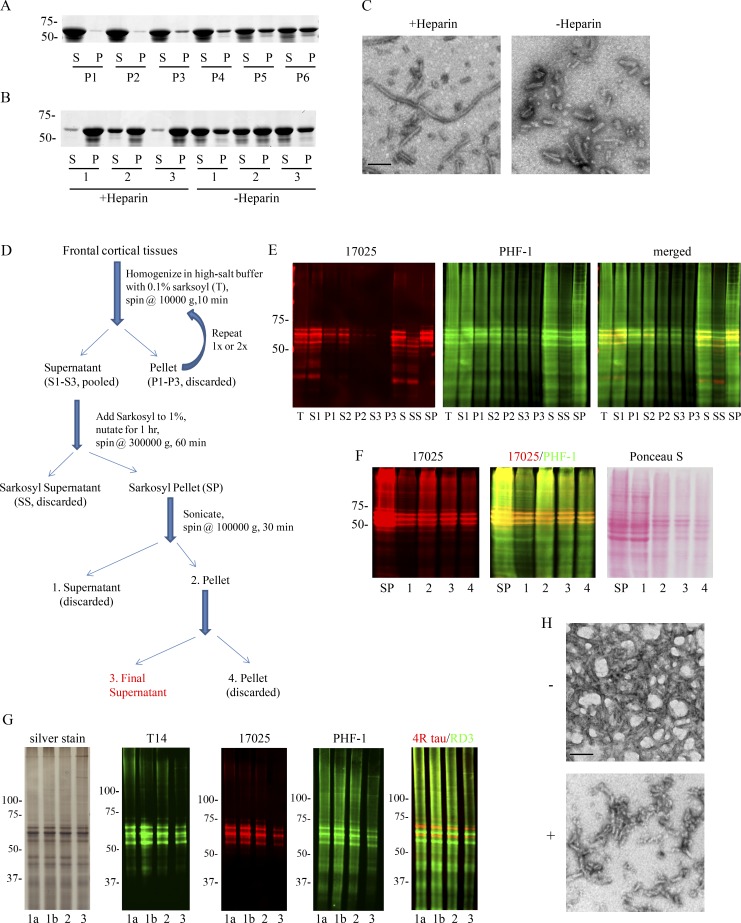
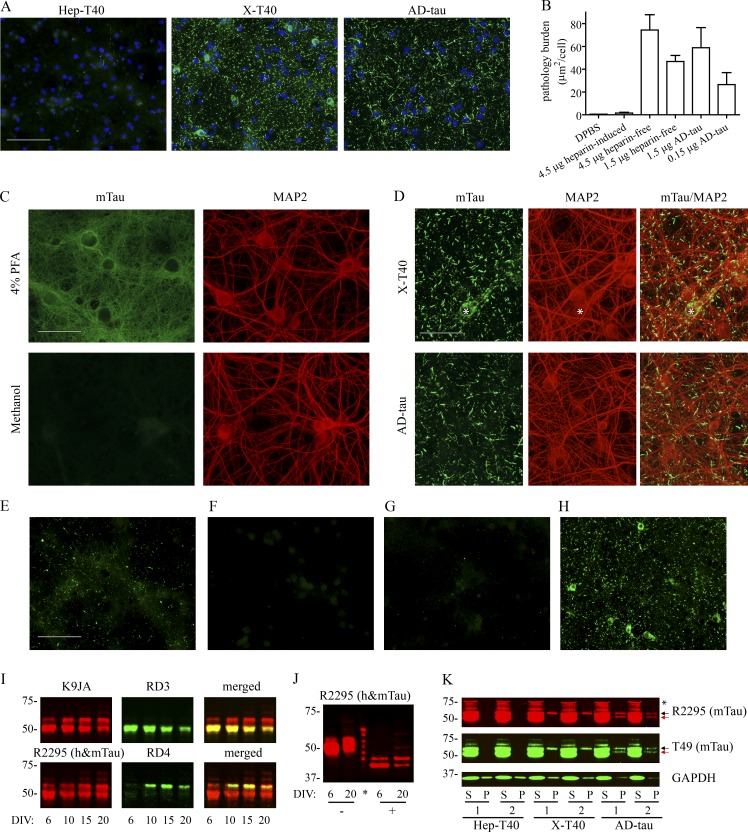
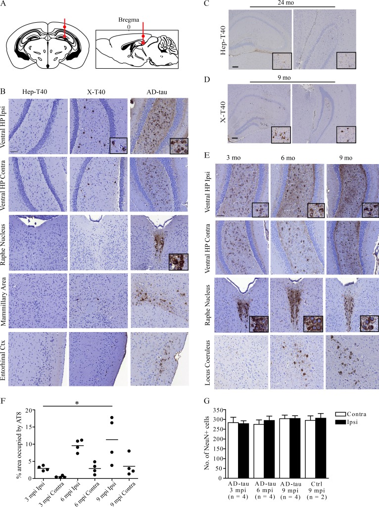
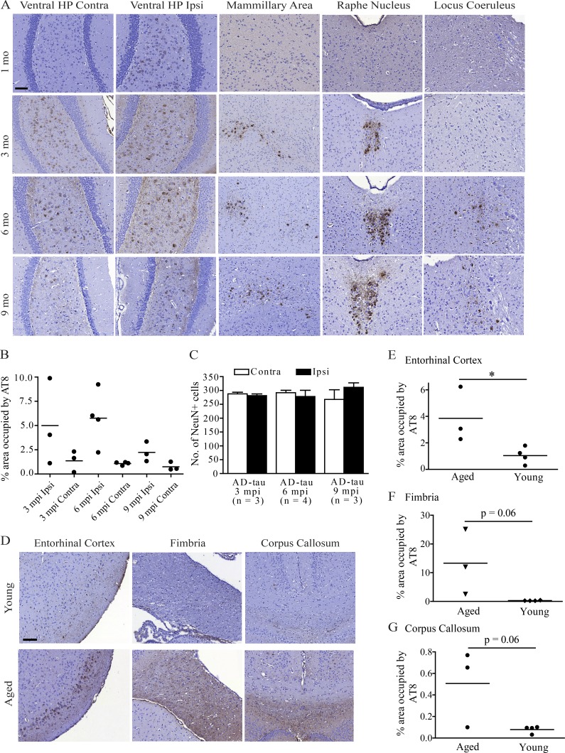
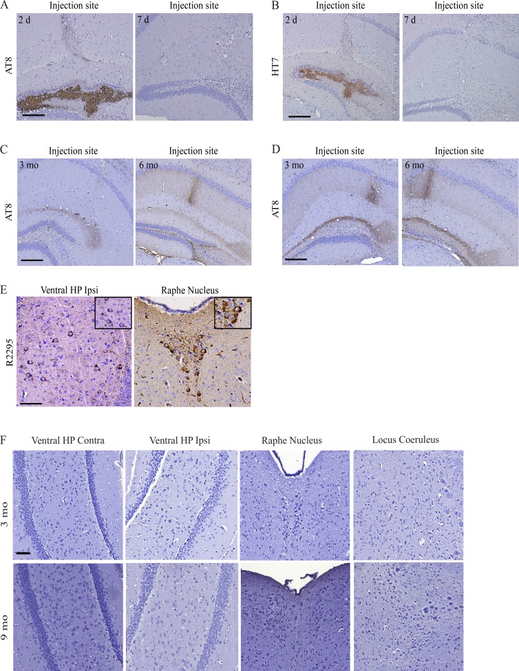
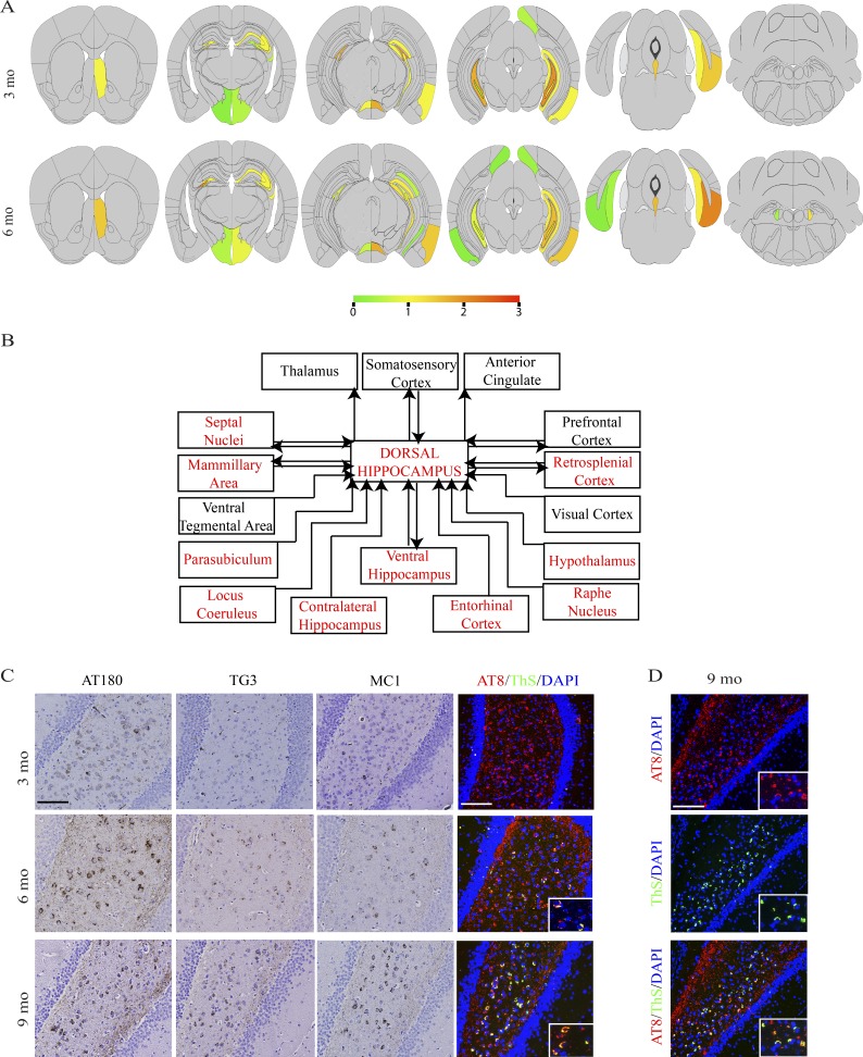
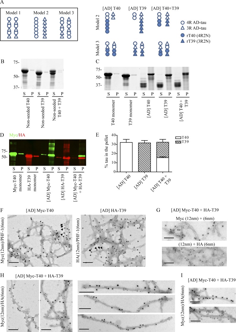
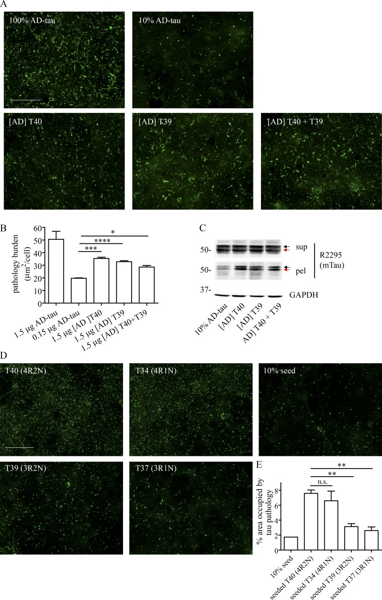
Filamentous tau aggregates are hallmark lesions in numerous neurodegenerative diseases, including Alzheimer’s disease (AD). Cell culture and animal studies showed that tau fibrils can undergo cell-to-cell transmission and seed aggregation of soluble tau, but this phenomenon was only robustly demonstrated in models overexpressing tau. In this study, we found that intracerebral inoculation of tau fibrils purified from AD brains (AD-tau), but not synthetic tau fibrils, resulted in the formation of abundant tau inclusions in anatomically connected brain regions in nontransgenic mice. Recombinant human tau seeded by AD-tau revealed unique conformational features that are distinct from synthetic tau fibrils, which could underlie the differential potency in seeding physiological levels of tau to aggregate. Therefore, our study establishes a mouse model of sporadic tauopathies and points to important differences between tau fibrils that are generated artificially and authentic ones that develop in AD brains.
Overview
The claims and statistics of this paper haven’t been checked yet.
Run the check — free, no account needed. It reads the open-access text and checks the sources it cites.
Integrity signals
Registry
Cites
Statistics
Figures
Verification
Claims the paper makes
The claims and statistics of this paper haven’t been checked yet.
Run the check — free, no account needed. It reads the open-access text and checks the sources it cites.
Claims other papers make about this one
Each is checked against this paper’s own text: does it say what the citing paper says it does?
The source backs part of the claim, not all of it — or backs it for a different population, condition or comparison.
In the citing paper
“Methanol fixation has previously been used to remove soluble proteins, including tau (20, 46).”
From Modeling late-onset Alzheimer’s disease neuropathology via direct neuronal reprogramming (2024) · cites this paper as [46]
Experimental — accuracy not yet measured
Self-check
Percentages, sums and reported differences re-computed from the paper’s own numbers.
The self-check needs the paper’s full text.
Citations
Cites
Amyloid oligomers exacerbate tau pathology in a mouse model of tauopathy
Selenica, M.-L. B., Brownlow, M., Jimenez, J. P., Lee, D. C., Pena, G., Dickey, C. A., Gordon, M. N., & Morgan, D. (2013) · Neurodegenerative Diseases · doi:10.1159/000337230 · Cited by 10,359
A protein factor essential for microtubule assembly
Weingarten, M. D., Lockwood, A. H., Hwo, S. Y., & Kirschner, M. W. (1975) · Proceedings of the National Academy of Sciences · doi:10.1073/pnas.72.5.1858 · Cited by 3,089
A mesoscale connectome of the mouse brain
Oh, S. W., Harris, J. A., Ng, L., Winslow, B., Cain, N., Mihalas, S., Wang, Q., Lau, C., Kuan, L., Henry, A. M., et al. (2014) · Nature · doi:10.1038/nature13186 · Cited by 2,612
Luk, K. C., Kehm, V., Carroll, J., Zhang, B., O’Brien, P., Trojanowski, J. Q., & Lee, V. M.-Y. (2012) · Science · doi:10.1126/science.1227157 · Cited by 2,563
Self-citationGoedert, M., Spillantini, M. G., Jakes, R., Rutherford, D., & Crowther, R. A. (1989) · Neuron · doi:10.1016/0896-6273(89)90210-9 · Cited by 2,492
Lee, V. M.-Y., Goedert, M., & Trojanowski, J. Q. (2001) · Annual Review of Neuroscience · doi:10.1146/annurev.neuro.24.1.1121 · Cited by 2,480
Self-citationTransmission and spreading of tauopathy in transgenic mouse brain
Clavaguera, F., Bolmont, T., Crowther, R. A., Abramowski, D., Frank, S., Probst, A., Fraser, G., Stalder, A. K., Beibel, M., Staufenbiel, M., et al. (2009) · Nature Cell Biology · doi:10.1038/ncb1901 · Cited by 1,803
Self-Propagating, Molecular-Level Polymorphism in Alzheimer's ß-Amyloid Fibrils
Petkova, A. T., Leapman, R. D., Guo, Z., Yau, W.-M., Mattson, M. P., & Tycko, R. (2005) · Science · doi:10.1126/science.1105850 · Cited by 1,687
Enhanced neurofibrillary degeneration in transgenic mice expressing mutant tau and APP
Lewis, J., Dickson, D. W., Lin, W.-L., Chisholm, L., Corral, A., Jones, G., Yen, S.-H., Sahara, N., Skipper, L., Yager, D., et al. (2001) · Science · doi:10.1126/science.1058189 · Cited by 1,611
Formation of Neurofibrillary Tangles in P301L Tau Transgenic Mice Induced by Aβ42 Fibrils
Götz, J., Chen, F., van Dorpe, J., & Nitsch, R. M. (2001) · Science · doi:10.1126/science.1062097 · Cited by 1,526
Cited by
Sorted by citations
Alzheimer Disease: An Update on Pathobiology and Treatment Strategies
Long, J. M., & Holtzman, D. M. (2019) · Cell · doi:10.1016/j.cell.2019.09.001 · Cited by 3,204
Cryo-EM structures of tau filaments from Alzheimer’s disease
Fitzpatrick, A. W. P., Falcon, B., He, S., Murzin, A. G., Murshudov, G., Garringer, H. J., Crowther, R. A., Ghetti, B., Goedert, M., & Scheres, S. H. W. (2017) · Nature · doi:10.1038/nature23002 · Cited by 2,061
The proteostasis network and its decline in ageing
Hipp, M. S., Kasturi, P., & Hartl, F. U. (2019) · Nature Reviews Molecular Cell Biology · doi:10.1038/s41580-019-0101-y · Cited by 1,642
Kim, S., Kwon, S.-H., Kam, T.-I., Panicker, N., Karuppagounder, S. S., Lee, S., Lee, J. H., Kim, W. R., Kook, M., Foss, C. A., et al. (2019) · Neuron · doi:10.1016/j.neuron.2019.05.035 · Cited by 1,397
Protein misfolding, aggregation, and conformational strains in neurodegenerative diseases
Soto, C., & Pritzkow, S. (2018) · Nature Neuroscience · doi:10.1038/s41593-018-0235-9 · Cited by 1,180
Structures of filaments from Pick’s disease reveal a novel tau protein fold
Falcon, B., Zhang, W., Murzin, A. G., Murshudov, G. N., Garringer, H. J., Vidal, R., Crowther, R. A., Ghetti, B. F., Scheres, S. H. W., & Goedert, M. (2018) · Nature · doi:10.1038/s41586-018-0454-y · Cited by 837
He, Z., Guo, J. L., McBride, J. D., Narasimhan, S., Kim, H., Changolkar, L., Zhang, B., Gathagan, R. J., Yue, C., Dengler, C., et al. (2018) · Nature Medicine · doi:10.1038/nm.4443 · Cited by 645
Self-citationProtein transmission in neurodegenerative disease
Peng, C., Trojanowski, J. Q., & Lee, V. M.-Y. (2020) · Nature Reviews Neurology · doi:10.1038/s41582-020-0333-7 · Cited by 625
Self-citationLRP1 is a master regulator of tau uptake and spread
Rauch, J. N., Luna, G., Guzman, E., Audouard, M., Challis, C., Sibih, Y. E., Leshuk, C., Hernandez, I., Wegmann, S., Hyman, B. T., et al. (2020) · Nature · doi:10.1038/s41586-020-2156-5 · Cited by 541
Propagation and spread of pathogenic protein assemblies in neurodegenerative diseases
Jucker, M., & Walker, L. C. (2018) · Nature Neuroscience · doi:10.1038/s41593-018-0238-6 · Cited by 450
Stancu, I.-C., Cremers, N., Vanrusselt, H., Couturier, J., Vanoosthuyse, A., Kessels, S., Lodder, C., Brône, B., Huaux, F., Octave, J.-N., et al. (2019) · Acta Neuropathologica · doi:10.1007/s00401-018-01957-y · Cited by 447
Glial contributions to neurodegeneration in tauopathies
Leyns, C. E. G., & Holtzman, D. M. (2017) · Molecular Neurodegeneration · doi:10.1186/s13024-017-0192-x · Cited by 409
Posttranslational Modifications Mediate the Structural Diversity of Tauopathy Strains
Arakhamia, T., Lee, C. E., Carlomagno, Y., Kumar, M., Duong, D. M., Wesseling, H., Kundinger, S. R., Wang, K., Williams, D., Deture, M. A., et al. (2020) · Cell · doi:10.1016/j.cell.2020.01.027 · Cited by 410
Rodent models for Alzheimer disease
Götz, J., Bodea, L., & Goedert, M. (2018) · Nature reviews. Neuroscience · doi:10.1038/s41583-018-0054-8 · Cited by 422
What is the evidence that tau pathology spreads through prion-like propagation?
Mudher, A., Colin, M., Dujardin, S., Medina, M., Dewachter, I., Alavi Naini, S. M., Mandelkow, E.-M., Mandelkow, E., Buée, L., Goedert, M., et al. (2017) · Acta Neuropathologica Communications · doi:10.1186/s40478-017-0488-7 · Cited by 384
Biological aging processes underlying cognitive decline and neurodegenerative disease
Gonzales, M. M., Garbarino, V. R., Pollet, E., Palavicini, J. P., Kellogg, D. L., Kraig, E., & Orr, M. E. (2022) · Journal of Clinical Investigation · doi:10.1172/jci158453 · Cited by 426
Narasimhan, S., Guo, J. L., Changolkar, L., Stieber, A., McBride, J. D., Silva, L. V., He, Z., Zhang, B., Gathagan, R. J., Trojanowski, J. Q., et al. (2017) · The Journal of Neuroscience · doi:10.1523/jneurosci.1230-17.2017 · Cited by 374
Self-citationTREM2 function impedes tau seeding in neuritic plaques
Leyns, C. E. G., Gratuze, M., Narasimhan, S., Jain, N., Koscal, L. J., Jiang, H., Manis, M., Colonna, M., Lee, V. M. Y., Ulrich, J. D., et al. (2019) · Nature Neuroscience · doi:10.1038/s41593-019-0433-0 · Cited by 332
Self-citationUemura, N., Yagi, H., Uemura, M. T., Hatanaka, Y., Yamakado, H., & Takahashi, R. (2018) · Molecular Neurodegeneration · doi:10.1186/s13024-018-0257-5 · Cited by 328
Mechanisms of secretion and spreading of pathological tau protein
Brunello, C. A., Merezhko, M., Uronen, R.-L., & Huttunen, H. J. (2020) · Cellular and Molecular Life Sciences · doi:10.1007/s00018-019-03349-1 · Cited by 304
Alzheimer’s disease brain-derived extracellular vesicles spread tau pathology in interneurons
Ruan, Z., Pathak, D., Venkatesan Kalavai, S., Yoshii-Kitahara, A., Muraoka, S., Bhatt, N., Takamatsu-Yukawa, K., Hu, J., Wang, Y., Hersh, S., et al. (2021) · Brain · doi:10.1093/brain/awaa376 · Cited by 283
Mechanisms of Cell-to-Cell Transmission of Pathological Tau: A Review
Gibbons, G. S., Lee, V. M. Y., & Trojanowski, J. Q. (2019) · JAMA Neurology · doi:10.1001/jamaneurol.2018.2505 · Cited by 257
Self-citationTau burden and the functional connectome in Alzheimer’s disease and progressive supranuclear palsy
Cope, T., Rittman, T., Borchert, R. J., Jones, P. S., Vatansever, D., Allinson, K. S. J., Passamonti, L., Rodríguez, P. V., Bevan-Jones, W. R., O'Brien, J. T., et al. (2017) · Brain · doi:10.1093/brain/awx347 · Cited by 251
Porta, S., Xu, Y., Restrepo, C. R., Kwong, L. K., Zhang, B., Brown, H. J., Lee, E. B., Trojanowski, J. Q., & Lee, V. M.-Y. (2018) · Nature Communications · doi:10.1038/s41467-018-06548-9 · Cited by 248
Self-citationSaito, T., Mihira, N., Matsuba, Y., Sasaguri, H., Hashimoto, S., Narasimhan, S., Zhang, B., Murayama, S., Higuchi, M., Lee, V. M. Y., et al. (2019) · Journal of Biological Chemistry · doi:10.1074/jbc.ra119.009487 · Cited by 248
Self-citationIdentification of the Tau phosphorylation pattern that drives its aggregation
Despres, C., Byrne, C., Qi, H., Cantrelle, F., Huvent, I., Chambraud, B., Baulieu, É.-É., Jacquot, Y., Landrieu, I., Lippens, G. M., et al. (2017) · Proceedings of the National Academy of Sciences · doi:10.1073/pnas.1708448114 · Cited by 263
MEG3 activates necroptosis in human neuron xenografts modeling Alzheimer’s disease
Balusu, S., Horré, K., Thrupp, N., Craessaerts, K., Snellinx, A., Serneels, L., T’Syen, D., Chrysidou, I., Arranz, A. M., Sierksma, A. S. R., et al. (2023) · Science · doi:10.1126/science.abp9556 · Cited by 246
Dural lymphatics regulate clearance of extracellular tau from the CNS
Patel, T. K., Habimana-Griffin, L., Gao, X., Xu, B. J., Achilefu, S., Alitalo, K. K., McKee, C. A., Sheehan, P. W., Musiek, E. S., Xiong, C., et al. (2019) · Molecular Neurodegeneration · doi:10.1186/s13024-019-0312-x · Cited by 220
Activated microglia mitigate Aβ-associated tau seeding and spreading
Gratuze, M., Chen, Y., Parhizkar, S., Jain, N., Strickland, M. R., Serrano, J. R., Colonna, M., Ulrich, J. D., & Holtzman, D. M. (2021) · Journal of Experimental Medicine · doi:10.1084/jem.20210542 · Cited by 215
Goedert, M., & Spillantini, M. G. (2017) · Molecular Brain · doi:10.1186/s13041-017-0298-7 · Cited by 213
Amyloid-β and Tau at the Crossroads of Alzheimer’s Disease
Gallardo, G., & Holtzman, D. M. (2019) · Advances in experimental medicine and biology · doi:10.1007/978-981-32-9358-8_16 · Cited by 210
Updates on mouse models of Alzheimer’s disease
Zhong, M. Z., Peng, T., Duarte, M. L., Wang, M., & Cai, D. (2024) · Molecular Neurodegeneration · doi:10.1186/s13024-024-00712-0 · Cited by 189
CSF tau microtubule binding region identifies tau tangle and clinical stages of Alzheimer’s disease
Horie, K., Barthélemy, N. R., Sato, C., & Bateman, R. J. (2021) · Brain · doi:10.1093/brain/awaa373 · Cited by 180
Mechanisms of Pathogenic Tau and Aβ Protein Spreading in Alzheimer’s Disease
d’Errico, P., & Meyer‐Luehmann, M. (2020) · Frontiers in Aging Neuroscience · doi:10.3389/fnagi.2020.00265 · Cited by 190
Transmission of tauopathy strains is independent of their isoform composition
He, Z., McBride, J. D., Xu, H. W., Changolkar, L. N., Kim, S., Zhang, B., Narasimhan, S., Gibbons, G. S., Guo, J., Kozak, M., et al. (2020) · Nature Communications · doi:10.1038/s41467-019-13787-x · Cited by 183
Self-citationNachman, E., Wentink, A. S., Madiona, K., Bousset, L., Katsinelos, T., Allinson, K. S. J., Kampinga, H. H., McEwan, W. A., Jahn, T. R., Melki, R., et al. (2020) · Journal of Biological Chemistry · doi:10.1074/jbc.ra120.013478 · Cited by 174
Limorenko, G., & Lashuel, H. A. (2021) · Chemical Society Reviews · doi:10.1039/d1cs00127b · Cited by 168
Passive immunotherapies targeting Aβ and tau in Alzheimer's disease
Plotkin, S. S., & Cashman, N. R. (2020) · Neurobiology of Disease · doi:10.1016/j.nbd.2020.105010 · Cited by 161
Tau accelerates α-synuclein aggregation and spreading in Parkinson’s disease
Pan, L., Li, C., Meng, L., Tian, Y., He, M., Yuan, X., Zhang, G., Zhang, Z., Xiong, J., Chen, G., et al. (2022) · Brain · doi:10.1093/brain/awac171 · Cited by 154
Katsumoto, A., Takeuchi, H., & Tanaka, F. (2019) · Frontiers in Neurology · doi:10.3389/fneur.2019.00980 · Cited by 151
Dam, T., Boxer, A. L., Golbe, L. I., Höglinger, G. U., Morris, H. R., Litvan, I., Lang, A. E., Corvol, J., Aiba, I., Grundman, M., et al. (2021) · Nature Medicine · doi:10.1038/s41591-021-01455-x · Cited by 152
Chronic TREM2 activation exacerbates Aβ-associated tau seeding and spreading
Jain, N., Lewis, C. A., Ulrich, J. D., & Holtzman, D. M. (2023) · Journal of Experimental Medicine · doi:10.1084/jem.20220654 · Cited by 146
Cell-to-Cell Transmission of Tau and α-Synuclein
Uemura, N., Uemura, M. T., Luk, K. C., Lee, V. M.-Y., & Trojanowski, J. Q. (2020) · Trends in Molecular Medicine · doi:10.1016/j.molmed.2020.03.012 · Cited by 149
Self-citationExperimental evidence for the age dependence of tau protein spread in the brain
Wegmann, S., Bennett, R. E., Delorme, L., Robbins, A. B., Hu, M., MacKenzie, D., Kirk, M. J., Schiantarelli, J., Tunio, N., Amaral, A. C., et al. (2019) · Science Advances · doi:10.1126/sciadv.aaw6404 · Cited by 144
Neurodegenerative Disease Tauopathies
Creekmore, B. C., Watanabe, R., & Lee, E. B. (2023) · Annual Review of Pathology Mechanisms of Disease · doi:10.1146/annurev-pathmechdis-051222-120750 · Cited by 155
Autosomal dominant VCP hypomorph mutation impairs disaggregation of PHF-tau
Darwich, N. F., Phan, J. M., Kim, B., Suh, E. R., Papatriantafyllou, J. D., Changolkar, L. N., Nguyen, A. T., O’Rourke, C. M., He, Z., Porta, S., et al. (2020) · Science · doi:10.1126/science.aay8826 · Cited by 132
Self-citationStrang, K. H., Croft, C. L., Sorrentino, Z. A., Chakrabarty, P., Golde, T. E., & Giasson, B. I. (2018) · Journal of Biological Chemistry · doi:10.1074/jbc.m117.815357 · Cited by 124
Induction of a transmissible tau pathology by traumatic brain injury
Zanier, E. R., Bertani, I., Sammali, E., Pischiutta, F., Chiaravalloti, M. A., Vegliante, G., Masone, A., Corbelli, A., Smith, D. H., Menon, D. K., et al. (2018) · Brain · doi:10.1093/brain/awy193 · Cited by 136
Neuronal APOE4 removal protects against tau-mediated gliosis, neurodegeneration and myelin deficits
Koutsodendris, N., Blumenfeld, J., Agrawal, A., Traglia, M., Grone, B., Zilberter, M., Yip, O., Rao, A., Nelson, M. R., Hao, Y., et al. (2023) · Nature Aging · doi:10.1038/s43587-023-00368-3 · Cited by 132
Cofactors are essential constituents of stable and seeding-active tau fibrils
Fichou, Y., Lin, Y., Rauch, J. N., Vigers, M., Zeng, Z., Srivastava, M., Keller, T. J., Freed, J. H., Kosik, K. S., & Han, S. (2018) · Proceedings of the National Academy of Sciences · doi:10.1073/pnas.1810058115 · Cited by 128
Amyloid-β pathology enhances pathological fibrillary tau seeding induced by Alzheimer PHF in vivo
Vergara, C., Houben, S., Suain, V., Yilmaz, Z., Decker, R. D., Dries, V. V., Boom, A., Mansour, S., Leroy, K., Ando, K., et al. (2018) · Acta Neuropathologica · doi:10.1007/s00401-018-1953-5 · Cited by 131
The prion model for progression and diversity of neurodegenerative diseases
Stopschinski, B. E., & Diamond, M. I. (2017) · The Lancet Neurology · doi:10.1016/s1474-4422(17)30037-6 · Cited by 133
APOE3ch alters microglial response and suppresses Aβ-induced tau seeding and spread
Chen, Y., Song, S., Parhizkar, S., Lord, J., Zhu, Y., Strickland, M. R., Wang, C., Park, J., Tabor, G. T., Jiang, H., et al. (2024) · Cell · doi:10.1016/j.cell.2023.11.029 · Cited by 120
Prokop, S., Miller, K. R., Labra, S. R., Pitkin, R. M., Hoxha, K., Narasimhan, S., Changolkar, L., Rosenbloom, A., Lee, V. M.-Y., & Trojanowski, J. Q. (2019) · Acta Neuropathologica · doi:10.1007/s00401-019-02048-2 · Cited by 119
Self-citationCell Death and NeurodegenerationPreprint
Andreone, B. J., Larhammar, M., & Lewcock, J. W. (2019) · Cold Spring Harbor Perspectives in Biology · doi:10.1101/cshperspect.a036434 · Cited by 125
Jiang, S., & Bhaskar, K. (2020) · Frontiers in Molecular Neuroscience · doi:10.3389/fnmol.2020.586731 · Cited by 119
Progressive supranuclear palsy: Advances in diagnosis and management
Coughlin, D. G., & Litvan, I. (2020) · Parkinsonism & Related Disorders · doi:10.1016/j.parkreldis.2020.04.014 · Cited by 118
α-Synuclein modulates tau spreading in mouse brains
Bassil, F., Meymand, E. S., Brown, H. J., Xu, H. W., Cox, T. O., Pattabhiraman, S., Maghames, C. M., Wu, Q., Zhang, B., Trojanowski, J. Q., et al. (2020) · The Journal of Experimental Medicine · doi:10.1084/jem.20192193 · Cited by 109
Self-citationTau filaments are tethered within brain extracellular vesicles in Alzheimer’s disease
Fowler, S. L., Behr, T. S., Turkes, E., O'Brien, D. P., Cauhy, P. M., Rawlinson, I., Edmonds, M. J., Foiani, M. S., Schaler, A. W., Crowley, G., et al. (2024) · Nature Neuroscience · doi:10.1038/s41593-024-01801-5 · Cited by 118
Human tau pathology transmits glial tau aggregates in the absence of neuronal tau
Narasimhan, S., Changolkar, L. N., Riddle, D. M., Kats, A. S., Stieber, A., Weitzman, S. A., Zhang, B., Li, Z., Roberson, E. D., Trojanowski, J. Q., et al. (2019) · The Journal of Experimental Medicine · doi:10.1084/jem.20190783 · Cited by 109
Self-citationRole of Microglial Cells in Alzheimer’s Disease Tau Propagation
Španić, E., Horvat, L. L., Hof, P. R., & Šimić, G. (2019) · Frontiers in Aging Neuroscience · doi:10.3389/fnagi.2019.00271 · Cited by 111
Trzeciakiewicz, H., Ajit, D., Tseng, J.-H., Chen, Y., Ajit, A., Tabassum, Z., Lobrovich, R., Peterson, C., Riddick, N. V., Itano, M. S., et al. (2020) · Nature Communications · doi:10.1038/s41467-020-19317-4 · Cited by 102
Self-citationPrion-like Spreading in Tauopathies
Ayers, J. I., Giasson, B. I., & Borchelt, D. (2017) · Biological Psychiatry · doi:10.1016/j.biopsych.2017.04.003 · Cited by 106
Propagation of Tau Pathology: Integrating Insights From Postmortem and In Vivo Studies
Vogels, T., Leuzy, A., Cicognola, C., Ashton, N. J., Smolek, T., Novák, M., Blennow, K., Zetterberg, H., Hromádka, T., Žilka, N., et al. (2019) · Biological Psychiatry · doi:10.1016/j.biopsych.2019.09.019 · Cited by 99
Shulman, M. B., Kong, J., O'Gorman, J. T., Ratti, E., Rajagovindan, R., Viollet, L., Huang, E., Sharma, S., Racine, A. M., Czerkowicz, J., et al. (2023) · Nature Aging · doi:10.1038/s43587-023-00523-w · Cited by 100
Gibbons, G. S., Banks, R. A., Kim, B., Changolkar, L., Riddle, D. M., Leight, S. N., Irwin, D. J., Trojanowski, J. Q., & Lee, V. M. Y. (2018) · Journal of Neuropathology & Experimental Neurology · doi:10.1093/jnen/nly010 · Cited by 94
Self-citationAge-related diseases as vicious cycles
Belikov, A. V. (2018) · Ageing Research Reviews · doi:10.1016/j.arr.2018.11.002 · Cited by 95
Ten Years of Tau-Targeted Immunotherapy: The Path Walked and the Roads Ahead
Novak, P., Kontsekova, E., Zilka, N., & Novak, M. (2018) · Frontiers in Neuroscience · doi:10.3389/fnins.2018.00798 · Cited by 87
“Models based on seeding tau pathology with exogenous material sometimes use recombinant tau fibrils, but human tauon strains are best replicated by utilizing genuine brain homogenates from human disease (Guo et al., 2016).”
BackgroundMethodCytosolic antibody receptor TRIM21 is required for effective tau immunotherapy in mouse models
Mukadam, A. S., Miller, L. V. C., Smith, A. E., Vaysburd, M., Sakya, S. A., Sanford, S., Keeling, S., Tuck, B. J., Katsinelos, T., Green, C., et al. (2023) · Science · doi:10.1126/science.abn1366 · Cited by 84
Tauopathies: Deciphering Disease Mechanisms to Develop Effective Therapies
Silva, M. C., & Haggarty, S. J. (2020) · International Journal of Molecular Sciences · doi:10.3390/ijms21238948 · Cited by 89
Uemura, N., Uemura, M. T., Lo, A., Bassil, F., Zhang, B., Luk, K. C., Lee, V. M. ‐., Takahashi, R., & Trojanowski, J. Q. (2019) · Journal of Neuropathology & Experimental Neurology · doi:10.1093/jnen/nlz070 · Cited by 86
Self-citationFate and propagation of endogenously formed Tau aggregates in neuronal cells
Chastagner, P., Loría, F., Vargas, J., Tois, J., Diamond, M. I., Okafo, G. N., Brou, C., & Zurzolo, C. (2020) · EMBO Molecular Medicine · doi:10.15252/emmm.202012025 · Cited by 80
Involvement of Oligodendrocytes in Tau Seeding and Spreading in Tauopathies
Ferrer, I., Aguiló, M., Carmona, M., Andrés‐Benito, P., Torrejón-Escribano, B., Garcia‐Esparcia, P., & del Rı́o, J. A. (2019) · Frontiers in Aging Neuroscience · doi:10.3389/fnagi.2019.00112 · Cited by 78
Modeling late-onset Alzheimer’s disease neuropathology via direct neuronal reprogramming
Sun, Z., Kwon, J.-S., Ren, Y., Chen, S., Walker, C. K., Lu, X., Cates, K., Karahan, H., Sviben, S., Fitzpatrick, J. A. J., et al. (2024) · Science · doi:10.1126/science.adl2992 · Cited by 77
“Methanol fixation has previously been used to remove soluble proteins, including tau (20, 46).”
All (animal) models (of neurodegeneration) are wrong. Are they also useful?
Ransohoff, R. M. (2018) · The Journal of Experimental Medicine · doi:10.1084/jem.20182042 · Cited by 83
Aging‐related tau astrogliopathy (ARTAG): not only tau phosphorylation in astrocytes
Ferrer, I., Aguiló, M., González, I. L., Lucena, D. D., Roig, A., Carmona, M., Llorens, F., Garcia‐Esparcia, P., Martínez-Maldonado, A., Frau‐Méndez, M. A., et al. (2018) · Brain Pathology · doi:10.1111/bpa.12593 · Cited by 77
Gomes, L. A., Uytterhoeven, V., Lopez-Sanmartin, D., Tomé, S. O., Tousseyn, T., Vandenberghe, R. R., Vandenbulcke, M., Arnim, C. A. F. V., Verstreken, P., & Thal, D. R. (2021) · Acta Neuropathologica · doi:10.1007/s00401-020-02251-6 · Cited by 77
Fluent molecular mixing of Tau isoforms in Alzheimer’s disease neurofibrillary tangles
Dregni, A. J., Duan, P., Xu, H. W., Changolkar, L. N., Mammeri, N. E., Lee, V. M. ‐., & Hong, M. (2022) · Nature Communications · doi:10.1038/s41467-022-30585-0 · Cited by 75
Self-citationAstrocyte-secreted glypican-4 drives APOE4-dependent tau hyperphosphorylation
Saroja, S. R., Gorbachev, K., Julia, T., Goate, A. M., & Pereira, A. C. (2022) · Proceedings of the National Academy of Sciences · doi:10.1073/pnas.2108870119 · Cited by 75
Propagation of tau and α-synuclein in the brain: therapeutic potential of the glymphatic system
Lopes, D. M., Llewellyn, S. K., & Harrison, I. F. (2022) · Translational Neurodegeneration · doi:10.1186/s40035-022-00293-2 · Cited by 75
The NLRP3 inflammasome modulates tau pathology and neurodegeneration in a tauopathy model
Stancu, I. C., Lodder, C., Botella Lucena, P., Vanherle, S., Gutiérrez de Ravé, M., Terwel, D., Bottelbergs, A., & Dewachter, I. (2022) · Glia · doi:10.1002/glia.24160 · Cited by 72
Ultrastructural and biochemical classification of pathogenic tau, α-synuclein and TDP-43
Tarutani, A., Adachi, T., Akatsu, H., Hashizume, Y., Hasegawa, K., Saito, Y., Robinson, A., Mann, D. M. A., Yoshida, M., Murayama, S., et al. (2022) · Acta Neuropathologica · doi:10.1007/s00401-022-02426-3 · Cited by 74
Tang, S. J., Fesharaki‐Zadeh, A., Takahashi, H., Nies, S. H., Smith, L. M., Luo, A., Chyung, A. S., Chiasseu, M., & Strittmatter, S. M. (2020) · Acta Neuropathologica Communications · doi:10.1186/s40478-020-00976-9 · Cited by 74
Scialò, C., Cecco, E. D., Manganotti, P., & Legname, G. (2019) · Viruses · doi:10.3390/v11030261 · Cited by 69
Cornblath, E. J., Li, H. L., Changolkar, L., Zhang, B., Brown, H. J., Gathagan, R. J., Olufemi, M. F., Trojanowski, J. Q., Bassett, D. S., Lee, V. M. Y., et al. (2021) · Science Advances · doi:10.1126/sciadv.abg6677 · Cited by 66
Self-citationPrion-like mechanisms in Alzheimer disease
Walker, L. C. (2018) · Handbook of clinical neurology · doi:10.1016/b978-0-444-63945-5.00016-7 · Cited by 67
Wang, C., Nambiar, A., Strickland, M. R., Lee, C., Parhizkar, S., Moore, A., Musiek, E. S., Ulrich, J. D., & Holtzman, D. M. (2023) · Journal of Clinical Investigation · doi:10.1172/jci169131 · Cited by 70
TDP-43 pathology is associated with increased tau burdens and seeding
Tomé, S. O., Tsaka, G., Ronisz, A., Ospitalieri, S., Gawor, K., Gomes, L. A., Otto, M., Arnim, C. A. F. V., Damme, P. V., Bosch, L. V. D., et al. (2023) · Molecular Neurodegeneration · doi:10.1186/s13024-023-00653-0 · Cited by 74
The prion-like propagation hypothesis in Alzheimer's and Parkinson's disease
Duyckaerts, C. J., Clavaguera, F., & Potier, M. (2019) · Current Opinion in Neurology · doi:10.1097/wco.0000000000000672 · Cited by 71
Capano, L. S., Sato, C., Ficulle, E., Yu, A., Horie, K., Kwon, J.-S., Burbach, K. F., Barthélemy, N. R., Fox, S. G., Karch, C. M., et al. (2022) · Cell Stem Cell · doi:10.1016/j.stem.2022.04.018 · Cited by 65
Delizannis, A. T., Nonneman, A., Tsering, W., Bondt, A. D., den Wyngaert, I. V., Zhang, B., Meymand, E. S., Olufemi, M. F., Koivula, P. P., Maimaiti, S., et al. (2021) · Acta Neuropathologica Communications · doi:10.1186/s40478-021-01251-1 · Cited by 71
Self-citationThe Enigma of Tau Protein Aggregation: Mechanistic Insights and Future Challenges
Zheng, H., Sun, H., Cai, Q., & Tai, H. (2024) · International Journal of Molecular Sciences · doi:10.3390/ijms25094969 · Cited by 69
Characterization of tau binding by gosuranemab
Sopko, R., Golonzhka, O., Arndt, J. W., Quan, C., Czerkowicz, J., Cameron, A., Smith, B., Yogapriya, M., Gibbons, G. S., Kim, S., et al. (2020) · Neurobiology of Disease · doi:10.1016/j.nbd.2020.105120 · Cited by 65
Self-citationMorrone, C. D., Raghuraman, R., Hussaini, S. A., & Yu, W. H. (2023) · Molecular Neurodegeneration · doi:10.1186/s13024-023-00617-4 · Cited by 68
Ferrer, I. (2018) · Progress in Neurobiology · doi:10.1016/j.pneurobio.2018.07.004 · Cited by 65
Neurotoxic tau oligomers after single versus repetitive mild traumatic brain injury
Bittar, A., Bhatt, N., Hasan, T. F., Montalbano, M., Puangmalai, N., McAllen, S., Ellsworth, A., Carretero Murillo, M., Taglialatela, G., Lucke-Wold, B., et al. (2019) · Brain Communications · doi:10.1093/braincomms/fcz004 · Cited by 60
Microglia: Friend and foe in tauopathy
Odfalk, K. F., Bieniek, K. F., & Hopp, S. C. (2022) · Progress in Neurobiology · doi:10.1016/j.pneurobio.2022.102306 · Cited by 60
Krishaa, L., Ng, T. K. S., Wee, H. N., & Ching, J. (2023) · Mechanisms of Ageing and Development · doi:10.1016/j.mad.2023.111787 · Cited by 64
Ferrer, I., Andrés‐Benito, P., Zelaya, M. V., Aguirre, M. E. E., Carmona, M., Ausín, K., Lachén‐Montes, M., Fernández‐Irigoyen, J., Santamaría, E., & del Río, J. A. (2020) · Acta Neuropathologica · doi:10.1007/s00401-019-02122-9 · Cited by 58
Alzheimer’s disease brain contains tau fractions with differential prion-like activities
Li, L., Shi, R., Gu, J., Tung, Y. C., Zhou, Y., Zhou, D., Wu, R., Chu, D., Jin, N., Deng, K., et al. (2021) · Acta Neuropathologica Communications · doi:10.1186/s40478-021-01127-4 · Cited by 60
Metadata
- Authors
- Guo, J. L., Narasimhan, S., Changolkar, L., He, Z., Stieber, A., Zhang, B., Gathagan, R. J., Iba, M., McBride, J. D., Trojanowski, J. Q., & Lee, V. M. Y.
- Affiliations
- University of Pennsylvania (US)
- Corresponding author
- Lee, Virginia M.Y.
- ORCID
- Venue
- Journal of Experimental Medicine
- Publisher
- Rockefeller University Press
- Published
- 2016-10-17
- Volume
- 213
- Issue
- 12
- Pages
- 2635-2654
- ISSN
- 0022-1007 (ISSN-L)
- 1540-9538
- PMC
- PMC5110027
- OpenAlex
- W2534587007
- Cited by
- 440
- References
- 60
- Citations by year
- 2026: 30 · 2025: 35 · 2024: 53 · 2023: 50 · 2022: 47 · 2021: 62 · 2020: 57 · 2019: 45 · 2018: 40 · 2017: 20
- FWCI
- 28.18
- Type
- journal article
- Language
- en
- Open access
- bronze
- doi.org
- Locations
- The Journal of Experimental Medicine (publishedVersion)
- Europe PMC (PubMed Central) (publishedVersion, cc-by-nc-sa)
- PubMed Central (publishedVersion, cc-by-nc-sa)
- PubMed (publishedVersion)
- Funders
- National Institutes of Health (AG10124, AG17586)
- CurePSP
- BrightFocus Foundation (A2014005F)
- Topics
- Life Sciences › Neuroscience › Cellular and Molecular Neuroscience › Alzheimer's disease research and treatments
- Health Sciences › Medicine › Psychiatry and Mental health › Dementia and Cognitive Impairment Research
- Life Sciences › Biochemistry, Genetics and Molecular Biology › Molecular Biology › Prion Diseases and Protein Misfolding
- Keywords
- tau pathology, Alzheimer's disease, tau fibrils, cell-to-cell transmission, intracerebral injection, tau inclusions
- MeSH terms
- Aging, Alzheimer Disease, Animals, Brain, Heparin, Mice, Inbred C57BL, Mice, Transgenic, Neurons, Phosphorylation, Protein Conformation, Tissue Extracts, Neurofibrillary Tangles, tau Proteins, Protein Isoforms, Tauopathies, Protein Aggregates
About these checks
Registry, retraction, Cites and statistics results are computed, not judged, with one exception: a printed reference that no index lists is read once by a model before it counts as a citation error. Claims are extracted by a model and kept only when their quote appears verbatim in the text; each cited source is then checked, with a model’s help, for whether it says what the claim relies on. Those verdicts can be wrong — read the claim’s quote and the cited source before relying on one.
Statistics and arithmetic are recomputed, not judged.
Confidence bands are shown only for fields calibrated on our annotated test set. None is calibrated yet, so no band is shown.
What counts as a claim ›How we measure confidence ›How we check this ›
Are you an author, or do you see a problem with this page? Request a review
