

The DOI matches its Crossref record; 60 of the 60 references it cites were found in a registry or an index.
Unique pathological tau conformers from Alzheimer’s brains transmit tau pathology in nontransgenic mice
Open accessAbstract
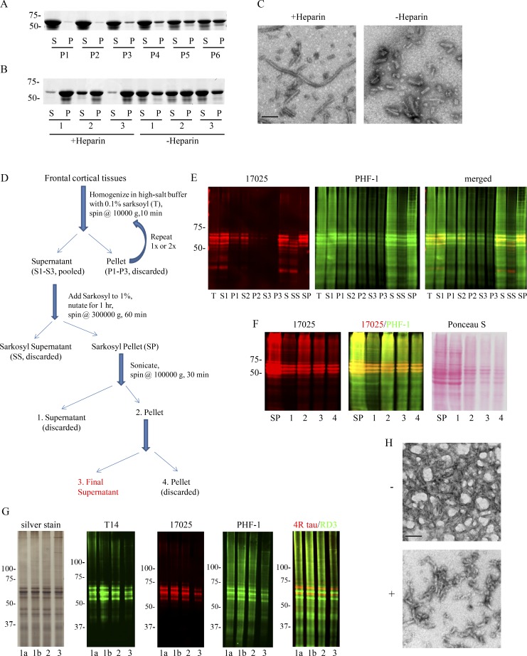
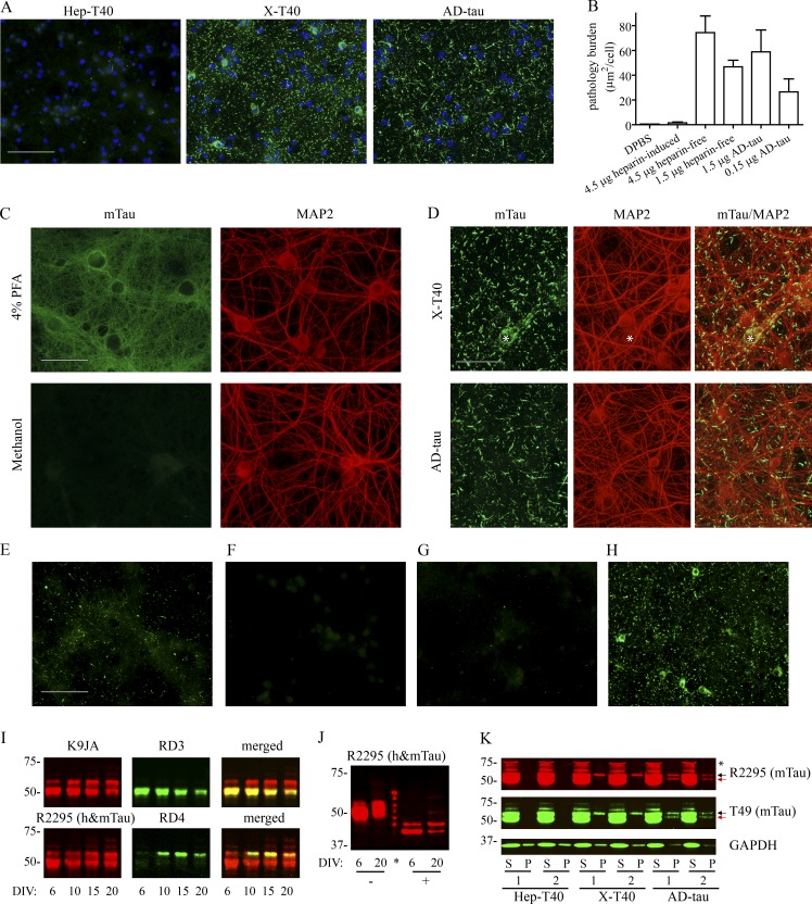
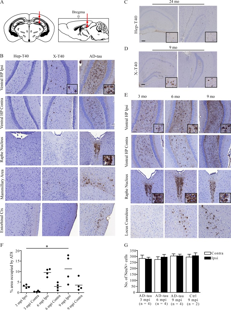
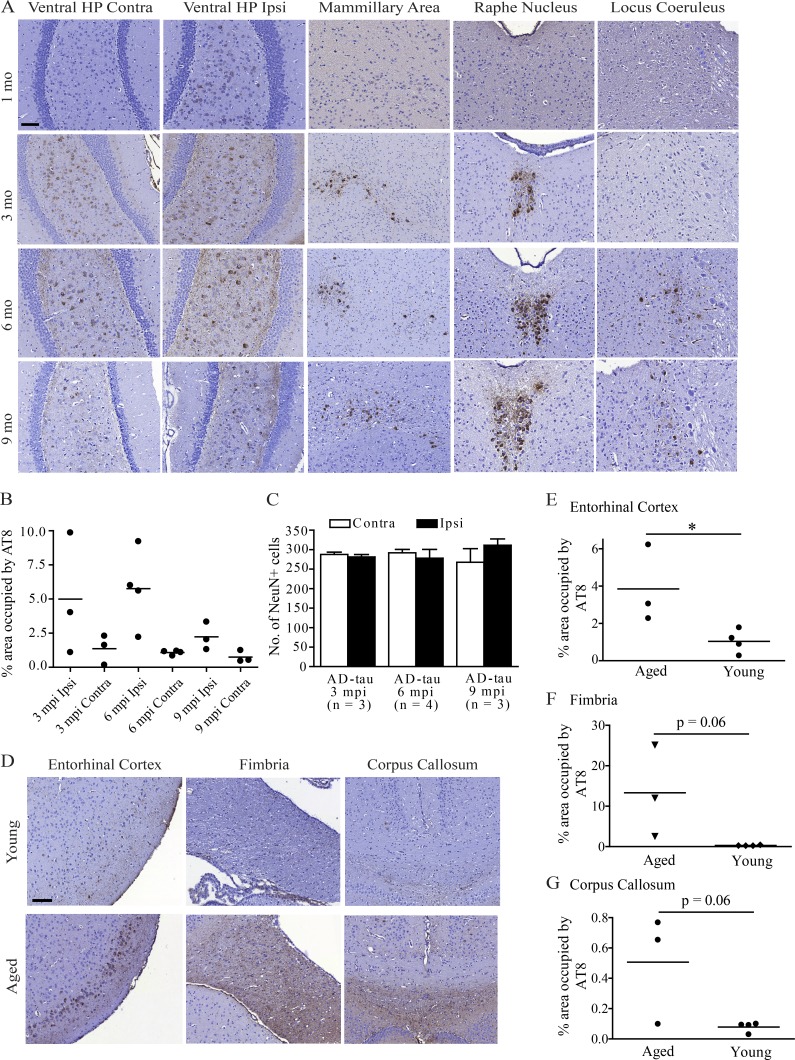
Filamentous tau aggregates are hallmark lesions in numerous neurodegenerative diseases, including Alzheimer’s disease (AD). Cell culture and animal studies showed that tau fibrils can undergo cell-to-cell transmission and seed aggregation of soluble tau, but this phenomenon was only robustly demonstrated in models overexpressing tau. In this study, we found that intracerebral inoculation of tau fibrils purified from AD brains (AD-tau), but not synthetic tau fibrils, resulted in the formation of abundant tau inclusions in anatomically connected brain regions in nontransgenic mice. Recombinant human tau seeded by AD-tau revealed unique conformational features that are distinct from synthetic tau fibrils, which could underlie the differential potency in seeding physiological levels of tau to aggregate. Therefore, our study establishes a mouse model of sporadic tauopathies and points to important differences between tau fibrils that are generated artificially and authentic ones that develop in AD brains.
Overview
The claims and statistics of this paper haven’t been checked yet.
Run the check — free, no account needed. It reads the open-access text and checks the sources it cites.
Integrity signals
Registry
Cites
Statistics
Figures
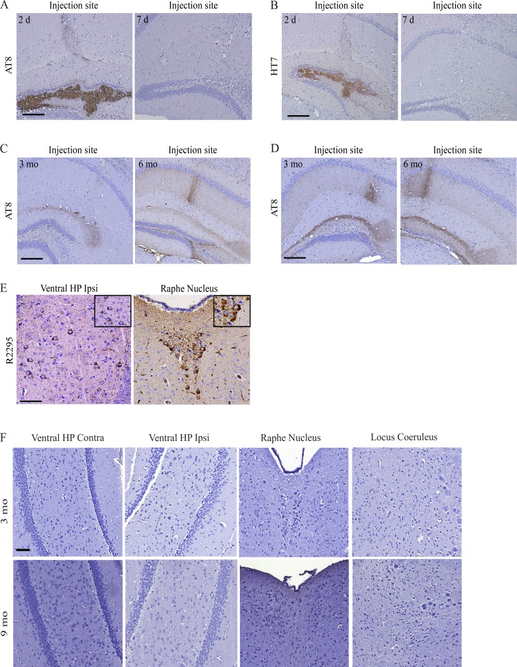
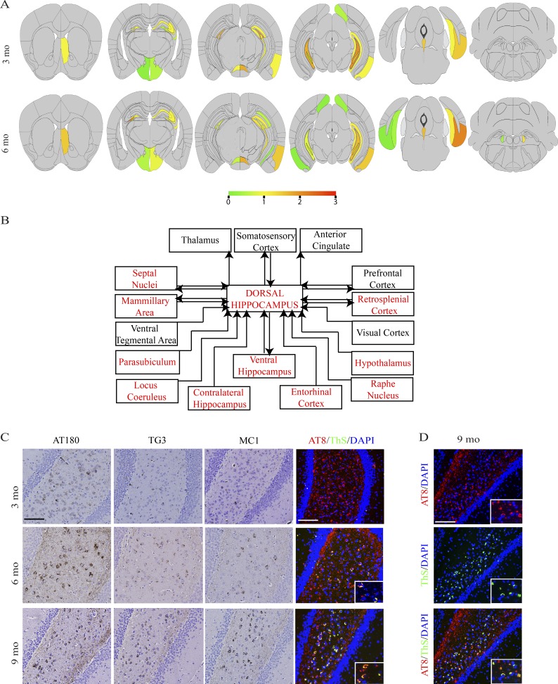
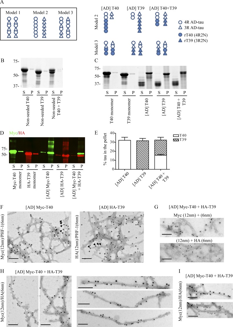
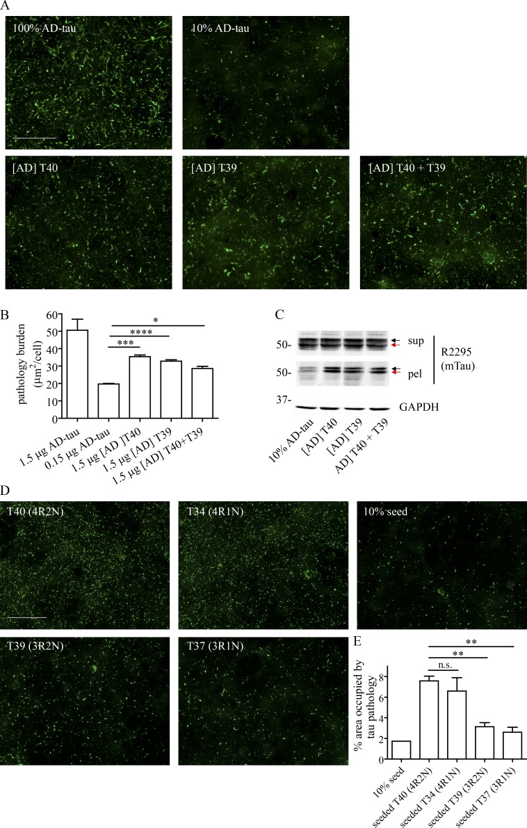
Verification
Claims the paper makes
The claims and statistics of this paper haven’t been checked yet.
Run the check — free, no account needed. It reads the open-access text and checks the sources it cites.
Claims other papers make about this one
Each is checked against this paper’s own text: does it say what the citing paper says it does?
The source backs part of the claim, not all of it — or backs it for a different population, condition or comparison.
In the citing paper
“Methanol fixation has previously been used to remove soluble proteins, including tau (20, 46).”
From Modeling late-onset Alzheimer’s disease neuropathology via direct neuronal reprogramming (2024) · cites this paper as [46]
Experimental — accuracy not yet measured
Self-check
Percentages, sums and reported differences re-computed from the paper’s own numbers.
The self-check needs the paper’s full text.
Citations
Cites
Amyloid oligomers exacerbate tau pathology in a mouse model of tauopathy
Selenica, M.-L. B., Brownlow, M., Jimenez, J. P., Lee, D. C., Pena, G., Dickey, C. A., Gordon, M. N., & Morgan, D. (2013) · Neurodegenerative Diseases · doi:10.1159/000337230 · Cited by 10,359
A protein factor essential for microtubule assembly
Weingarten, M. D., Lockwood, A. H., Hwo, S. Y., & Kirschner, M. W. (1975) · Proceedings of the National Academy of Sciences · doi:10.1073/pnas.72.5.1858 · Cited by 3,089
A mesoscale connectome of the mouse brain
Oh, S. W., Harris, J. A., Ng, L., Winslow, B., Cain, N., Mihalas, S., Wang, Q., Lau, C., Kuan, L., Henry, A. M., et al. (2014) · Nature · doi:10.1038/nature13186 · Cited by 2,612
Luk, K. C., Kehm, V., Carroll, J., Zhang, B., O’Brien, P., Trojanowski, J. Q., & Lee, V. M.-Y. (2012) · Science · doi:10.1126/science.1227157 · Cited by 2,563
Self-citationGoedert, M., Spillantini, M. G., Jakes, R., Rutherford, D., & Crowther, R. A. (1989) · Neuron · doi:10.1016/0896-6273(89)90210-9 · Cited by 2,492
Lee, V. M.-Y., Goedert, M., & Trojanowski, J. Q. (2001) · Annual Review of Neuroscience · doi:10.1146/annurev.neuro.24.1.1121 · Cited by 2,480
Self-citationTransmission and spreading of tauopathy in transgenic mouse brain
Clavaguera, F., Bolmont, T., Crowther, R. A., Abramowski, D., Frank, S., Probst, A., Fraser, G., Stalder, A. K., Beibel, M., Staufenbiel, M., et al. (2009) · Nature Cell Biology · doi:10.1038/ncb1901 · Cited by 1,803
Self-Propagating, Molecular-Level Polymorphism in Alzheimer's ß-Amyloid Fibrils
Petkova, A. T., Leapman, R. D., Guo, Z., Yau, W.-M., Mattson, M. P., & Tycko, R. (2005) · Science · doi:10.1126/science.1105850 · Cited by 1,687
Enhanced neurofibrillary degeneration in transgenic mice expressing mutant tau and APP
Lewis, J., Dickson, D. W., Lin, W.-L., Chisholm, L., Corral, A., Jones, G., Yen, S.-H., Sahara, N., Skipper, L., Yager, D., et al. (2001) · Science · doi:10.1126/science.1058189 · Cited by 1,611
Formation of Neurofibrillary Tangles in P301L Tau Transgenic Mice Induced by Aβ42 Fibrils
Götz, J., Chen, F., van Dorpe, J., & Nitsch, R. M. (2001) · Science · doi:10.1126/science.1062097 · Cited by 1,526
A68: a Major Subunit of Paired Helical Filaments and Derivatized Forms of Normal Tau
Lee, V. M.-Y., Balin, B. J., Otvos, L., & Trojanowski, J. Q. (1991) · Science · doi:10.1126/science.1899488 · Cited by 1,418 · cited 1×
“Compared with the traditional purification method using sucrose gradient (Lee et al., 1999), our new protocol resulted in much higher yields of PHF-tau (15–30 µg tau per gram of gray matter using our new protocol vs. 1–5 µg tau per gram of gray matter using the traditional protocol) with remarkable…”
MethodSelf-citationPropagation of Tau Misfolding from the Outside to the Inside of a Cell
Frost, B., Jacks, R. L., & Diamond, M. I. (2009) · Journal of Biological Chemistry · doi:10.1074/jbc.m808759200 · Cited by 1,252
Luk, K. C., Kehm, V. M., Zhang, B., O’Brien, P., Trojanowski, J. Q., & Lee, V. M. ‐. (2012) · The Journal of Experimental Medicine · doi:10.1084/jem.20112457 · Cited by 1,133
Self-citationα-Synuclein strains cause distinct synucleinopathies after local and systemic administration
Peelaerts, W., Bousset, L., Van der Perren, A., Moskalyuk, A., Pulizzi, R., Giugliano, M., Van den Haute, C., Melki, R., & Baekelandt, V. (2015) · Nature · doi:10.1038/nature14547 · Cited by 1,130
Goedert, M., Jakes, R., Spillantini, M. G., Hasegawa, M., Smith, M. J., & Crowther, R. A. (1996) · Nature · doi:10.1038/383550a0 · Cited by 1,036
Distinct Tau Prion Strains Propagate in Cells and Mice and Define Different Tauopathies
Sanders, D. W., Kaufman, S. K., DeVos, S. L., Sharma, A. M., Mirbaha, H., Li, A., Barker, S. J., Foley, A. C., Thorpe, J. R., Serpell, L. C., et al. (2014) · Neuron · doi:10.1016/j.neuron.2014.04.047 · Cited by 986
Cleveland, D. W., Hwo, S.-Y., & Kirschner, M. W. (1977) · Journal of Molecular Biology · doi:10.1016/0022-2836(77)90213-3 · Cited by 915 · cited 2×
“Tau is a microtubule (MT)-associated protein predominantly expressed in neuronal axons with a primary function of promoting assembly and stability of MTs (Weingarten et al., 1975; Cleveland et al., 1977b).”
BackgroundPhysical and chemical properties of purified tau factor and the role of tau in microtubule assembly
Cleveland, D. W., Hwo, S. Y., & Kirschner, M. W. (1977) · Journal of Molecular Biology · doi:10.1016/0022-2836(77)90214-5 · Cited by 881
Prion-like spreading of pathological α-synuclein in brain
Masuda-Suzukake, M., Nonaka, T., Hosokawa, M., Oikawa, T., Arai, T., Akiyama, H., Mann, D. M. A., & Hasegawa, M. (2013) · Brain · doi:10.1093/brain/awt037 · Cited by 867
Heparan sulfate proteoglycans mediate internalization and propagation of specific proteopathic seeds
Holmes, B. B., DeVos, S. L., Kfoury, N., Li, M., Jacks, R., Yanamandra, K., Ouidja, M. O., Brodsky, F. M., Marasa, J., Bagchi, D. P., et al. (2013) · Proceedings of the National Academy of Sciences · doi:10.1073/pnas.1301440110 · Cited by 859
Brain homogenates from human tauopathies induce tau inclusions in mouse brain
Clavaguera, F., Akatsu, H., Fraser, G. M., Crowther, R. A., Frank, S., Hench, J., Probst, A., Winkler, D. T., Reichwald, J., Staufenbiel, M., et al. (2013) · Proceedings of the National Academy of Sciences · doi:10.1073/pnas.1301175110 · Cited by 792
Post-translational modifications of tau protein: Implications for Alzheimer's disease
Martin, L., Latypova, X., & Terro, F. (2011) · Neurochemistry International · doi:10.1016/j.neuint.2010.12.023 · Cited by 683
Distinct α-Synuclein Strains Differentially Promote Tau Inclusions in Neurons
Guo, J. L., Covell, D. J., Daniels, J. P., Iba, M., Stieber, A., Zhang, B., Riddle, D. M., Kwong, L. K., Xu, Y., Trojanowski, J. Q., et al. (2013) · Cell · doi:10.1016/j.cell.2013.05.057 · Cited by 680
Self-citationIba, M., Guo, J. L., McBride, J. D., Zhang, B., Trojanowski, J. Q., & Lee, V. M.-Y. (2013) · The Journal of Neuroscience · doi:10.1523/jneurosci.2642-12.2013 · Cited by 664
Self-citationCell-to-cell transmission of pathogenic proteins in neurodegenerative diseases
Guo, J. L., & Lee, V. M. Y. (2014) · Nature Medicine · doi:10.1038/nm.3457 · Cited by 641
Self-citationSeeding of Normal Tau by Pathological Tau Conformers Drives Pathogenesis of Alzheimer-like Tangles
Guo, J. L., & Lee, V. M.-Y. (2011) · Journal of Biological Chemistry · doi:10.1074/jbc.m110.209296 · Cited by 636
Self-citationTrans-cellular Propagation of Tau Aggregation by Fibrillar Species
Kfoury, N., Holmes, B. B., Jiang, H., Holtzman, D. M., & Diamond, M. I. (2012) · Journal of Biological Chemistry · doi:10.1074/jbc.m112.346072 · Cited by 552
Kampers, T., Friedhoff, P., Biernat, J., Mandelkow, E.-M., & Mandelkow, E. (1996) · FEBS Letters · doi:10.1016/s0014-5793(96)01386-5 · Cited by 548
Wu, J. W., Herman, M., Liu, L., Simoes, S., Acker, C. M., Figueroa, H., Steinberg, J. I., Margittai, M., Kayed, R., Zurzolo, C., et al. (2013) · Journal of Biological Chemistry · doi:10.1074/jbc.m112.394528 · Cited by 537
Alzheimer brain-derived tau oligomers propagate pathology from endogenous tau
Lasagna-Reeves, C. A., Castillo-Carranza, D. L., Sengupta, U., Guerrero-Munoz, M. J., Kiritoshi, T., Neugebauer, V., Jackson, G. R., & Kayed, R. (2012) · Scientific Reports · doi:10.1038/srep00700 · Cited by 502
Paumier, K. L., Luk, K. C., Manfredsson, F. P., Kanaan, N. M., Lipton, J. W., Collier, T. J., Steece‐Collier, K., Kemp, C. J., Celano, S. L., Schulz, E., et al. (2015) · Neurobiology of Disease · doi:10.1016/j.nbd.2015.06.003 · Cited by 411
Self-citationPurified and synthetic Alzheimer’s amyloid beta (Aβ) prions
Stöhr, J., Watts, J. C., Mensinger, Z. L., Oehler, A., Grillo, S. K., DeArmond, S. J., Prusiner, S. B., & Giles, K. (2012) · Proceedings of the National Academy of Sciences · doi:10.1073/pnas.1206555109 · Cited by 375
Neurodegenerative Diseases: Expanding the Prion Concept
Walker, L. C., & Jucker, M. (2015) · Annual Review of Neuroscience · doi:10.1146/annurev-neuro-071714-033828 · Cited by 361
Seeded Aggregation and Toxicity of α-Synuclein and Tau
Nonaka, T., Watanabe, S., Iwatsubo, T., & Hasegawa, M. (2010) · Journal of Biological Chemistry · doi:10.1074/jbc.m110.148460 · Cited by 348
A nucleated assembly mechanism of Alzheimer paired helical filaments
Friedhoff, P., von Bergen�, M., Mandelkow�, E., Davies, P., & Mandelkow�, E. (1998) · Proceedings of the National Academy of Sciences · doi:10.1073/pnas.95.26.15712 · Cited by 340
Boluda, S., Iba, M., Zhang, B., Raible, K. M., Lee, V. M. ‐., & Trojanowski, J. Q. (2014) · Acta Neuropathologica · doi:10.1007/s00401-014-1373-0 · Cited by 268
Self-citationConformation Determines the Seeding Potencies of Native and Recombinant Tau Aggregates
Falcon, B., Cavallini, A., Angers, R. C., Glover, S. J., Murray, T. K., Barnham, L., Jackson, S., O'Neill, M. J., Isaacs, A. M., Hutton, M. L., et al. (2014) · Journal of Biological Chemistry · doi:10.1074/jbc.m114.589309 · Cited by 265
Anionic Micelles and Vesicles Induce Tau Fibrillization in Vitro
Chirita, C. N., Necula, M., & Kuret, J. (2003) · Journal of Biological Chemistry · doi:10.1074/jbc.m301663200 · Cited by 244
Structural Principles of Tau and the Paired Helical Filaments of Alzheimer's Disease
Mandelkow, E., Von Bergen, M., Biernat, J., & Mandelkow, E. (2007) · Brain Pathology · doi:10.1111/j.1750-3639.2007.00053.x · Cited by 243
Amyloid accelerates tau propagation and toxicity in a model of early Alzheimer’s disease
Pooler, A. M., Polydoro, M., Maury, E. A., Nicholls, S. B., Reddy, S. M., Wegmann, S., William, C., Saqran, L., Cagsal-Getkin, O., Pitstick, R., et al. (2015) · Acta Neuropathologica Communications · doi:10.1186/s40478-015-0199-x · Cited by 236
Iba, M., McBride, J. D., Guo, J. L., Zhang, B., Trojanowski, J. Q., & Lee, V. M.-Y. (2015) · Acta Neuropathologica · doi:10.1007/s00401-015-1458-4 · Cited by 223
Self-citationPeeraer, E., Bottelbergs, A., Van Kolen, K., Stancu, I.-C., Vasconcelos, B., Mahieu, M., Duytschaever, H., Ver Donck, L., Torremans, A., Sluydts, E., et al. (2015) · Neurobiology of Disease · doi:10.1016/j.nbd.2014.08.032 · Cited by 214
Self-citationTaniguchi-Watanabe, S., Arai, T., Kametani, F., Nonaka, T., Masuda-Suzukake, M., Tarutani, A., Murayama, S., Saito, Y., Arima, K., Yoshida, M., et al. (2016) · Acta Neuropathologica · doi:10.1007/s00401-015-1503-3 · Cited by 200
Characterization of Two VQIXXK Motifs for Tau Fibrillization in Vitro
Li, W., & Lee, V. M. ‐. (2006) · Biochemistry · doi:10.1021/bi061422+ · Cited by 197
Self-citationWeissmann, C., Enari, M., Klöhn, P., Rossi, D., & Flechsig, E. (2002) · Proceedings of the National Academy of Sciences · doi:10.1073/pnas.172403799 · Cited by 161
Stancu, I.-C., Vasconcelos, B., Ris, L., Wang, P., Villers, A., Peeraer, E., Buist, A., Terwel, D., Baatsen, P., Oyelami, T., et al. (2015) · Acta Neuropathologica · doi:10.1007/s00401-015-1413-4 · Cited by 143
von Bergen�, M., Barghorn, S., Müller, S. A., Pickhardt, M., Biernat, J., Mandelkow, E., Davies, P., Aebi, U., & Mandelkow�, E. (2006) · Biochemistry · doi:10.1021/bi052530j · Cited by 141
Differential Incorporation of Tau Isoforms in Alzheimer's Disease
Espinoza, M., de Silva, R., Dickson, D. W., & Davies, P. (2008) · Journal of Alzheimer's Disease · doi:10.3233/jad-2008-14101 · Cited by 119
Guo, J., & Lee, V. M. ‐. (2013) · FEBS Letters · doi:10.1016/j.febslet.2013.01.051 · Cited by 117
Self-citationThree Repeat Isoforms of Tau Inhibit Assembly of Four Repeat Tau Filaments
Adams, S. J., Deture, M. A., McBride, M., Dickson, D. W., & Petrucelli, L. (2010) · PLoS ONE · doi:10.1371/journal.pone.0010810 · Cited by 110
Variations in filament conformation dictate seeding barrier between three- and four-repeat Tau
Dinkel, P. D., Siddiqua, A., Huynh, H., Shah, M., & Margittai, M. (2011) · Biochemistry · doi:10.1021/bi2004685 · Cited by 104
Quantitative Characterization of Heparin Binding to Tau Protein
Zhu, H., Fernández, C., Fan, J.-B., Shewmaker, F. P., Chen, J., Minton, A. P., & Liang, Y. (2009) · Journal of Biological Chemistry · doi:10.1074/jbc.m109.035691 · Cited by 102
Morozova, O. A., March, Z. M., Robinson, A. S., & Colby, D. W. (2013) · Biochemistry · doi:10.1021/bi400866w · Cited by 92
De novo generation of prion strains
Colby, D. W., & Prusiner, S. B. (2011) · Nature Reviews Microbiology · doi:10.1038/nrmicro2650 · Cited by 88
Lee, E. B., Skovronsky, D., Abtahian, F., Doms, R. W., & Lee, V. M. ‐. (2003) · Journal of Biological Chemistry · doi:10.1074/jbc.m210105200 · Cited by 78
Self-citationConformational basis for asymmetric seeding barrier in filaments of three- and four-repeat tau
Siddiqua, A., Luo, Y., Meyer, V., Swanson, M. A., Yu, X., Wei, G., Zheng, J., Eaton, G. R., Ma, B., Nussinov, R., et al. (2012) · Journal of the American Chemical Society · doi:10.1021/ja303498q · Cited by 75
Weissmann, C., Enari, M., Klöhn, P., Rossi, D., & Flechsig, E. (2002) · The Journal of Infectious Diseases · doi:10.1086/344575 · Cited by 75
Furukawa, Y., Kaneko, K., & Nukina, N. (2011) · Journal of Biological Chemistry · doi:10.1074/jbc.m111.248963 · Cited by 73
Three- and Four-repeat Tau Coassemble into Heterogeneous Filaments
Siddiqua, A., & Margittai, M. (2010) · Journal of Biological Chemistry · doi:10.1074/jbc.m110.185728 · Cited by 67
[6] Purification of paired helical filament tau and normal tau from human brain tissue
Lee, V. M. ‐., Wang, J., & Trojanowski, J. Q. (1999) · Methods in enzymology on CD-ROM/Methods in enzymology · doi:10.1016/s0076-6879(99)09008-4 · Cited by 52
Self-citation
Cited by
Sorted by citations
Alzheimer Disease: An Update on Pathobiology and Treatment Strategies
Long, J. M., & Holtzman, D. M. (2019) · Cell · doi:10.1016/j.cell.2019.09.001 · Cited by 3,204
Cryo-EM structures of tau filaments from Alzheimer’s disease
Fitzpatrick, A. W. P., Falcon, B., He, S., Murzin, A. G., Murshudov, G., Garringer, H. J., Crowther, R. A., Ghetti, B., Goedert, M., & Scheres, S. H. W. (2017) · Nature · doi:10.1038/nature23002 · Cited by 2,061
The proteostasis network and its decline in ageing
Hipp, M. S., Kasturi, P., & Hartl, F. U. (2019) · Nature Reviews Molecular Cell Biology · doi:10.1038/s41580-019-0101-y · Cited by 1,642
Kim, S., Kwon, S.-H., Kam, T.-I., Panicker, N., Karuppagounder, S. S., Lee, S., Lee, J. H., Kim, W. R., Kook, M., Foss, C. A., et al. (2019) · Neuron · doi:10.1016/j.neuron.2019.05.035 · Cited by 1,397
Protein misfolding, aggregation, and conformational strains in neurodegenerative diseases
Soto, C., & Pritzkow, S. (2018) · Nature Neuroscience · doi:10.1038/s41593-018-0235-9 · Cited by 1,180
Metadata
- Authors
- Guo, J. L., Narasimhan, S., Changolkar, L., He, Z., Stieber, A., Zhang, B., Gathagan, R. J., Iba, M., McBride, J. D., Trojanowski, J. Q., & Lee, V. M. Y.
- Affiliations
- University of Pennsylvania (US)
- Corresponding author
- Lee, Virginia M.Y.
- ORCID
- Venue
- Journal of Experimental Medicine
- Publisher
- Rockefeller University Press
- Published
- 2016-10-17
- Volume
- 213
- Issue
- 12
- Pages
- 2635-2654
- ISSN
- 0022-1007 (ISSN-L)
- 1540-9538
- PMC
- PMC5110027
- OpenAlex
- W2534587007
- Cited by
- 440
- References
- 60
- Citations by year
- 2026: 30 · 2025: 35 · 2024: 53 · 2023: 50 · 2022: 47 · 2021: 62 · 2020: 57 · 2019: 45 · 2018: 40 · 2017: 20
- FWCI
- 28.18
- Type
- journal article
- Language
- en
- Open access
- bronze
- doi.org
- Locations
- The Journal of Experimental Medicine (publishedVersion)
- Europe PMC (PubMed Central) (publishedVersion, cc-by-nc-sa)
- PubMed Central (publishedVersion, cc-by-nc-sa)
- PubMed (publishedVersion)
- Funders
- National Institutes of Health (AG10124, AG17586)
- CurePSP
- BrightFocus Foundation (A2014005F)
- Topics
- Life Sciences › Neuroscience › Cellular and Molecular Neuroscience › Alzheimer's disease research and treatments
- Health Sciences › Medicine › Psychiatry and Mental health › Dementia and Cognitive Impairment Research
- Life Sciences › Biochemistry, Genetics and Molecular Biology › Molecular Biology › Prion Diseases and Protein Misfolding
- Keywords
- tau pathology, Alzheimer's disease, tau fibrils, cell-to-cell transmission, intracerebral injection, tau inclusions
- MeSH terms
- Aging, Alzheimer Disease, Animals, Brain, Heparin, Mice, Inbred C57BL, Mice, Transgenic, Neurons, Phosphorylation, Protein Conformation, Tissue Extracts, Neurofibrillary Tangles, tau Proteins, Protein Isoforms, Tauopathies, Protein Aggregates
About these checks
Registry, retraction, Cites and statistics results are computed, not judged, with one exception: a printed reference that no index lists is read once by a model before it counts as a citation error. Claims are extracted by a model and kept only when their quote appears verbatim in the text; each cited source is then checked, with a model’s help, for whether it says what the claim relies on. Those verdicts can be wrong — read the claim’s quote and the cited source before relying on one.
Statistics and arithmetic are recomputed, not judged.
Confidence bands are shown only for fields calibrated on our annotated test set. None is calibrated yet, so no band is shown.
What counts as a claim ›How we measure confidence ›How we check this ›
Are you an author, or do you see a problem with this page? Request a review
